Luciérnagas (Coleoptera: Lampyridae) del sur de México y descripción de 39 especies nuevas
Fireflies (Coleoptera: Lampyridae) of southern Mexico and description of 39 new species
Santiago Zaragoza-Caballero a, Sara López-Pérez b, *, Mireya González-Ramírez e, Geovanni M. Rodríguez-Mirón b, Viridiana Vega-Badillo c, Ishwari G. Gutiérrez-Carranza d, Daniel Edwin Domínguez-León d, e, Paulina Cifuentes-Ruiz f, Miriam Aquino-Romero g, Anel Teresa Mercedes Núñez-Monroy a, Oscar Pérez-Flores h
a Universidad Nacional Autónoma de México, Departamento de Zoología, Instituto de Biología, Apartado postal 70-153, 04510 Ciudad de México, México
b Universidad Nacional Autónoma de México, Facultad de Estudios Superiores Zaragoza, Museo de Zoología, Colección Coleopterológica, Av. Batalla del 5 de mayo s/n, Col. Ejército de Oriente, 09230 Ciudad de México, México
c Instituto de Ecología A.C., Colección entomológica IEXA “Dr. Miguel Ángel Morón Ríos”, Carretera Antigua a Coatepec 351, El Haya, 91073 Xalapa, Veracruz, México
d IUCN Species Survival Commission-Firefly Specialist Group, Rue Mauverney 28 1196, Gland, Suiza
e Universidad Nacional Autónoma de México, Facultad de Estudios Superiores Zaragoza, Av. Batalla del 5 de mayo s/n, Col. Ejército de Oriente, 09230 Ciudad de México, México.
f Universidad Nacional Autónoma de México, Escuela Nacional Preparatoria 8 Miguel E. Schulz, Colegio de Biología, Av. Lomas de Plateros s/n, Merced Gómez, 01600 Ciudad de México, México
g University of Georgia, Entomology Department, 136 Cedar St., Bldg. C, Athens, GA 30602, EUA
h Universidad Nacional Autónoma de México, Escuela Nacional de Estudios Superiores Unidad Morelia, Laboratorio Nacional de Análisis y Síntesis Ecológica, Antigua Carretera a Pátzcuaro Núm. 8701, Exhacienda de San José de la Huerta, 58190 Morelia, Michoacán, México
*Autor para correspondencia: slopez.p@hotmail.com (S. López-Pérez)
Recibido: 01 febrero 2026; aceptado: 23 abril 2026
Resumen
Se presenta información sobre luciérnagas registradas en Chiapas, Guerrero y Oaxaca, región que se denominó sur. Además, se provee una descripción breve de aquellas presentes en esta región y que no se reportan en trabajos previos. Se describen 39 especies nuevas de Photinus Laporte. Cada especie nueva está ilustrada dorsal y ventralmente, así como los genitales masculinos. Se presentan registros nuevos para: Aspisoma aelianum (Gorham), Cratomorphus picipennis Gorham, Lucidota discolor Gorham, Photinus ahuizotli Zaragoza-Caballero y Domínguez-León, P. anagabrielae Zaragoza-Caballero y Gutiérrez-Carranza, P. hoffmanni Zaragoza-Caballero y Gutiérrez-Carranza, P. juanrulfoi Zaragoza-Caballero y Domínguez-León, P. kaedii Zaragoza-Caballero y González-Ramírez, P. paracongruus Zaragoza-Caballero, P. silveirai Zaragoza-Caballero y González-Ramírez, P. tenuilineatus Zaragoza-Caballero, P. toledoi Zaragoza-Caballero, P. perezi Zaragoza-Caballero, Photuris fulvipes (Blanchard in Brullé) y Pyropyga extensa Green. Después de la revisión del ejemplar de Photinus consimilis Green depositado en la Colección Nacional de Insectos del Instituto de Biología, UNAM, se concluye que corresponde a una hembra de P. tenuilineatus Zaragoza-Caballero. Con la descripción de las especies nuevas de esta región, Chiapas tiene un registro de 65 especies, Guerrero 41 y Oaxaca 79, la diversidad de luciérnagas en México asciende a 346.
Palabras clave: Diversidad; Lampyridae; Especies en riesgo; Conservación; Photinus consimilis
Abstract
Fireflies from Chiapas, Guerrero, and Oaxaca are documented, an area which is designated as the southern region. Information on species in the region and brief descriptions of those not previously treated are provided. Thirty-nine new species belonging to the genus Photinus Laporte are described. Each new species is illustrated in dorsal and ventral views, as well as the male genitalia. New state records for Aspisoma aelianum (Gorham), Cratomorphus picipennis Gorham, Lucidota discolor Gorham, Photinus ahuizotli Zaragoza-Caballero and Domínguez-León, P. anagabrielae Zaragoza-Caballero and Gutiérrez-Carranza, P. hoffmanni Zaragoza-Caballero and Gutiérrez-Carranza, P. juanrulfoi Zaragoza-Caballero and Domínguez-León, P. kaedii Zaragoza-Caballero and González-Ramírez, P. paracongruus Zaragoza-Caballero, P. silveirai Zaragoza-Caballero and González-Ramírez, P. tenuilineatus Zaragoza-Caballero, P. toledoi Zaragoza-Caballero, P. perezi Zaragoza-Caballero, Photuris fulvipes (Blanchard in Brullé), and Pyropyga extensa Green are provided. After examining the specimen of Photinus consimilis Green deposited in the National Insect Collection of the Institute of Biology, UNAM, it was concluded that it corresponds to a female of P. tenuilineatus Zaragoza-Caballero. With the description of the new species from this region, the state of Chiapas has a record of 65 species, Guerrero 41, and Oaxaca 79, the diversity of fireflies in Mexico rises to 346.
Keywords: Diversity; Lampyridae; Species at risk; Conservation; Photinus consimilis
Introducción
La familia Lampyridae Latreille, 1817 se caracteriza por presentar cuerpo poco esclerosado, paralelo u ovalado, generalmente aplanado; longitud variada. Coloraciones variadas que van desde el negro, castaño, rojizo y amarillento. La cabeza está completa o parcialmente cubierta por el pronoto, hipognata. Los ojos usualmente no emarginados (emarginados en Luciolinae). Antenas variables, pueden ser filiformes, serradas, flabeladas o biflabeladas, se conforma de 7-62 antenómeros (Nunes et al., 2020). Pronoto de forma variada, márgenes generalmente redondos, base aproximadamente de la misma longitud que la base de los élitros. Élitros lisos, algunos con pubescencia, generalmente cubriendo todo el abdomen con excepción de las hembras neoténicas. Alas membranosas presentes, reducidas o en algunas especies ausentes. Fórmula tarsal 5-5-5, uñas simples, dentadas o bífidas. Órgano fotógeno presente en uno o ambos sexos generalmente en los ventritos V, VI y VII, ausente en especies diurnas. Edeago trilobado, de formas variadas, con o sin excresencias dorsolaterales (Branham, 2010).
Las luciérnagas presentan una distribución amplia en el mundo, ocupando diversos hábitats, desde bosques húmedos y secos hasta zonas áridas (Lewis et al., 2020; Zaragoza-Caballero y Ramírez-García, 2009). Sus larvas pueden ser acuáticas, semiacuáticas o terrestres y presentan un órgano fotógeno (Branham, 2010). Su alimentación se basa en caracoles, lombrices y otros invertebrados pequeños (Lewis et al., 2020). En México, hasta 2025 se habían descrito 307 especies, las cuales se distribuyen por todo el país; sin embargo, las condiciones ambientales son determinantes en la distribución de la riqueza estatal. En Zacatecas y Tlaxcala solo se han registrado 2 y 3 especies, respectivamente, en comparación con los estados de Veracruz, Oaxaca y Chiapas que concentran en conjunto más de 50% de las especies conocidas en México (Zaragoza-Caballero, López-Pérez et al., 2020; Zaragoza-Caballero et al., 2023, 2024).
Actualmente, las luciérnagas se encuentran amenazadas por diversos factores como la contaminación lumínica, el cambio climático, la pérdida de hábitat, el cambio de uso de suelo y el uso de pesticidas que afectan a estos insectos en alguna etapa de su desarrollo (Lewis et al., 2020). La pérdida y degradación del hábitat pueden ser perjudiciales principalmente para aquellas que cuentan con hembras no voladoras, que son más vulnerables al pisoteo o la destrucción del hábitat debido a su limitada capacidad de movimiento (Fallon et al., 2021).
Recientemente, Fallon et al. (2021) evaluaron los riesgos de extinción de las luciérnagas utilizando los criterios de la Lista Roja de la Unión Internacional para la Conservación de la Naturaleza (UICN por sus siglas en inglés), a partir de la cual catalogaron a 18 especies de 132 distribuidas en EU y Canadá como vulnerables, en peligro y en peligro crítico. Algunas de las especies evaluadas también se distribuyen en México; sin embargo, para las especies que habitan en el país es evidente la falta de información sobre su distribución, hábitat e historia natural. En la mayoría de los casos solo se cuenta con la información proveniente de la descripción original de las especies (Zaragoza-Caballero, López-Pérez et al., 2020; Zaragoza-Caballero et al., 2023, 2024).
En México hasta finales del siglo XX se conocían 91 especies de 17 géneros, al año 2025 se contabilizaron 307 especies, lo cual denota el déficit linneano de las luciérnagas en el territorio mexicano. El conocimiento taxonómico de las luciérnagas en México ha tenido un gran avance en los últimos años. A partir del denominado “Proyecto luciérnagas de México” (Zaragoza-Caballero, Cifuentes-Ruiz et al., 2020) se han descrito poco más de 100 especies nuevas de 23 estados del territorio mexicano. Además de las descripciones, se han presentado claves taxonómicas para la separación de subfamilias, géneros y especies del género Photinus Laporte. Asimismo, la mayoría de las especies previamente descritas han sido ilustradas mediante fotografías, no solo en sus vistas dorsales y ventrales, sino también diferentes vistas del aparato reproductor masculino, indispensable en la separación de especies.
Los insectos están pasando por una crisis global (Kehoe et al., 2021; Montgomery et al., 2020) y para realizar planes de conservación, es fundamental conocer qué especies se encuentran registradas en cierta zona; además de generar información sobre su historia natural. En el presente trabajo se contribuye al conocimiento taxonómico de los estados de Chiapas, Guerrero y Oaxaca y se describen 39 especies nuevas para la ciencia. Se incorporan fotografías de las especies nuevas en su hábito dorsal y ventral, así como del aparato reproductor masculino. Además, se presenta una breve descripción de las especies que se han registrado con anterioridad en estos estados y que no se han incluido en Zaragoza-Caballero, López-Pérez et al. (2020) y Zaragoza-Caballero et al. (2023, 2024). Así mismo se dan a conocer nuevos registros estatales para 15 especies.
Materiales y métodos
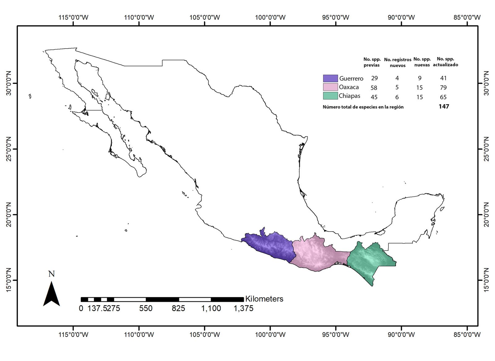
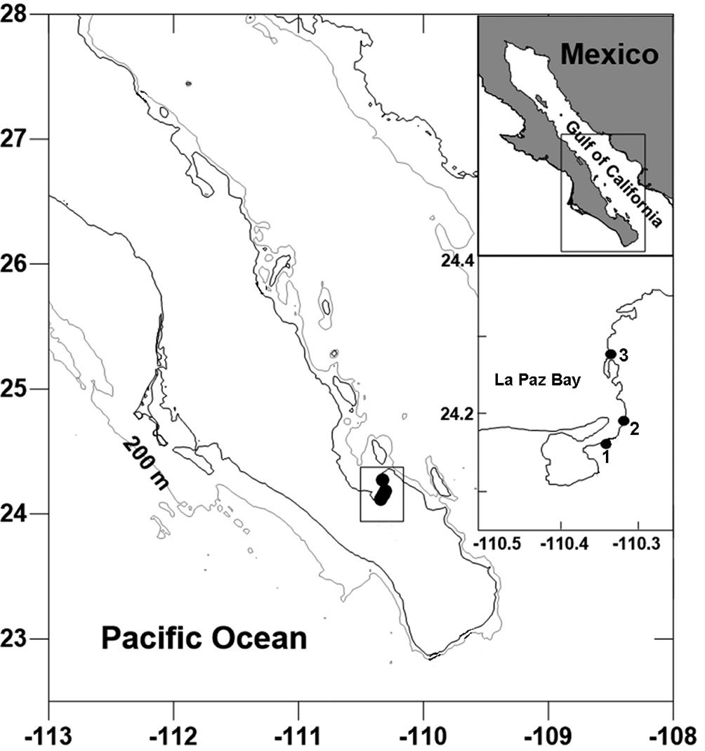
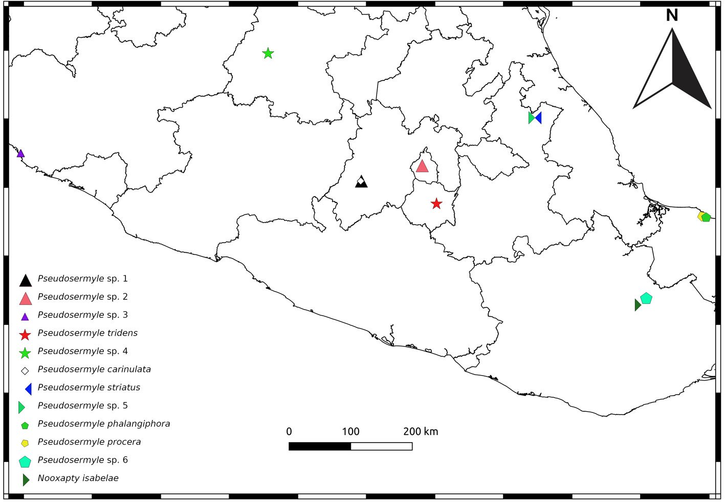
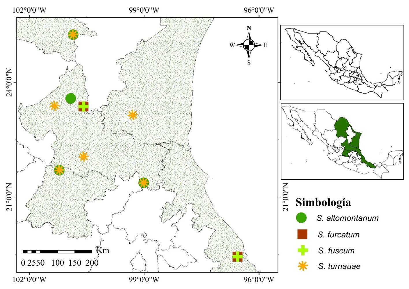
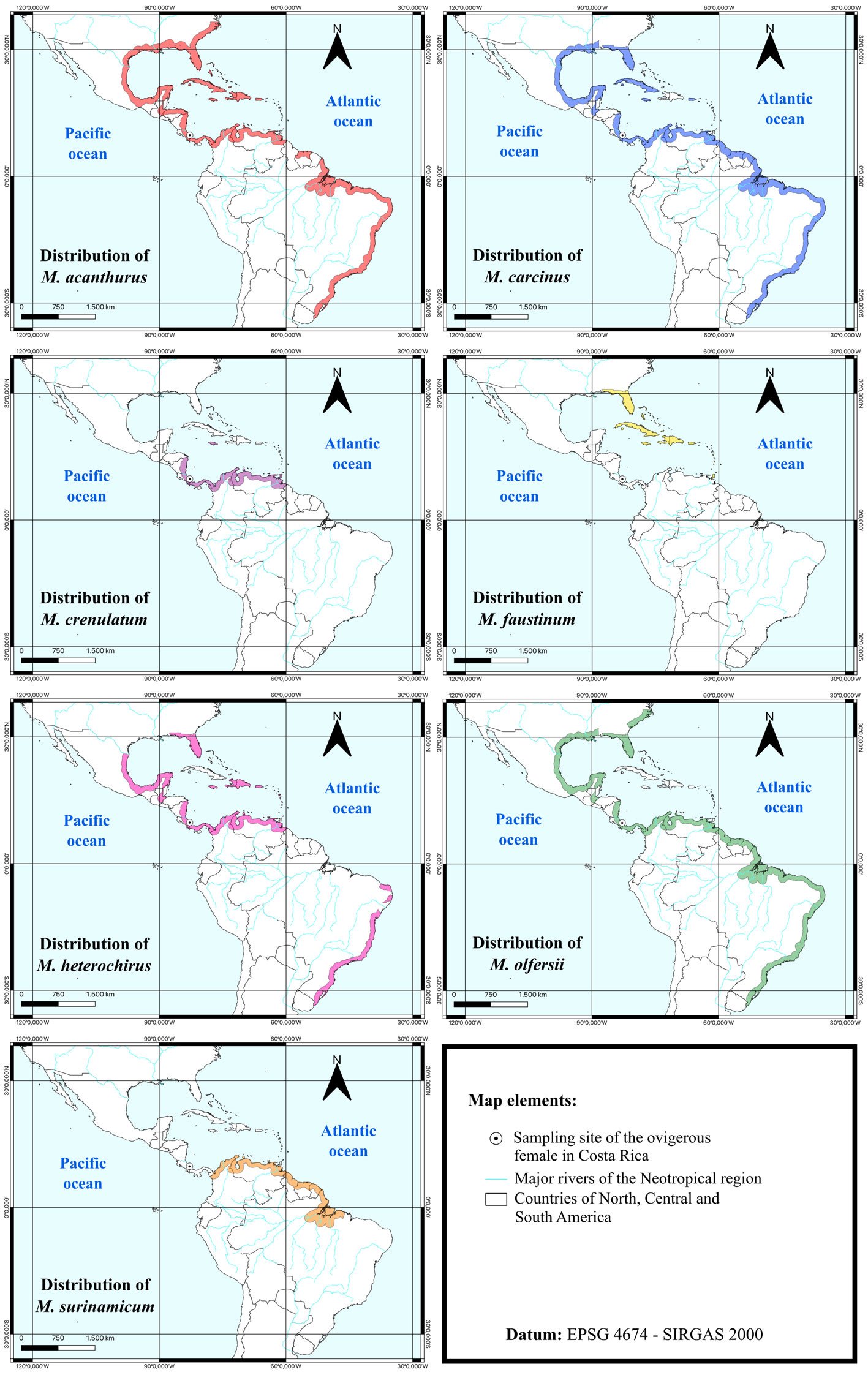
El presente estudio se limita a las especies distribuidas en Chiapas, Guerrero y Oaxaca, que en conjunto se denominó región sur (fig. 1) (Zaragoza-Caballero, Cifuentes-Ruiz et al., 2020). Todos los ejemplares utilizados en el presente estudio se encuentran depositados en la Colección Nacional de Insectos (CNIN) del Instituto de Biología de la Universidad Nacional Autónoma de México. Los ejemplares tipo quedan depositados en la misma colección.
Para las especies previamente registradas en la región y que no se incluyen en Zaragoza-Caballero, López-Pérez et al. (2020, 2023), Zaragoza-Caballero et al. (2024) se ilustra el hábito en vista dorsal y ventral. Las especies previamente registradas por Zaragoza-Caballero (2015, 2017), Zaragoza-Caballero y Navarrete-Heredia (2014), Zaragoza-Caballero y Gutiérrez-Carranza (2018), Ferreira (2024) y Gutiérrez-Carranza et al. (2025) no se ilustran en este trabajo ya que estas publicaciones cuentan con fotografías de buena calidad. Lucidota bicellonycha McDermott, 1958, Photinus moestus (Perty, 1830), P. nigridorsis Gorham, 1881 P. ovatus Gorham, 1881, P. salvini Gorham, 1881 y Cratomorphus elevatus Olivier, 1896 no se ilustran en fotografías debido a la falta de material; las diagnosis y datos de estas especies se recuperaron de las descripciones originales. Para el resto de las especies que se registran en la región, pero no se han abordado en trabajos recientes y de acceso libre, se presenta una descripción breve realizada a partir de ejemplares depositados en la CNIN y descripciones originales. Para las especies nuevas se ilustra su hábito dorsal y ventral, así como los genitales masculinos. Para la revisión de los ejemplares, la separación del edeago en machos y la toma de medidas se utilizó un microscopio estereoscopio Carl Zeiss™ Discovery V8; las medidas se expresan en mm.
La información del resumen taxonómico se estandarizó mostrando primero el estado donde se registra la especie, la localidad y posteriormente la fecha; la información restante se presenta de acuerdo con la etiqueta. Para separar información de distintas etiquetas se utilizó “|”. Las fotografías digitales se tomaron en el Laboratorio de Microscopía y Fotografía de la Biodiversidad del Instituto de Biología de la Universidad Nacional Autónoma de México (UNAM), con un microscopio estereoscópico Carl Zeiss™ Axio Zoom V16, equipado con una cámara Zeiss Axiocam MRc5 y con el software ZEN™ = Zeiss Efficient Navigation pro-2012 y con una cámara Canon EOS M6 Mark II, empleando 2 lentes Ultra Macro: Laowa 65 mm f/2.8 2X y Laowa 25 mm f/2.8 2.5-5X; y un riel automatizado StackShot Macro Reil, las imágenes capturadas fueron apiladas con el programa Zerene Stacker v.1.04. Todas las fotografías fueron editadas en Adobe Photoshop v. 22.5.0. A todos los ejemplares de las especies nuevas se les extrajo el aparato reproductor masculino, siguiendo el protocolo de Gutiérrez-Carranza (2023).
La distribución se toma a partir de las descripciones originales y otras fuentes como Cicero et al. (2021), Gorham (1881), Pérez-Hernández et al. (2022), Zaragoza-Caballero (1988), Zaragoza-Caballero, López-Pérez et al. (2020, 2023), Zaragoza-Caballero et al. (2024), además de ejemplares depositados en la CNIN. El arreglo supraespecífico sigue a Bouchard et al. (2011) y Martín et al. (2019), las especies aparecen en orden alfabético. Se adiciona la distribución conocida y nuevos registros de cada especie.
Descripciones
Previo a este trabajo, en la región sur de México se registraban 94 especies (Ferreira, 2024; Gutiérrez-Carranza et al., 2023, 2025; Lima et al., 2021; Pérez-Hernández et al., 2022; Zaragoza-Caballero et al., 2023). Photinus consimilis Green, 1956 fue reportado para Oaxaca por Pérez-Hernández et al. (2022) a partir de un ejemplar colectado en Oaxaca y depositado en la CNIN, el cual al ser revisado en este trabajo se determinó como hembra de P. tenuilineatus Zaragoza-Caballero, 1996 conocida anteriormente solo para Veracruz. Photinus consimilis se distribuye solo en Canadá y EUA. Con esta contribución se adicionan 15 registros estatales nuevos y 39 especies nuevas, por lo que la riqueza de luciérnagas de la región aumentó a 147 (fig. 1) y el total de especies mexicanas a 346.
Subfamilia Psilocladinae McDermott, 1964
Psilocladus Blanchard, 1845
Psilocladus cancinoi Gutiérrez-Carranza, Zaragoza-Caballero y Keller, 2025
Diagnosis: en Gutiérrez-Carranza et al. (2025)
Distribución: Chiapas.
Subfamilia Chespiritoinae Ferreira, Keller y Branham, 2020
Chespirito Ferreira, Keller y Branham, 2020
Diagnosis: en Zaragoza-Caballero et al. (2023).
Chespirito zaragozai Ferreira, Keller y Branham, 2020
Diagnosis: en Zaragoza-Caballero et al. (2023).
Distribución: Chiapas.
Xacoxoxotlani Ferreira, 2024
Xacoxoxotlani chiapensis Ferreira, 2024
Diagnosis: en Ferreira (2024).
Distribución: Chiapas.
Subfamilia Lampyrinae Rafinesque, 1815
Tribu Cratomorphini Olivier, 1911
Aspisoma Laporte, 1833
Aspisoma aegrota (Gorham, 1880)
Diagnosis: en Zaragoza-Caballero, López-Pérez et al. (2020).
Distribución: Chiapas, Hidalgo, Jalisco, Michoacán, Morelos, Oaxaca, Puebla, San Luis Potosí, Tamaulipas y Veracruz.
Aspisoma aelianum (Gorham, 1884)
Diagnosis: en Zaragoza-Caballero et al. (2023).
Distribución: Jalisco, Nayarit y San Luis Potosí. Nuevo registro: Oaxaca (CNIN). Material examinado: México, Oaxaca, San Pedro Mixtepec, 15 km S. Río Grande, 10-1989, F. A. Noguera, A. Rodríguez.
Aspisoma depictum (Gorham, 1880)
Diagnosis: en Zaragoza-Caballero et al. (2023).
Distribución: Campeche, Chiapas, Guerrero, Michoacán, Oaxaca, Quintana Roo, Tamaulipas y Veracruz.
Aspisoma diaphana (Gorham, 1880)
Diagnosis: en Zaragoza-Caballero, López-Pérez et al. (2020).
Distribución: Oaxaca, Puebla y Veracruz.
Aspisoma pulchellum (Gorham, 1880)
Diagnosis: en Zaragoza-Caballero, López-Pérez et al. (2020).
Distribución: Chiapas, Estado de México, Tabasco y Veracruz.
Aspisomoides Zaragoza-Caballero, 1995
Diagnosis: en Zaragoza-Caballero, López-Pérez et al. (2020) y Zaragoza-Caballero et al. (2023).
Aspisomoides bilineatum (Gorham, 1880)
Diagnosis en Zaragoza-Caballero, López-Pérez et al. (2020).
Distribución: Guerrero, Jalisco, Michoacán, Morelos, Nayarit, Oaxaca, San Luis Potosí, Sinaloa, Sonora, Tabasco, Tamaulipas y Veracruz
Aspisomoides costatum (Gorham, 1880)
Diagnosis: en Zaragoza-Caballero et al. (2023).
Distribución: Chiapas, Durango, Guerrero, Oaxaca, Tabasco y Veracruz.
Cratomorphus Motschulsky, 1853
Diagnosis: en Zaragoza-Caballero (2023).
Cratomorphus anitae Zaragoza-Caballero, 1996
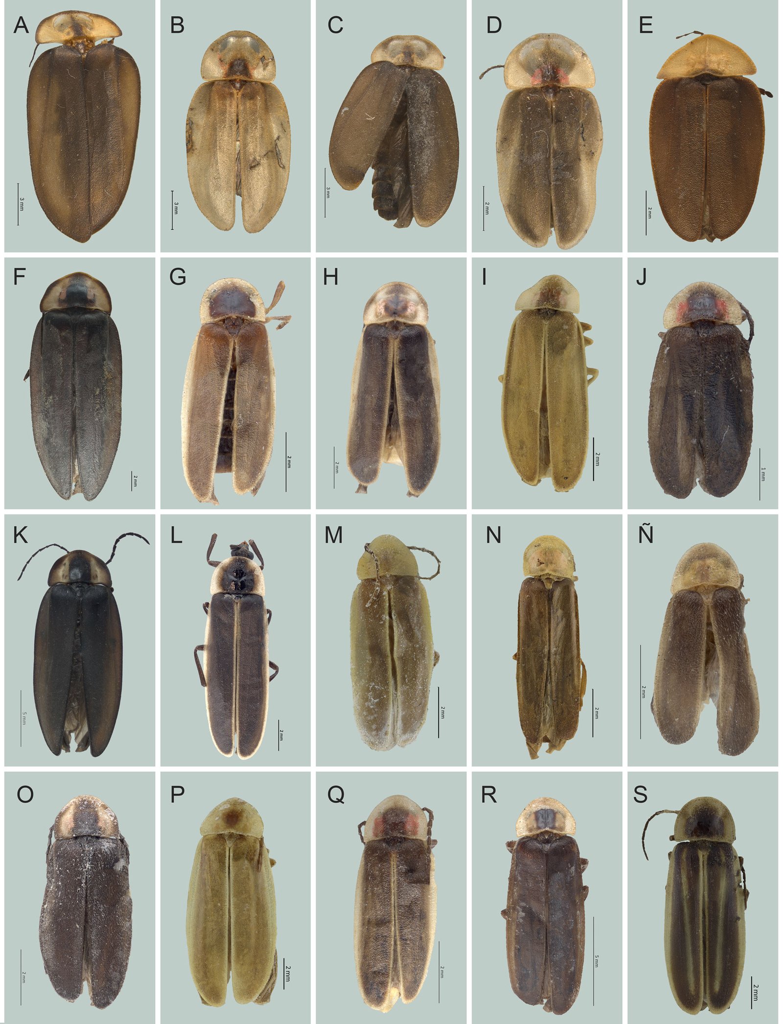
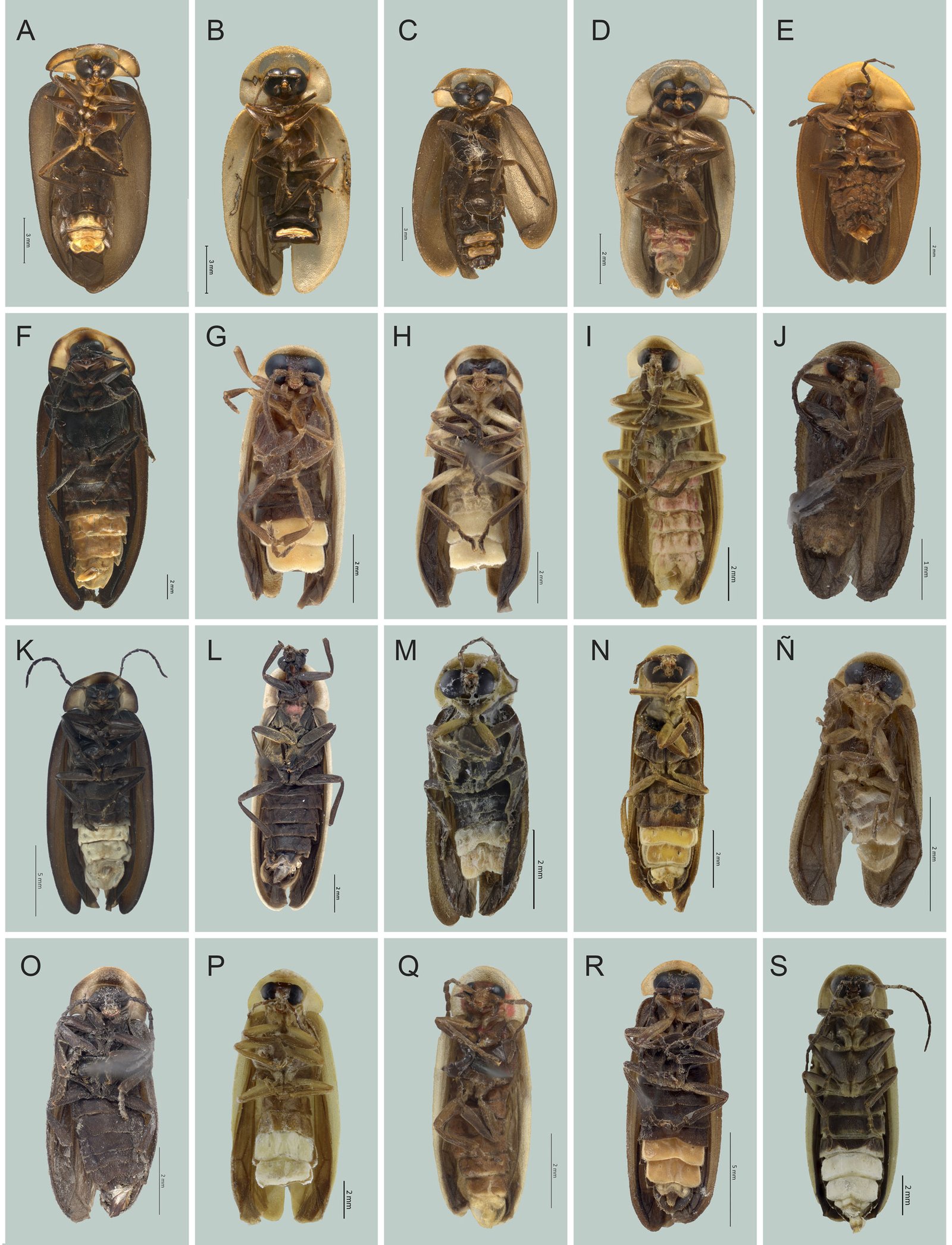
Figs. 2A, 3A
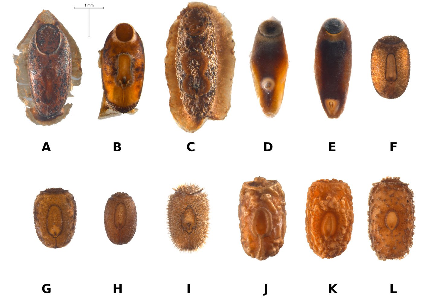
Largo 18.14 mm; ancho 7.95 mm. Coloración castaño oscura con excepción de pronoto, prosterno, pro y mesocoxas, parte basal de los fémures, los 3 últimos esternitos y el pigidio, amarillentos; pronoto con el margen y una mancha morena en la parte media. Pronoto casi triangular, más ancho que largo, con el disco ligeramente convexo, lados explanados, margen posterior sinuoso, el anterior redondeado, integumento chagrinado, pilosidad procumbente. Élitros aovados, de 3 veces y media tan largos como anchos, casi planos, integumento chagrinado. Aparato luminoso presente en la parte media de los esternitos 5-6. Edeago alargado, lóbulos laterales soldados en su base dorsal al lóbulo medio; lóbulo medio con el borde basal esclerosado, el ventral más angosto, membranoso, saco interno no visible, orificio medio abierto en el tercio distal.
Distribución: Chiapas.
Cratomorphus ayalai Zaragoza-Caballero, 1996
Figs. 2B, 3B
Largo 16.93 mm; ancho 7.60. Coloración castaño claro, con una mancha oscura en la parte media basal en el pronoto, cabeza y abdomen negro. Pronoto más ancho que largo, con el disco convexo, parte anterior ligeramente deflexionada, lados posteriores explanados, borde posterior casi recto, ángulos posteriores redondeados, integumento gruesamente chagrinado, pilosidad procumbente. Élitros aovados, 4 veces más largos que anchos, con 2 costillas apenas marcadas, integumento finamente chagrinado. Aparato luminoso ocupando gran parte de los esternitos 5-6. Edeago alargado, con los lóbulos laterales de forma triangular y ápice recurvado, lóbulo medio curvado con el ápice dilatado, borde dorsal esclerosado, el ventral formado por 2 quillas, orificio medio casi terminal.
Distribución: Chiapas.
Cratomorphus concolor (Perty, 1830)
Diagnosis: en Zaragoza-Caballero, López-Pérez et al. (2020) y Zaragoza-Caballero et al. (2023).
Distribución: Campeche, Chiapas, Estado de México, Hidalgo, Puebla, Quintana Roo, San Luis Potosí, Tabasco, Veracruz y Yucatán.
Cratomorphus elevatus Olivier, 1896
Cuerpo ovoide, de color castaño; cabeza negra; pronoto amarillo pálido, con una mancha basal oscura, carenado longitudinal; escutelo triangular amarillento; élitros cafés con márgenes pálidos, con 3 costillas. Abdomen lustroso, pubescente, brillante, negro, último segmento triangular; pigidio ojival.
Distribución: Oaxaca.
Cratomorphus leoneli Lima, Lima da Silveira, Vasconcelos-da Fonseca y Zaragoza-Caballero, 2021
Diagnosis: en Lima et al. (2021).
Distribución: Campeche, Chiapas y Oaxaca.
Cratomorphus picipennis Gorham, 1881
Diagnosis: en Zaragoza-Caballero et al. (2023).
Distribución: Chiapas y Veracruz. Nuevo registro: Oaxaca (CNIN).
Material examinado: México, Oaxaca, 6.7 km SO entronque carr. Salina Cruz-Sta. Cruz Huatulco, Arroyo Xuchitl, 11-07-2005. México, Oaxaca, Parque Nal. Huatulco 0.5 km N Est. El Sabanal, 05-11-2005, S. Zaragoza. Oaxaca, Parque Nal. Huatulco Estación El Sabana, 04/11/2005, S. Zaragoza.
Cratomorphus ramirezi Zaragoza-Caballero, 1996
Figs. 2C, 3C
Diagnosis: Largo 14 mm; ancho 5.9 mm. Coloración negra con excepción del pronoto amarillento. Pronoto semicircular, más ancho que largo, con el disco convexo, parte anterior muy deflexionado, lados basales explanados, ángulos posteriores redondeados, integumento gruesamente chagrinado, pilosidad procumbente. Élitros aovados, un poco más de 3 veces tan largos como anchos, disco ligeramente convexo y con 2 costillas apenas marcadas que alcanzan casi la mitad del élitro, integumento finamente chagrinado. Aparato luminoso ocupando casi toda la superficie de los esternitos 5-6. Edeago alargado, con los lóbulos laterales casi triangulares, membranosos en su porción apical, dorsalmente soldados al lóbulo medio, lóbulo medio ancho, de ápice recurvado, región ventral membranosa, orificio medio terminal, saco interno no visible.
Distribución: Chiapas.
Cratomorphus rodriguezae Zaragoza-Caballero, 1996
Figs. 2D, 3D
Diagnosis: Largo 10.02 mm; ancho 4.88 mm. Color castaño oscuro, con excepción de pronoto, ápice del escutelo, margen de los élitros, coxas y base de los fémures que son amarillentos; pronoto con una mancha negra en la parte media basal, flanqueada por manchas rojizas. Pronoto semicircular, más ancho que largo, disco convexo, parte laterales planas, margen anterior ligeramente deflexionado, el posterior recto, ángulos posteriores redondeados, integumento gruesamente chagrinado, pilosidad procumbente. Élitros aovados más anchos hacia la base, casi 3 veces más largos que anchos, disco convexo. Aparato luminoso sobre los esternitos 5-6. Edeago corto, robusto en su base, lóbulos laterales triangulares, con una excrecencia semicircular en su cara interna, ápice recurvado, membranoso, soldados en su base dorsal al lóbulo medio, lóbulo medio ancho, corto, borde dorsal casi recto, el ventral con 2 quillas que limitan un área membranosa en donde se localiza el orificio medio que es terminal, saco interno no visible.
Distribución: Chiapas y Oaxaca.
Cratomorphus signativentris Olivier, 1895
Diagnosis: en Zaragoza-Caballero et al. (2023).
Distribución: Campeche, Chiapas, Oaxaca, Querétaro, Quintana Roo, San Luis Potosí, Tabasco y Veracruz.
Pyractomena Melsheimer, 1846
Diagnosis: en Zaragoza-Caballero, López-Pérez et al. (2020) y Zaragoza-Caballero et al. (2023).
Pyractomena striatella Gorham, 1880
Diagnosis: en Zaragoza-Caballero et al. (2024).
Distribución: Chiapas, Quintana Roo y Tabasco.
Tenaspis LeConte, 1881
Diagnosis: en Zaragoza-Caballero et al. (2023, 2024).
Tenaspis angularis (Gorham, 1880)
Figs. 2E, 3E
Largo 9.46 mm; ancho 5.40 mm. Cuerpo ovado, coloración castaño-rojiza, con excepción del pronoto, que es amarillento, y la base del pronoto con una mancha castaña transversal. Pronoto triangular, más ancho que largo, disco cóncavo, medianamente carinado, parte lateral plana, margen anterior y posterior ligeramente deflexionados, ángulos posteriores redondeados, integumento rugosamente punteado, pilosidad procumbente. Élitros aovados más anchos a la base, con una costilla longitudinal levemente marcada, casi 3 veces más largos que anchos (7.44 × 2.68 mm), disco convexo. Sin aparato luminoso. Edeago corto, robusto en su base, lóbulos laterales en vista dorsal soldados en su base, romboideos, y en vista lateral un tanto triangulares, con una angosta y larga prolongación, lóbulo medio dorsalmente cubierto por los lóbulos laterales en su mitad basal, mitad apical angosta, quitinizada, lateralmente membranosa, lo mismo que toda la región ventral, orificio medio terminal.
Distribución: Chiapas, Hidalgo y San Luis Potosí.
Tenaspis gonzalenzis Zaragoza-Caballero, 1995
Diagnosis: en Zaragoza-Caballero et al. (2024).
Distribución: Chiapas y Veracruz.
Tribu Photinini LeConte, 1881
Subtribu Lucidotina Lacordaire, 1857
Lucidota Laporte, 1833
Diagnosis: en Zaragoza-Caballero, López-Pérez et al. (2020).
Lucidota bella Gorham, 1880
Diagnosis: en Zaragoza-Caballero, López-Pérez et al. (2020).
Distribución: Campeche, Chiapas, Estado de México, Oaxaca, Tabasco y Veracruz.
Lucidota bicellonycha McDermott, 1958
Diagnosis: largo 8.9 mm; ancho 4 mm. Color castaño oscuro, patas rojizas a castaño oscuro. Pronoto ancho, con una línea negra longitudinal, ángulos posteriores proyectados hacia atrás del medio a la base, margen basal sinuado, superficie transparente, fuertemente punteada. Élitros ovalados, más anchos aproximadamente en la mitad de su longitud, con una costilla poco visible, márgenes explanados bastante anchos en la base, estrechándose rápidamente. Sin órgano luminoso. Edeago con lóbulo medio aplanado, aparentemente tubular, curvo hacia arriba; lóbulos laterales subrectangulares presentan una corta proyección cerca de la base en el borde dorsal, y se prolongan hacia adelante en una punta estrecha.
Distribución: Oaxaca.
Lucidota discolor Gorham, 1881
Diagnosis: en Zaragoza-Caballero et al. (2024).
Distribución: Veracruz. Nuevo registro: Chiapas. Material examinado: México, Chiapas, Ocozocoautla de Espinosa, 3 km O Laguna Bélgica, 16°53’47.18” N, 93°27’9.50” O, 05 al 20-07-2003, V.H. Toledo, A.M. Corona.
Subtribu Photinina LeConte, 1881
Ankonophallus Zaragoza-Caballero y Navarrete-Heredia, 2014
Diagnosis: en Zaragoza-Caballero y Navarrete-Heredia (2014).
Ankonophallus consueloae Zaragoza-Caballero y Gutiérrez-Carranza, 2018
Diagnosis: en Zaragoza-Caballero y Gutiérrez-Carranza (2018).
Distribución: Chiapas.
Ankonophallus faciater Zaragoza-Caballero y Navarrete-Heredia, 2014
Diagnosis: en Zaragoza-Caballero y Navarrete-Heredia (2014).
Distribución: Guerrero.
Photinus Laporte, 1833
Diagnosis: en Zaragoza-Caballero, López-Pérez et al. (2020) y Zaragoza-Caballero et al. (2023).
Photinus acontrerasi Zaragoza-Caballero y Aquino-Romero sp. nov.
http://zoobank.org/urn:lsid:zoobank.org:act:8F6AB3EC-B0A2-4F37-972A-67A281ED3873
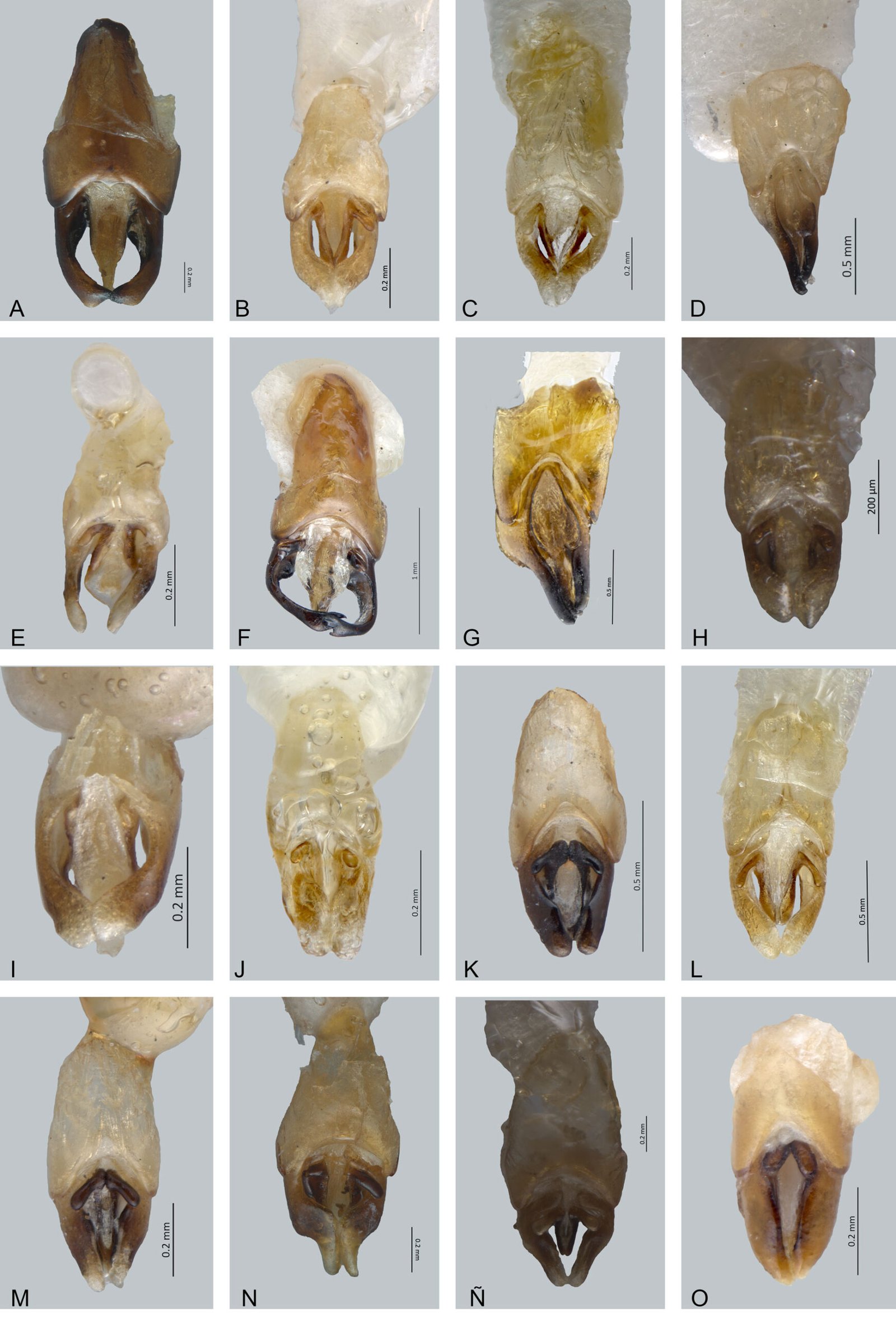
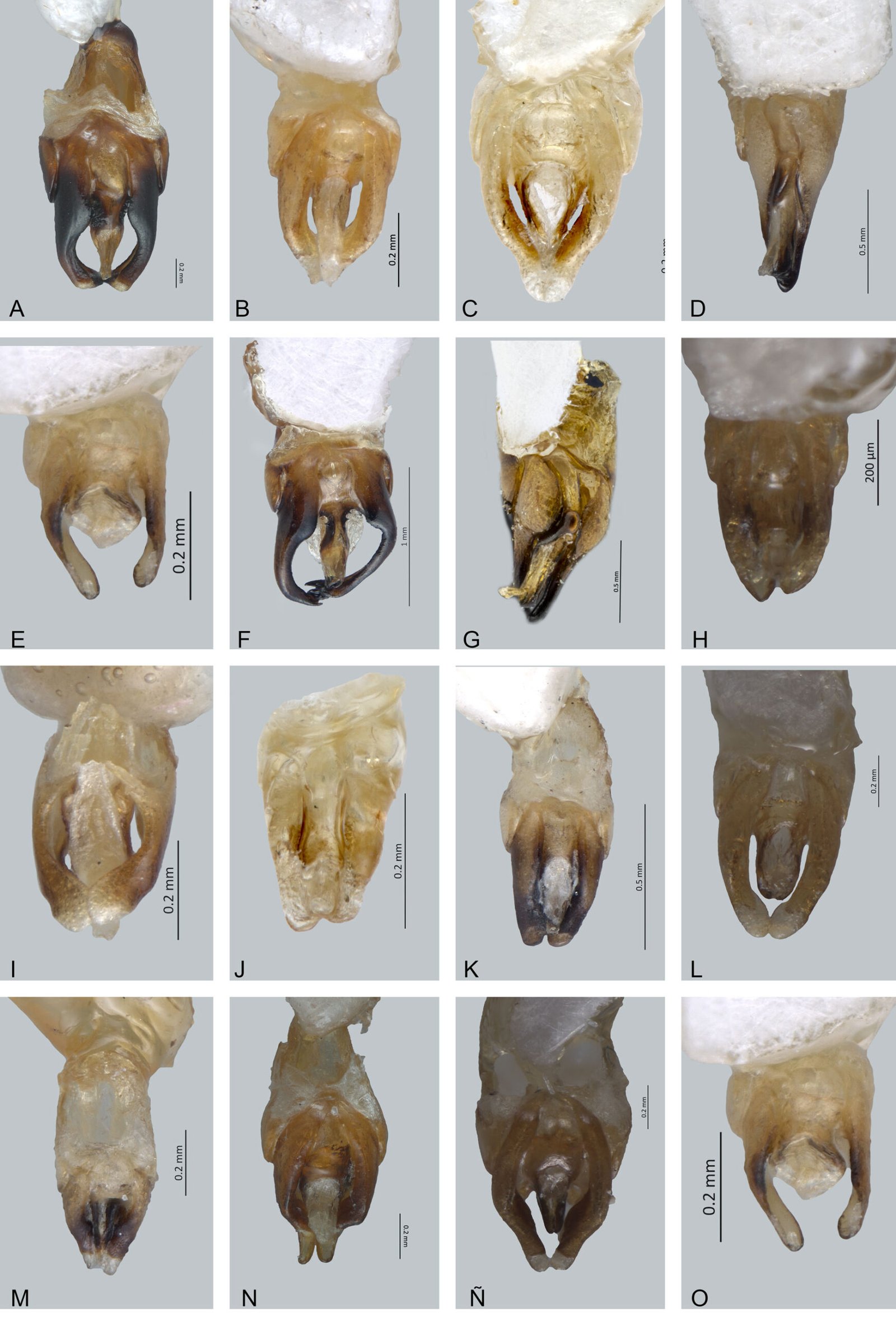
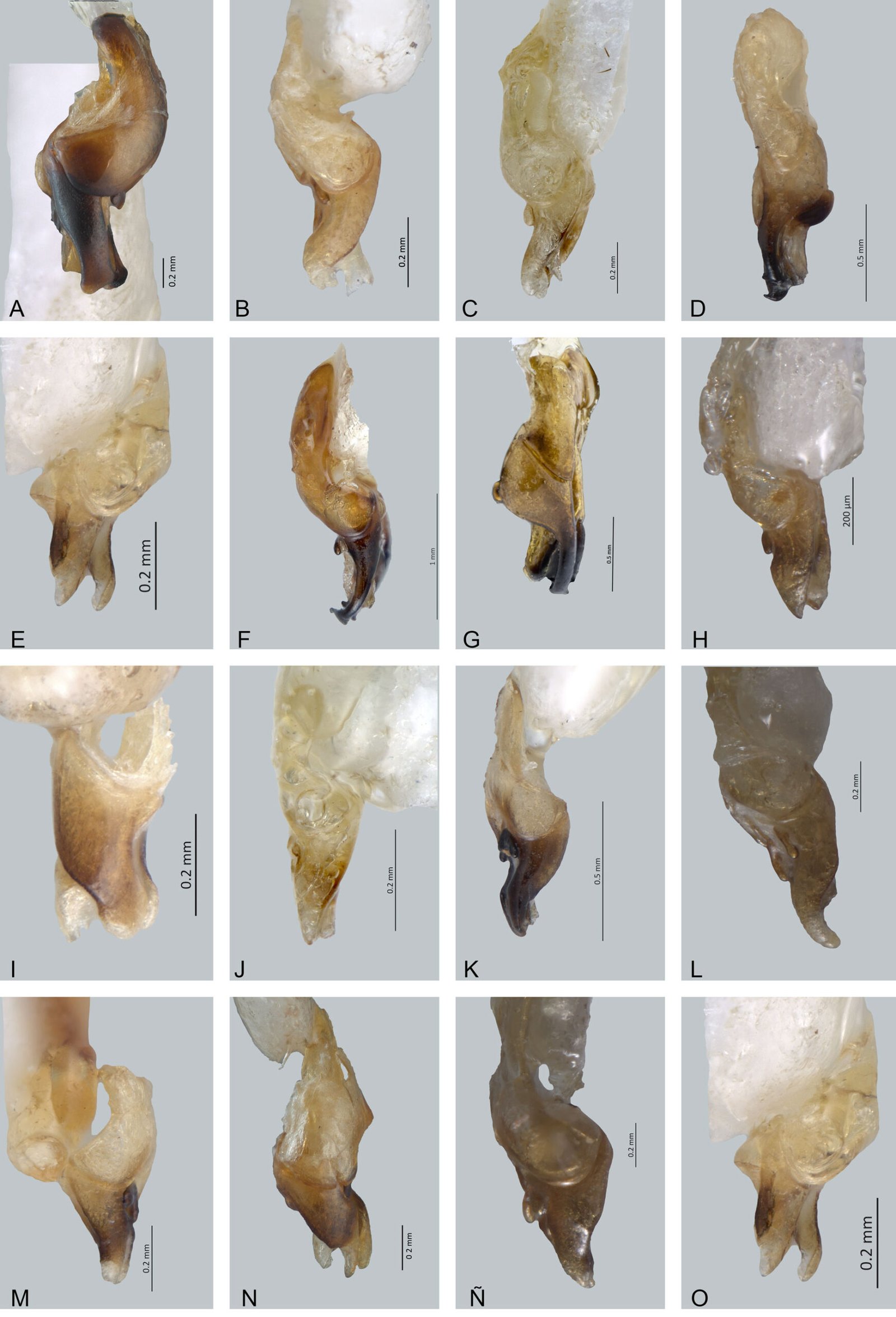
Figs. 2F, 3F, 4A, 5A, 6A
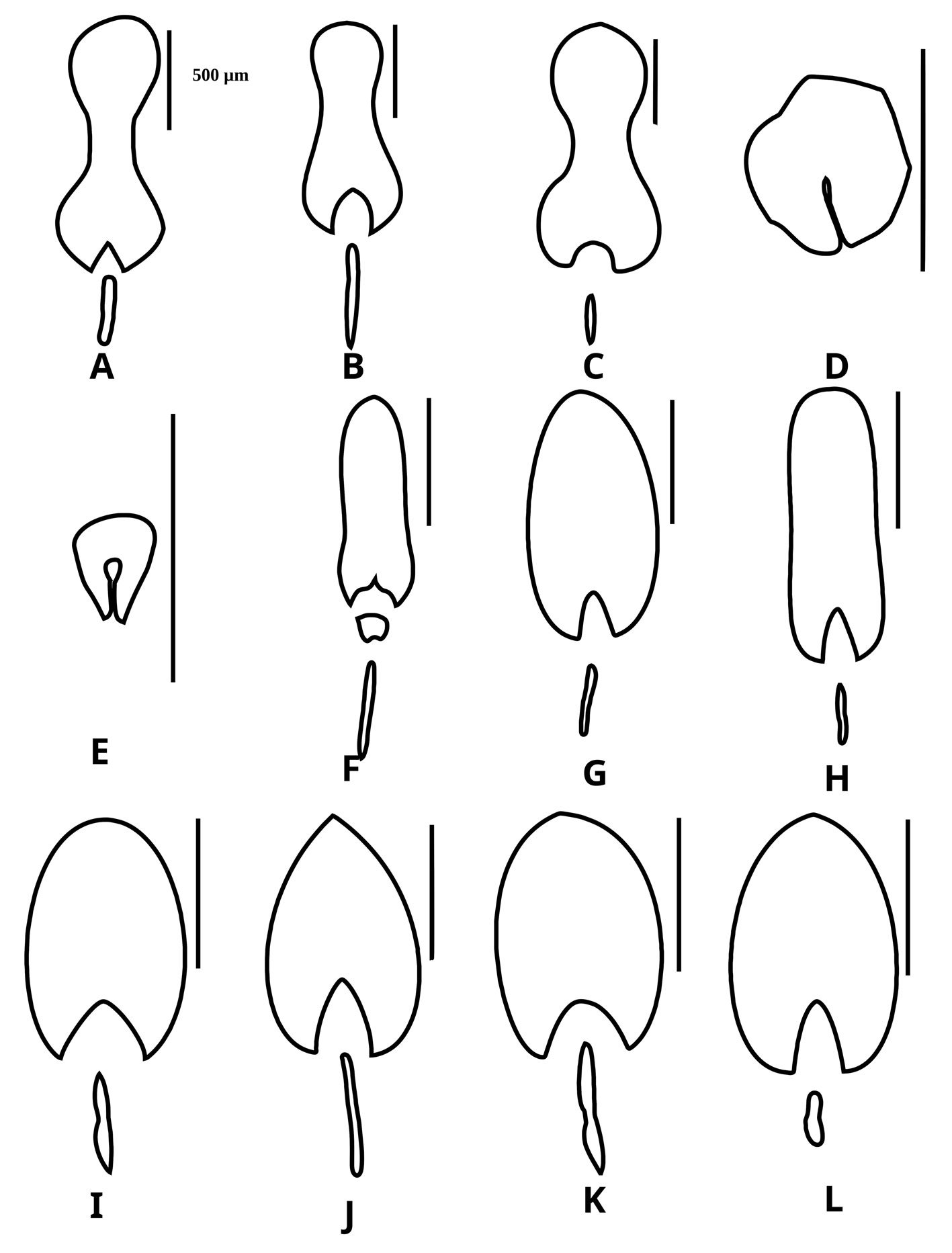
Macho. Largo 23.81 mm, ancho 9.27 mm; cuerpo semi paralelo; de color negro, pronoto cremoso con márgenes laterales negros y una mancha negra más o menos paralela basalmente ensanchada, proyectada en 2 pequeñas líneas del mismo color; aparato luminoso ocupando los esternitos 5-6. Cabeza: espacio interocular (0.88 mm), convexo, divergente, integumento opaco, chagrinado, con diminutas sedas; frente vertical, distancia interantenal (0.18 mm), más angosta que fosita antenal (0.46 mm); ojos finamente facetados, semiesféricos, más largos (1.13 mm) que anchos (0.52 mm); antenas comprimidas, largas, rebasando coxas posteriores, escapo de 1.09 mm de longitud, claviforme, un poco más largo que los 2 siguientes antenitos juntos (1.14 mm), segundo corto (0.38 mm), tercero de 0.76 mm, cuarto-décimo 0.84 mm, undécimo 0.92 mm; sutura frontoclipeal membranosa, cóncava; clípeo cuadrado con sedas cortas y largas en el borde anterior recto; mandíbulas falcadas; palpómero maxilar apical fusiforme, robusto; labial securiforme. Tórax: pronoto más ancho (7.18 mm) que largo (4.63 mm), ojival, borde posterior ondulado, ángulos posteriores obtusos, disco brillante, convexo, lados ampliamente explanados, con poros glandulares ordenados en el margen; escutelo espatulado, con borde posterior romo, superficie brillante, diminutamente punteada y pilosidad decumbente; élitros poco más de 4 veces más largos (19.18 mm) que anchos (4.54 mm), superficie rugosamente punteada, opaca, con 2 tipos de pilosidad decumbente, una más larga; epipleura angosta; espiráculos respiratorios mesotorácicos membranosos; patas cortas, pro y mesopatas semejantes entre sí, las posteriores un poco más largas, fémures comprimidos, casi fusiformes, tibias acanaladas, apicalmente dilatadas, borde externo no crenulado, tarsómeros lateralmente comprimidos, primer metatarsómero más largo (0.95 mm) que los 2 siguientes juntos (0.87 mm), cuarto bífido, cubriendo la mitad del quinto, uñas simples. Abdomen: últimos tergitos con ángulos posteriores agudos, pigidio convexo y lateralmente angulado; esternitos 5-6 más largos que los precedentes, con puntuaciones estigmatiformes, sexto y séptimo esternito con borde posterior cóncavo, el octavo espatulado; edeago (figs. 4A, 5A, 6A) con pieza basal simétrica, más larga (1.20 mm) que lóbulos laterales (1.05 mm), con borde dorsal anterior convexo, posterior con una proyección triangular en la concavidad; lóbulo medio un poco más de 2.5 tan largo (0.76 mm), como ancho (0.30 mm), casi cónico, con el ápice no rebasando los lóbulos laterales, membranoso en vista dorsal y ventralmente quitinoso, parte media, con estructuras de apariencia molar en parte media, ápice membranoso, orificio medio apicalmente abierto, excrecencias dorsobasales simétricas, lobuladas, divergentes y separadas en su base; lóbulos laterales interiormente excavados y con ápice en vista dorsal truncada, paralelos en vista lateral, ventralmente dilatados, curvados con el ápice truncado, sin modificaciones para la recepción de las excrecencias del lóbulo medio. Hembra desconocida.
Resumen taxonómico
Etimología: especie dedicada a Atilano Contreras Ramos, destacado entomólogo dedicado al estudio de insectos acuáticos.
Material examinado: holotipo macho. México, Chiapas, El Triunfo, 8-VII-1993, Col. S. Zaragoza. | COL. TIP-03981.
Comentarios taxonómicos
Photinus acontrerasi sp. nov. difiere de P. sarukhani sp. nov. que alcanza una talla de 17.27 mm, con el borde posterior del esternito 4 de color cremoso, el borde posterior de la pieza basal cóncavo, lóbulos laterales con ápice redondeado, en vista lateral acuminados. Photinus acontrerasi alcanza una talla de 23.81 mm, el borde posterior del esternito 4 de color cremoso solo en la parte media, el borde posterior de la pieza basal cóncavo con una proyección media triangular, lóbulos laterales con ápice truncado, en vista lateral casi paralelos.
Photinus acremophallus Zaragoza-Caballero y Gutiérrez-Carranza, 2018
Diagnosis: en Zaragoza-Caballero y Gutiérrez-Carranza (2018).
Distribución: Campeche, Chiapas, Oaxaca y Quintana Roo.
Photinus advenus Olivier, 1907
Diagnosis: en Zaragoza-Caballero et al. (2023).
Distribución: Michoacán, Oaxaca, San Luis Potosí y Veracruz.
Photinus ahuizotli Zaragoza-Caballero y Domínguez-León, 2020 en Zaragoza-Caballero, López-Pérez et al. (2020).
Diagnosis: en Zaragoza-Caballero, López-Pérez et al. (2020).
Distribución: Estado de México. Nuevo registro: Guerrero (CNIN). Material examinado (2): México, Guerrero 32 km O Iguala, 15-IX-1982, elev 1,350 m | J. a: Powell, J. A. Chemsak, at light.
Photinus alanisae Zaragoza-Caballero y Vega-Badillo sp. nov.
http://zoobank.org/urn:lsid:zoobank.org:act:8DA8ED71-23F7-4E80-813E-A3C7C429C752
Figs. 2G, 3G, 4B, 5B, 6B
Macho. Largo 8.57 mm, ancho 3.00 mm; cuerpo semiparalelo; de color castaño, con excepción del pronoto, sutura elitral, márgenes laterales de los élitros, coxas, trocánter, fémur que son ambarinos, pronoto con una mancha castaña casi rectangular; aparato luminoso ocupando los esternitos 5-6. Cabeza: espacio interocular (0.66 mm), cóncavo, paralelo, integumento brillante, chagrinado, con diminutas sedas; frente vertical, distancia interantenal (0.10 mm), más angosta que la fosita antenal (0.21 mm); ojos finamente facetados, semiesféricos, más largos (0.91 mm) que anchos (0.75 mm); antenas comprimidas, largas, alcanzando las coxas posteriores, escapo de 0.43 mm de longitud, en forma de clava, un poco más corto que los 2 siguientes antenitos juntos (0.51 mm), segundo corto (0.16 mm), tercero y cuarto de 0.35 mm, quinto-décimo (0.29 mm), undécimo 0.40 mm; sutura frontoclipeal membranosa, cóncava; clípeo trapezoidal, borde anterior recto, superficie espaciadamente punteada, con sedas largas y erectas; mandíbulas falcadas, con sedas basales; palpómero maxilar apical securiforme, robusto; labial fusiforme. Tórax: pronoto más ancho (2.37 mm) que largo (1.56 mm), semicircular, borde posterior recto, bordes laterales casi paralelos en su mitad basal, ángulos posteriores rectos, disco brillante, convexo, lados angostamente explanados, con poros glandulares ordenados en el margen, más abundantes en la parte anterior del disco; escutelo casi triangular, con el borde posterior romo, superficie brillante, diminutamente punteada y pilosidad decumbente; élitros 4.2 veces más largos (5.31 mm) que anchos (1.25 mm), superficie rugosamente punteada, brillante, con pilosidad decumbente; epipleura reducida; espiráculos respiratorios mesotorácicos tubulares; patas cortas, pro y mesopatas semejantes entre sí, las posteriores un poco más largas, fémures comprimidos, casi fusiformes, tibias acanaladas, apicalmente dilatadas, borde externo crenulado, tarsómeros lateralmente no comprimidos, primer metatarsómero más largo (0.35 mm) que los 2 siguientes juntos (0.28 mm), el cuarto escotado, cubriendo casi todo el quinto, uñas simples. Abdomen: penúltimo tergito con los ángulos posteriores agudos, pigidio bicóncavo; esternitos 5-6 más cortos que los precedentes, con puntuaciones estigmatiformes, séptimo esternito con el borde posterior escotado, el octavo espatulado; edeago (figs. 4B, 5B, 6B) con la pieza basal simétrica, más larga (0.57 mm) que los lóbulos laterales (0.46 mm), con el borde dorsal posterior cóncavo, el proximal convexo; lóbulo medio basalmente cilíndrico, 5 veces tan largo (0.26 mm) como ancho (0.05 mm), porción apical trunca, sin alcanzar el ápice de los lóbulos laterales, parte dorsal y ventral quitinosas, ápice membranoso, orificio medio terminal, excrecencias dorsobasales largas, simétricas, lobuladas, divergentes, con una base ancha esclerotizada que se extiende hasta el lóbulo medio; lóbulos laterales interiormente excavados y, en vista dorsal con el ápice redondeado, ventralmente casi paralelos, lateralmente en forma de S con el ápice transparente, con modificaciones para la recepción de las excrecencias del lóbulo medio. Hembra desconocida.
Resumen taxonómico
Etimología: especie dedicada a la estudiante Alanis Ninette Veliz Ojeda, que inicia sus estudios de coleópteros de la familia Lampyridae.
Material examinado: holotipo macho. México, Chiapas, Tuxtla Gutiérrez, El Zapotal, 2 min S Tuxtla Gutiérrez. 15-VII-1956. J.W. MacSwain, D.D. Linsdale col. | COL.TIP-03982 | Paratipos: 4 machos. (1) Mismos datos que el holotipo | COL.TIP-03983. (2) México, Chiapas, Tuxtla Gutiérrez, El Zapotal, 2 min S Tuxtla Gutiérrez. 10-VII-1957. J.A. Chemsak, B.J. Rannells col. | COL. TIP-03984-85. (1) México, Chiapas, Tuxtla Gutiérrez, SE Tuxtla Gutiérrez el Jobo. 908 m snm. 13-VIII-2002. A.M. Corona, V.H. Toledo cols. | QR 51900 | COL.TIP-03986.
Comentarios taxonómicos
Especie morfológicamente similar a Photinus marthae Zaragoza-Caballero y Cifuentes-Ruiz, 2023 y a Photinus reynosoi sp. nov. de las cuales se distingue por la forma y tamaño de la pieza basal del edeago.
Photinus anagabrielae Zaragoza-Caballero y Gutiérrez-Carranza, 2020 en Zaragoza-Caballero, López-Pérez et al. (2020).
Diagnosis: en Zaragoza-Caballero, López-Pérez et al. (2020).
Distribución: Puebla y Veracruz. Nuevo registro: Chiapas (CNIN). Material examinado: México, Chiapas, Carr. 202 cascadas de Welib ja 216 m snm, 17°22’431” N, 91°48’013” O, 7-VII-2009, L.F. de la Mora. México, Chiapas, Simojovel, VIII/1-16/58 | J.A. Chemsak collector.
Photinus albolimbatus Gemminger, 1870
Diagnosis: en Zaragoza-Caballero, López-Pérez et al. (2020) y Zaragoza-Caballero et al. (2023).
Distribución: Ciudad de México, Durango, Hidalgo, Oaxaca, Puebla y Veracruz.
Photinus aliciae Zaragoza-Caballero, 2005
Diagnosis: en Zaragoza-Caballero et al. (2023).
Distribución: Colima, Guerrero, Jalisco y Michoacán.
Photinus ater Gorham, 1881
Diagnosis: en Zaragoza-Caballero et al. (2023).
Distribución: Ciudad de México, Durango, Guanajuato, Jalisco, Michoacán, Oaxaca y Puebla.
Photinus avendanoae Zaragoza-Caballero y Aquino-Romero sp. nov.
http://zoobank.org/urn:lsid:zoobank.org:act:694068D5-04CA-4A44-BF16-8F7833A5DD0C
Figs. 2H, 3H, 4C, 5C, 6C
Macho. Largo 9.09 mm, ancho 3.72 mm; cuerpo semi paralelo; cuerpo cremoso; disco pronotal con ángulos externos mínimamente más oscuros, centro con 2 manchas cafés ambarinas, una sobre de otra, la superior más difusa, con 2 manchas de color rosa pálido, a un lado de la mancha más oscura; escutelo castaño, más claro que los élitros; élitros cafés en el centro, con los márgenes del mismo color que el resto del cuerpo; fémures con la base castaña, así como tibias y tarsómeros; aparato luminoso blanquecino, ocupando los esternitos 5-6. Cabeza: espacio interocular (0.85 mm) cóncavo, integumento brilloso, chagrinado, pilosidad ligera, decumbente; distancia interantenal (0.20 mm) ligeramente más amplia que fosita antenal (0.16 mm); ojos finamente facetados, semiesféricos, una tercera parte más largos (0.94 mm) que anchos (0.64 mm); antenas filiformes largas, que sobrepasan metacoxas; escapo (0.79 mm) claviforme, ligeramente más corto que los 2 próximos antenitos juntos, segundo corto (0.20 mm) piriforme, tercero al décimo 0.50 mm, ligeramente aplanados, undécimo más largo (0.61 mm), cilíndrico; sutura frontoclipeal membranosa ligeramente emarginada; clípeo trapezoidal, con el borde anterior convexo, superficie rugosa, con puntuación densa y profunda, pilosidad densa, procumbente, con las sedas largas; mandíbulas falcadas, curvadas; palpómeros maxilar apical y labial securiformes. Tórax: pronoto más ancho (3.03 mm) que largo (2.29 mm), ojival, semicircular, con el borde posterior ligeramente cóncavo; disco brillante, convexo, con lados explanados y pilosidad densa y procumbente, poros glandulares ordenados en el margen; escutelo lingüiforme, con el borde anterior convexo, superficie brillante, rugosa, con pilosidad fina, decumbente; élitros casi 4 veces más largos (8.45 mm) que anchos (2.09 mm), superficie, brillosa, integumento rugoso, pilosidad corta, densa y procumbente; epipleura estrecha, ligeramente más angosta en la base; espiráculos respiratorios mesotorácicos tubulares; patas cortas, fémures fusiformes, metafémures ligeramente más estrechos y alargados; pro y mesotibias con la base sutilmente más ensanchada que las metatibias; tarsómeros estrechos, primer metatarsómero ligeramente más corto (0.27 mm) que los 2 siguientes juntos (0.32 mm), cuarto bífido, cubriendo parte del quinto, uñas simples. Abdomen: esternitos 5-6 el doble de largo (1.02 mm) que los precedentes (0.46 mm), sin puntuaciones estigmatiformes, octavo con la base romo; pigidio con el borde superior bisinuado; edeago (figs. 4C, 5C, 6C) con pieza basal simétrica, casi tan ancha (0.43 mm) como larga (0.50 mm), con bordes anteriores fuertemente convexos en vista dorsal, ángulo anterior cóncavo; lóbulo medio cuneiforme, mínimamente más ancho (0.17 mm) que largo (0.13 mm), estrecho hacia la mitad de la base, parte dorsal y ventral quitinosa, orificio medio ventralmente abierto, sin excrecencias dorsobasales; lóbulos laterales cilíndricos, estrechos, 5 veces más largos (0.35 mm) que anchos (0.06), con ápice romo, sin modificaciones para la recepción de las excrecencias del lóbulo medio. Hembra desconocida.
Resumen taxonómico
Etimología: especie dedicada a Roberta Avendaño Martínez “La Tita”, activista universitaria del Movimiento Estudiantil de 1968, egresada de la Facultad de Derecho de la Universidad Nacional Autónoma de México.
Material examinado: holotipo macho. México: Guerrero: Atoyac de Álvarez km 14, El Paraíso, Nueva Delhi, 19-VII-2005, 17°24’12” N, 100°11’37” O, 1,153 m snm, L. Cervantes J. Calonico | COL.TIP-03987. Paratipo 1 macho. México, Guerrero, Atoyac de Álvarez km 14, El Paraíso, Santiago La Unión, 19-VII-2005, 17°15’12” N, 100°20’32” O, L. Cervantes R. Mariño. | COL.TIP-03988.
Comentarios taxonómicos
Aunque a simple vista esta especie comparte características morfológicas similares a Photinus abrilae Zaragoza-Caballero y González-Ramírez, 2020, principalmente en la forma del edeago, difieren por el ancho de los lóbulos laterales, en P. avendanoae sp. nov. son un poco curvos, mientras que en P. abrilae no son curvos en vista lateral, ligeramente fusiformes.
Photinus benignoi Zaragoza-Caballero y Domínguez-León sp. nov.
http://zoobank.org/urn:lsid:zoobank.org:act:F41AA590-84F7-4AB6-B980-3D5A3931CF07
Figs. 2I, 3I, 4D, 5D, 6D
Macho. Largo 11.12 mm, ancho 4.02 mm, cuerpo semi paralelo; de color castaño claro, pronoto con márgenes laterales ambarinos y el centro con una mancha castaña que se extiende cerca del margen anterior, adicionalmente con 2 pequeñas manchas rosas en cada lado; aparato luminoso ocupando los esternitos 5-7. Cabeza: espacio interocular (0.68 mm), cóncavo, integumento brillante, chagrinado, con diminutas sedas; frente vertical, distancia interantenal (0.07 mm), más angosta que fosita antenal (0.28 mm); ojos divergentes, finamente facetados, semiesféricos, más largos (0.9 mm) que anchos (0.45 mm); antenas comprimidas, largas, rebasando las coxas posteriores, escapo de 0.76 mm de longitud, semitriangular, más corto que los 2 siguientes antenitos juntos (1 mm), segundo corto (0.24 mm), tercero 0.75 mm, cuarto-décimo 0.87 mm, undécimo 0.97 mm; sutura frontoclipeal membranosa, cóncava; clípeo trapezoidal, borde anterior recto, superficie espaciadamente punteada, con sedas largas y erectas; mandíbulas falcadas, con sedas basales; palpómero maxilar apical fusiforme, robusto; labial securiforme. Tórax: pronoto más ancho (3.22 mm) que largo (2.12 mm), ojival, borde posterior sinuado, ángulos posteriores obtusos, disco brillante, convexo, lados ampliamente explanados, con poros glandulares ordenados en el margen, más abundantes en parte anterior del disco; escutelo casi triangular, con borde posterior romo, superficie brillante, diminutamente punteada y pilosidad decumbente; élitros casi 5 veces más largos (9 mm) que anchos (2.01 mm), superficie rugosamente punteada, opaca, con 2 tipos de pilosidad decumbente, una un poco más larga; epipleura angosta; espiráculos respiratorios mesotorácicos tubulares; patas cortas, pro y meso-patas semejantes entre sí, posteriores un poco más largas, fémures comprimidos, casi fusiformes, tibias acanaladas, apicalmente dilatadas, borde externo no crenulado, tarsómeros lateralmente comprimidos, primer metatarsómero más largo (0.51 mm) que los 2 siguientes juntos (0.45 mm), cuarto bífido, cubriendo casi todo el quinto, uñas simples. Abdomen: pigidio convexo; esternitos 5-6 tan largos que los precedentes, sin puntuaciones estigmatiformes, 7 con borde posterior cóncavo, 8 no se encuentra en el ejemplar; edeago (figs. 4D, 5D, 6D) con pieza basal simétrica, más larga (0.85 mm) que ancha (0.63 mm), más corta que lóbulos laterales (0.92 mm), con borde dorsal posterior cóncavo, proximal convexo; lóbulo medio basalmente cilíndrico, un poco más de 7 veces tan largo (0.78 mm) como ancho (0.1 mm), porción apical acuminada, casi alcanzando ápice de los lóbulos laterales, parte dorsal membranosa, ventral quitinosa, orificio medio ventralmente abierto, excrecencias dorso basales ausentes; lóbulos laterales esbeltos, un poco más de 8 veces tan largos (0.92 mm) como anchos (0.11 mm), en vista lateral el ápice presenta un dentículo, ventralmente dilatados en la parte media. Hembra desconocida.
Resumen taxonómico
Etimología: especie dedicada al Dr. Benigno Gómez, gran promotor de la entomología cultural y la etnobiología; coordinador de Desarrollo Organizacional del Colegio de la Frontera Sur en Chiapas, México.
Material examinado: holotipo macho. México: Oaxaca: Pluma Hidalgo, 31-I-89. E. Barrera, A. Cadena, E. Ramírez cols. | COL.TIP-03989.
Comentarios taxonómicos
Especie morfológicamente similar desde la vista dorsal a Photinus plumbeus Gorham, 1880 de la cual se distingue por su tamaño corporal y por la forma particular del edeago. El tamaño corporal de P. plumbeus es menor con respecto a P. benignoi sp. nov.; adicionalmente los lóbulos laterales en esta última presentan un dentículo en el ápice. La forma del edeago es similar a la de Photinus perotensis Zaragoza-Caballero, 1996, sin embargo, P. benignoi es diferente en la morfología dorsal, tiene un tamaño corporal menor, mientras el lóbulo medio es más largo que los lóbulos laterales en P. perotensis y los dentículos de los lóbulos laterales apicales son menos marcados en P. benignoi sp. nov.
Photinus buenoi Zaragoza-Caballero y Pérez-Flores sp. nov.
http://zoobank.org/urn:lsid:zoobank.org:act:F702CE20-842A-40D4-BCCC-78EA67DC2C0E
Figs. 2J, 3J, 4E, 5E, 6E
Macho. Largo 4.38 mm, ancho 2.76 mm; cuerpo paralelo; integumento mayormente de color castaño; pronoto con los márgenes laterales anchos, amarillos, centralmente castaño con manchones rojizos a los lados; élitros con márgenes laterales castaño-amarillentos, el resto de la superficie castaña; coxas, trocánteres y mitad anterior de fémures de patas anteriores y medias amarillos; sin aparato luminoso. Cabeza: espacio interocular (0.18 mm), ligeramente deprimido, integumento brillante, con sedas cortas; frente casi vertical, distancia interantenal (0.01 mm), más angosta que la fosita antenal (0.03 mm); ojos divergentes, finamente facetados, semiesféricos, prominentes, más largos (0.19 mm) que anchos (0.13 mm); antenas comprimidas, largas, sobrepasando el ápice de las mecoxas, escapo de 0.14 mm de longitud, claviforme, más corto que los 2 siguientes antenitos juntos (0.23 mm), segundo corto (0.03 mm), tercero y quinto de 0.11 mm, cuarto de 0.12 mm, sexto-décimo 0.13 mm, undécimo 0.14 mm de largo; sutura frontoclipeal membranosa, cóncava; clípeo rectangular, borde anterior recto, superficie espaciadamente punteada, con sedas largas y erectas; mandíbulas falcadas, esbeltas, con sedas basales; palpómero maxilar apical securiforme, labial securiforme. Tórax: pronoto más ancho (1.38 mm) que largo (0.94 mm), ojival, borde posterior ligeramente cóncavo, ángulos posteriores casi rectos, disco brillante, convexo, lados ampliamente explanados, con poros glandulares ordenados en el margen, distribuidos heterogéneamente; escutelo lingüiforme, con el borde posterior truncado, superficie brillante, con puntuaciones toscas y pilosidad decumbente; élitros 3.5 veces más largos (3.43 mm) que anchos (0.96 mm), superficie rugosamente punteada, brillante, con 2 tipos de pilosidad decumbente, una más larga; epipleura reducida; espiráculos respiratorios mesotorácicos tubulares; patas largas, pro y meso-patas semejantes entre sí, las posteriores más largas, fémures comprimidos, algo fusiformes, tibias acanaladas, poco dilatadas apicalmente, borde externo no crenulado, tarsómeros comprimidos lateralmente, primer metatarsómero ligeramente más largo (0.07 mm) que los 2 siguientes juntos (0.05 mm), cuarto bífido, cubriendo más de la mitad del quinto, uñas simples. Abdomen: últimos tergitos con ángulos posteriores obtusos, pigidio convexo, con ángulos posteriores redondeados; esternitos 5-6 más cortos que los precedentes, sin puntuaciones estigmatiformes, 7 con el borde posterior ligeramente cóncavo, 8 espatulado; edeago (figs. 4E, 5E, 6E) con pieza basal simétrica, más larga (0.27 mm) que ancha (0.23 mm), apenas más larga que lóbulos laterales (0.26 mm), con borde dorsal posterior cóncavo, proximal convexo; lóbulo medio cuneiforme, apenas más largo (0.22 mm) que ancho (0.19 mm), con ápice alcanzando el tercio posterior de lóbulos laterales, parte dorsal y ventral membranosa, ápice membranoso, orificio medio ventralmente abierto, excrecencias dorso basales largas, asimétricas, lobuladas, divergentes, basalmente separadas; lóbulos laterales esbeltos, 2 veces tan largos (0.26 mm) como altos (0.13 mm), interiormente excavados, en vista dorsal con el ápice redondeado, ventralmente dilatados en la parte media, en vista lateral más altos en su base, sin modificaciones para la recepción de las excrecencias del lóbulo medio. Hembra desconocida.
Resumen taxonómico
Etimología: especie dedicada al Dr. Julián Bueno Villegas, reconocido investigador dedicado al estudio de los miriápodos neotropicales.
Material examinado: holotipo macho. México, Oaxaca, San Mateo, Yetla, 25-IX-1989, F. A. Noguera, A. Rodríguez | COL.TIP-03989. Paratipos 12 machos. (2) México, Oaxaca, Temascal, 30-VI-1964. A.C. Raske col. | QR 51997 | COL.TIP-03990-91. (1) México, Oaxaca, La Ventosa 48 min E., 21-VII-63. Doyen Collector | Photinus sp. (Laporte, 1833) | Colección del Instituto de Biología, UNAM, México D.F. | QR 51995 | COL.TIP-03992. (1) México, Chiapas: Solosuchiapa km 1, Solosuchiapa-Tuxtla Gutiérrez, 25-VI-2007, 17°241’434” N, 93°01’47” O, 104 m snm. C. Mayorga, G. Ortega, L. Cervantes | COL.TIP-03993. (2) México, Chiapas, Ocosingo Chajul, Reserva Montes Azules, 10-16-VII-87, F. Arias, R. Barba, L. Cervantes cols. | COL.TIP-03994-95. (2) México, Chiapas, Santo Domingo, 15 min SE Simojovel, 8-15-VII-58 | COL.TIP-03996-97. (1) México, Chiapas, Pijijiapan, km 28, 3-VII-1988, A. Cadena y L. Cervantes cols. | COL. TIP- 03998. (3) México: Veracruz: Acayucan 18 min S, 18-VII-63. J. Doyen Collector | Photinus sp. ♂ (Laporte, 1833) | Colección del Instituto de Biología, UNAM, México D.F. | QR 51925 | COL.TIP-03999-04001.
Comentarios taxonómicos
Esta especie es morfológicamente similar a Photinus aliciarodriguezae Zaragoza-Caballero y López-Pérez, 2023 y a P. zongolicaensis Zaragoza-Caballero y Domínguez-León, 2020, de las cuales se distingue por el tamaño y la forma del edeago. El tamaño de Photinus buenoi sp. nov. es menor en comparación a P. aliciarodriguezae y P. zongolicaensis. La forma del edeago en P. aliciarodriguezae es distintivamente más esbelta, con los lóbulos laterales delgados, en P. buenoi sp. nov. son más anchos los lóbulos laterales y el lóbulo medio es de menor tamaño que estos. De P. zongolicaensis se diferencia por los lóbulos laterales anchos, dilatados apicalmente, mientras que en P. buenoi sp. nov., son más esbeltos y apicalmente se estrechan, además, en este último el lóbulo medio es más ensanchado en la mitad posterior. La forma dorsal general también es similar a Photinus angieae Zaragoza-Caballero y Domínguez-León, 2023, Photinus apahtzii Zaragoza-Caballero y López-Pérez, 2023 y a Photinus aulophallus Zaragoza-Caballero y Gutiérrez-Carranza, 2018, pero se diferencia de todos los anteriores porque estos presentan aparato luminoso y P. buenoi sp. nov. no presenta aparato luminoso.
Photinus ciervophallus Zaragoza-Caballero y Rodríguez-Mirón sp. nov.
http://zoobank.org/urn:lsid:zoobank.org:act:5B46C900-3D0A-484E-85AD-9AD834D7D4B9
Figs. 2K, 3K, 4F, 5F, 6F
Macho. Largo 20.39 mm, ancho 7.44 mm; cuerpo ovoide; de color negro, pronoto con el margen negro y una ancha mancha paralela negra, lados cremosos; aparato luminoso ocupando los esternitos 5-6. Cabeza: espacio interocular (0.76 mm), plano, divergente, integumento brillante, chagrinado, con diminutas sedas; frente vertical, distancia interantenal (0.23 mm), más angosta que fosita antenal (0.28 mm); ojos finamente facetados, semiesféricos, más largos (1.67 mm) que anchos (0.62 mm); antenas comprimidas, largas, rebasando coxas posteriores, escapo de 0.84 mm de longitud, casi cilíndrico, un poco más corto que los 2 siguientes antenitos juntos (0.99 mm), segundo corto (0.28 mm), tercero-décimo 0.71 mm y, undécimo 0.78 mm; sutura frontoclipeal membranosa, cóncava; clípeo semicircular, rugosamente punteado, con largas sedas; mandíbulas falcadas; palpómero maxilar apical fusiforme, robusto; el labial securiforme. Tórax: pronoto más ancho (5.72 mm) que largo (3.63 mm), ojival, borde posterior recto, ángulos posteriores obtusos, disco brillante, convexo, lados ampliamente explanados, con poros glandulares ordenados en el margen, irregulares en parte anterior del disco; escutelo espatulado, con borde posterior romo, superficie brillante, rugosamente punteada; élitros más de 4 veces más largos (16.36 mm) que anchos (3.72 mm), superficie rugosamente punteada, opaca, con 2 tipos de pilosidad decumbente, una más larga; epipleura angosta; espiráculos respiratorios mesotorácicos tubulares; patas cortas, pro y mesopatas semejantes entre sí, posteriores un poco más largas, fémures comprimidos, casi fusiformes, tibias acanaladas, apicalmente dilatadas, borde externo no crenulado, tarsómeros lateralmente comprimidos, primer metatarsómero más largo (0.84 mm) que los 2 siguientes juntos (0.71 mm), el cuarto bífido, casi cubriendo al quinto, uñas simples. Abdomen: últimos tergitos con los ángulos posteriores agudos, pigidio convexo, con el borde posterior ondulado; esternitos 5-6 tan largos como los precedentes, con puntuaciones estigmatiformes, sexto y séptimo esternito con el borde posterior cóncavo, octavo espatulado con un surco longitudinal; edeago (figs. 4F, 5F, 6F) con pieza basal asimétrica, más larga (1.36 mm) que los lóbulos laterales (1.05 mm), con borde dorsal anterior un tanto triangular, posterior cóncavo, lóbulo medio un poco más de 3 veces tan largo (0.75 mm), como ancho (0.24 mm), casi cilíndrico, con ápice no rebasando los lóbulos laterales, parcialmente membranoso en vista dorsal y ventralmente quitinoso, angosto y con espinas en parte media, ápice membranoso, orificio medio apicalmente abierto, excrecencias dorso basales simétricas, acuminadas, divergentes y separadas en su base; lóbulos laterales muy curvados, con ápice dentado, en vista lateral acuminados, sin modificaciones para la recepción de las excrecencias del lóbulo medio. Hembra desconocida.
Resumen taxonómico
Etimología: el epíteto hace referencia a la forma de las astas de los ciervos que tienen los lóbulos laterales del
Material examinado: holotipo macho. México, Chiapas, Unión Juárez, Chiquihuites, 15°05’43.79” N; 92°05’57.61” O, 2,018 m snm, 8-X-2018, trampa de luz blanca y negra. Cols. Cancino-López R.J. y Luna-Luna A.M | COL.TIP-04002. Paratipos 3 machos. (1) Guatemala, Depto. of Guatemala Huerta, Puerta Parada. 4.5 km E. Guatemala City, M. Maldonado 11-X-1983 | COL.TIP-04003. (1) mismos datos, con fecha de 11-IX-83 | COL.TIP-04004. (1) mismos datos, con fecha de 22-IX-83 | COL.TIP-04005.
Comentarios taxonómicos
Únicamente 3 especies de lampíridos mexicanos, sobrepasan los 20 mm de longitud: Photinus congruus Chevrolat, 1834 (21.72 mm), P. meteoralis Gorham, 1881 (21.69 mm) y P. ciervophallus sp. nov. (20.39 mm). La primera tiene una forma y color diferente, el reproductor también difiere mucho de las otras 2. Los élitros de P. meteoralis son acuminados y la mancha negra pronotal es semicircular. En P. ciervophallus sp. nov. la mancha es paralela, y los élitros son aovados. Lo largo y la forma de astas de los lóbulos laterales, la hace totalmente diferente a todas las especies de Photinus.
Photinus cisteilae Zaragoza-Caballero, 2015
Diagnosis: en Zaragoza-Caballero (2015).
Distribución: Guerrero y Oaxaca.
Photinus congruus Chevrolat, 1834
Diagnosis: en Zaragoza-Caballero, López-Pérez et al. (2020).
Distribución: Chiapas, Oaxaca, Puebla, Quintana Roo y Veracruz.
Photinus corrusca (Linnaeus, 1767)
Diagnosis: en Zaragoza-Caballero, López-Pérez et al. (2020) y Zaragoza-Caballero (2023).
Distribución: Chiapas, Ciudad de México, Estado de México, Morelos, Oaxaca, Puebla, San Luis Potosí, Tamaulipas, Tlaxcala y Veracruz.
Photinus danieli Zaragoza-Caballero, 2017
Diagnosis: en Zaragoza-Caballero (2017).
Distribución: Oaxaca.
Photinus davilae Zaragoza-Caballero y Rodríguez-Mirón sp. nov.
http://zoobank.org/urn:lsid:zoobank.org:act:E1B01E3F-315D-441C-AF31-C7156F8A69B0
Figs. 2L, 3L, 4G, 5G, 6G
Macho. Largo 14.18 mm, ancho 4.18 mm; cuerpo paralelo; de color castaño; pronoto con lados amarillos, disco castaño; élitros cafés, lados amarillentos; sin aparato luminoso. Cabeza: espacio interocular (0.82 mm) transverso, deprimido, paralelo, integumento lustroso, finamente punteado, con sedas conspicuas, decumbentes; frente (0.16 mm), más angosta que la fosita antenal (0.24 mm); ojos finamente facetados, semiesféricos, más largos (0.46 mm) que anchos (0.38 mm); antenas comprimidas, escapo de 0.62 mm de longitud, subcónico, más corto que los 2 siguientes antenitos juntos (0.77 mm), segundo corto (0.2 mm), tercero de 0.57 mm, cuarto 0.6 mm, quinto 0.63 mm; sutura frontoclipeal membranosa, cóncava; clípeo trapezoidal, borde anterior recto, superficie espaciadamente punteada, con sedas largas y erectas; mandíbulas falcadas, con sedas basales; palpómero maxilar apical securiforme, robusto; palpómero labial triangular. Tórax: pronoto semicircular, más ancho (4.27 mm) que largo (2.82 mm), parte media del borde posterior recto, ángulos posteriores romos, disco brillante, con un surco, diminutamente punteado, lados explanados hacia arriba, con poros glandulares ordenados en el margen, más abundantes en la parte anterior del disco; escutelo subtriangular, con el borde posterior obtuso, diminutamente punteada y pilosidad decumbente; élitros 5.4 veces más largos (11.36 mm) que anchos (2.09 mm), superficie rugosamente punteada, lustroso, pilosidad decumbente amarilla; epipleura amplia en la base y reducida hacia el ápice; espiráculos respiratorios mesotorácicos tubulares; pro y mesopatas semejantes entre sí, posteriores un poco más largas, fémures comprimidos, casi fusiformes, tibias aplanadas, levemente dilatadas al ápice, borde externo crenulado, tarsómeros lateralmente comprimidos, primer metatarsómero más largo (0.44 mm) que los 2 siguientes juntos (0.37 mm), cuarto bífido, cubriendo casi todo el quinto, uñas simples. Abdomen: borde del pigidio convexo; esternitos 5-6 tan largos que los precedentes, sin puntuaciones estigmatiformes, 5-6 con borde posterior recto, 7 con borde posterior cóncavo con forma de “V”, 8 ojival; edeago (figs. 4G, 5G, 6G) con pieza basal simétrica, poco más larga (1.1 mm) que ancha (1 mm), más larga que lóbulos laterales (0.81 mm), con borde dorsal cóncavo; lóbulo medio, ventralmente quitinoso, dilatado basalmente, lanceolado, región anterior esbelta, porción apical redondeado, casi alcanzando ápice de los lóbulos laterales, parte dorsal membranosa, orificio medio ventralmente abierto, excrecencias dorso basales robustas, en forma de protuberancias connadas al lóbulo medio; lóbulos laterales dilatados en la base, subcuadrada (0.4 mm), 5 veces más anchos en la base que la parte apical, ápice ahusado, sin modificaciones para la recepción de las excrecencias del lóbulo medio. Hembra desconocida.
Resumen taxonómico
Etimología: dedicada a la Dra. Patricia Dolores Dávila Aranda, por sus aportes a la botánica mexicana y por su designación como primera secretaria general de la UNAM.
Material examinado: holotipo macho. México, Oaxaca, Pochutla, Hagia sofia, X-24-2012, J. Ramírez col. | COL.TIP-04006. Paratipo. Un macho. México, Oaxaca, 4 min al sur de Ixtepec, X-26-1966 | Ray F. Smith col. | COL.TIP-04007.
Comentarios taxonómicos
Especie cercana a Photinus benignoi sp. nov. de la que se distingue por diferencias en el tamaño, forma del edeago y patrón de coloración. P. davilae sp. nov. tiene una longitud de 14.18 mm y ancho 4.18 mm, la pieza basal del edeago es tan larga como ancha, ápice del lóbulo lateral ahusado, el color del pronoto y élitros cafés oscuro y los márgenes son amarillos. Photinus benignoi sp. nov. tiene una longitud de 11.12 mm, ancho 4.02 mm, la pieza basal del edeago es más larga que ancha, el ápice del lóbulo lateral tiene un dentículo, el pronoto y los élitros son amarillos y los tonos más claros en los márgenes.
Photinus dispersus Zaragoza-Caballero y Pérez-Flores sp. nov.
http://zoobank.org/urn:lsid:zoobank.org:act:F0B80697-A67B-415A-9357-2C3F3A8DF383
Figs. 2M, 3M, 4H, 5H, 6H
Macho. Largo 7.67 mm, ancho 2.86 mm; cuerpo paralelo; integumento mayormente de color castaño; pronoto con los márgenes laterales amarillos, centralmente castaños; élitros con márgenes laterales castaño-amarillentos, el resto de la superficie castaña; patas anteriores castaño amarillentas en algunas zonas; aparato luminoso ocupando los esternitos 5-6. Cabeza: espacio interocular (0.48 mm), deprimido, integumento opaco, chagrinado, con diminutas sedas; frente vertical, distancia interantenal (0.08 mm), más angosta que fosita antenal (0.14 mm); ojos divergentes, finamente facetados, esféricos, prominentes, ligeramente más largos (0.56 mm) que anchos (0.53 mm); antenas comprimidas, largas, alcanzando ápice de coxas posteriores, escapo de 0.28 mm de longitud, claviforme, un poco más corto que los 2 siguientes antenitos juntos (0.36 mm), segundo corto (0.08 mm), tercero de 0.21 mm, cuarto de 0.28 mm, quinto-décimo 0.26 mm, undécimo 0.30 mm de largo; sutura frontoclipeal membranosa, cóncava; clípeo trapezoidal, borde anterior recto, superficie espaciadamente punteada, con sedas largas y erectas; mandíbulas esbeltas, falcadas, con sedas basales; palpómero maxilar apical securiforme, robusto, labial securiforme. Tórax: pronoto más ancho (2.08 mm) que largo (1.74 mm), ojival, borde posterior ligeramente cóncavo, ángulos posteriores ligeramente obtusos, disco opaco, convexo, lados ampliamente explanados, con poros glandulares dispersos en margen, distribuidos heterogéneamente; escutelo espatulado, con borde posterior romo, superficie brillante, con puntuaciones toscas y pilosidad decumbente; élitros más de 4.5 veces más largos (5.94 mm) que anchos (1.23 mm), superficie rugosamente punteada, poco brillante, con 2 tipos de pilosidad decumbente, una más larga; epipleura reducida; espiráculos respiratorios mesotorácicos tubulares; patas largas, mesopatas ligeramente más largas que propatas, posteriores más largas que las 2 anteriores, fémures comprimidos, algo fusiformes, tibias acanaladas, dilatadas hacia el ápice, borde externo no crenulado, tarsómeros comprimidos lateralmente, primer metatarsómero más largo (0.18 mm) que los 2 siguientes juntos (0.11 mm), cuarto bífido, cubriendo más de la mitad del quinto, uñas simples. Abdomen: últimos tergitos con ángulos posteriores redondeados, pigidio convexo, con ángulos posteriores ondulados; esternitos 5-6 tan largos que los precedentes, con puntuaciones estigmatiformes, 7 con borde posterior ligeramente cóncavo, 8 espatulado; edeago (figs. 4H, 5H. 6H) con pieza basal simétrica, casi tan larga (0.33 mm) como ancha (0.27 mm), más larga que lóbulos laterales (0.29 mm), con borde dorsal posterior cóncavo, proximal convexo; lóbulo medio subcilíndrico, estrechándose hacia el ápice, más de 3 veces tan largo (0.24 mm) como ancho (0.07 mm), con ápice no rebasando lóbulos laterales, parte dorsal membranosa y ventralmente quitinoso, ápice membranoso, orificio medio apicalmente abierto, excrecencias dorso basales cortas, simétricas, lobuladas, divergentes, basalmente separadas; lóbulos laterales esbeltos, más de 2 veces tan largos (0.29 mm) como anchos (0.13 mm), interiormente excavados, en vista dorsal con ápice obtuso, ventralmente dilatados en parte media, en vista lateral ligeramente más altos en su base, sin modificaciones para la recepción de excrecencias del lóbulo medio. Hembra desconocida.
Resumen taxonómico
Etimología: el epíteto específico se refiere a la disposición de los poros glandulares en el pronoto.
Material examinado: holotipo macho. México, Guerrero, Xilitla, 8 km N Mezcala, 580 m, IX-17/23-1982 J. A. Powell, J. A. Chemsak, at light. | COL.TIP-04008.
Comentarios taxonómicos
Especie morfológicamente similar a Photinus cempoalli Zaragoza-Caballero y Zurita-García, 2020, de la cual se distingue por su tamaño corporal y por la forma del edeago. El tamaño corporal de P. cempoalli es mayor con respecto a P. dispersus sp. nov. La pieza basal del edeago es más corta que los lóbulos laterales en P. cempoalli, en P. dispersus sp. nov. es más larga que los lóbulos laterales. En vista lateral, en esta última los lóbulos laterales están evidentemente dilatados.
Photinus dugesi Zaragoza-Caballero, 1995
Diagnosis: en Zaragoza-Caballero, López-Pérez et al. (2020) y Zaragoza-Caballero (2023).
Distribución: Ciudad de México, Colima, Estado de México, Hidalgo, Morelos y Oaxaca.
Photinus ednae Zaragoza-Caballero y López-Pérez sp. nov.
http://zoobank.org/urn:lsid:zoobank.org:act:E1C9F63B-1339-4A5B-A6A0-19071A941C17
Figs. 2N, 3N, 4I, 5I, 6I
Macho. Largo 10.89 mm, ancho 2.72 mm; cuerpo paralelo; de color castaño, pronoto amarillento, disco con mancha roja, lados amarillentos; aparato luminoso ocupando esternitos 5-7. Cabeza: espacio interocular (0.91 mm) deprimido, casi paralelo, integumento opaco, finamente punteado, con sedas cortas esparcida; frente vertical, distancia interantenal (0.21 mm), más angosta que la fosita antenal (mm); ojos finamente facetados, semiesféricos, más largos (0.88 mm) que anchos (0.52 mm); escapo de 0.3 mm de longitud, cilíndrico, más corto que los 2 siguientes antenitos juntos (0.42 mm), segundo corto (0.13 mm), tercero de 0.29 mm, cuarto 0.29 mm, quinto 0.14 mm; sexto-noveno 0.2 mm, sutura frontoclipeal membranosa, cóncava; clípeo trapezoidal, borde anterior recto, superficie esparcidamente punteada, con sedas largas y erectas; mandíbulas falcadas, sin sedas basales; palpómero maxilar apical triangular, robusto; labial securiforme. Tórax: pronoto más ancho (2.45 mm) que largo (2.09 mm), ojival, borde posterior recto, ángulos posteriores obtusos, disco brillante, con un surco en el medio, lados explanados, con poros glandulares ordenados en el margen, más abundantes en la parte anterior del disco; escutelo casi rectangular, con el borde posterior truncado, superficie brillante, diminutamente punteada y pilosidad decumbente; élitros 6.4 veces más largos (8.8 mm) que anchos (1.36 mm), superficie rugosamente punteada, opaca, con 2 pilosidades decumbentes; epipleura angosta; espiráculos respiratorios mesotorácicos tubulares; pro y meso-patas semejantes entre sí, posteriores un poco más largas, fémures comprimidos, casi fusiformes, tibias aplanadas, apicalmente dilatadas, borde externo no crenulado, tarsómeros lateralmente comprimidos, primer metatarsómero más largo (0.18 mm) como los 2 siguientes juntos (0.14 mm), cuarto bífido, cubriendo casi todo el quinto, uñas simples. Abdomen: penúltimo tergito con ángulos posteriores romos; esternitos 5-6 más cortos que los precedentes, sin puntuaciones estigmatiformes, el 7 con borde posterior cóncavo, 8 espatulado; edeago (figs. 4I, 5I, 6I) con pieza basal simétrica, más corta (0.8 mm) que los lóbulos laterales (0.9 mm), con el borde dorsal posterior cóncavo; lóbulo medio más de 2 veces tan largo (0.18 mm) como ancho 12 (0.08 mm), cilíndrico, de igual longitud que lóbulos laterales (0.9 mm), membranoso en vista ventral y dorsalmente quitinoso, ápice membranoso, orificio medio ventralmente abierto, excrecencias dorso basales muy cortas; lóbulos laterales con ápice en vista dorsal redondeado, ventralmente dilatados en el ápice, en vista lateral curvos, con ápice redondo, tocándose. Hembra desconocida.
Resumen taxonómico
Etimología: especie dedicada a la Dra. Edna Naranjo García, curadora de la Colección Nacional de Moluscos del Instituto de Biología, UNAM.
Material examinado: holotipo macho. México, Oaxaca, Ocotlán, 8-VII-1952 | E. E. Gilbert, C. D. MacNell, col. | COL.TIP-04009 | Paratipo. Un macho. México, Oaxaca, Puerto Ángel, VII-20-1963, A. B. Lau,
Col. | COL.TIP-04010.
Comentarios taxonómicos
Photinus ednae sp. nov. es una especie con excrecencias muy pequeñas en el lóbulo medio; la especie más cercana es P. tuxtlaensis Zaragoza-Caballero. 1995 por presentar un hábito similar; sin embargo, la estructura del edeago es diferente, el lóbulo medio de P. tuxtlaensis es más delgado y largo, los lóbulos laterales no se juntan en el ápice.
Photinus elvirae Zaragoza-Caballero y Núñez-Monroy sp. nov.
http://zoobank.org/urn:zoobank.org:act:1CA50883-9A7E-4BB8-9C76-E7025B21F961
Figs. 2Ñ, 3Ñ, 4J, 5J, 6J
Macho. Largo 7.86 mm, ancho 2.76 mm. Cuerpo semi paralelo; de color castaño, pronoto cremoso, disco con una macula naranja; aparato luminoso ocupando los esternitos 6 y 7. Cabeza: espacio interocular (2.03 mm), cóncavo, divergente, integumento brillante, chagrinado, con sedas; frente vertical, distancia interantenal (1.18 mm), más angosta que la fosa antenal (1.63 mm); ojos finamente facetados, semiesféricos, más anchos (3.2 mm) que largos (2.13 mm); antenas comprimidas, largas, rebasando las metacoxas, escapo de 1.67 mm de longitud, en forma de clava, un poco más largo que los 2 siguientes antenitos juntos (1.29 mm), el segundo corto (0.46 mm), del tercero al décimo 0.83 mm; sutura frontoclipeal membranosa, emarginada; clípeo trapezoidal, membranoso, borde anterior recto, con sedas largas; mandíbulas falcadas, esbeltas, y angularmente arqueadas; palpómero maxilar apical fusiforme, robusto; el labial securiforme. Tórax: pronoto más ancho (1.85 mm) que largo (1.48 mm), semicircular, borde posterior convexo, ángulos posteriores rectos, disco brillante diminutamente punteada, convexo, lados ampliamente explanados, con poros glandulares ordenados en el margen; escutelo curvo, con el borde posterior romo, superficie brillante; élitros casi 4 veces más largos (5 mm) que anchos (1.38 mm), superficie punteada, brillosa, con pilosidad decumbente; epipleura angosta; espiráculos respiratorios mesotorácicos tubulares; patas cortas, pro y meso-patas semejantes entre sí, las posteriores un poco más largas, fémures comprimidos, casi fusiformes, tibias acanaladas, apicalmente dilatadas, borde externo no crenulado, tarsómeros lateralmente comprimidos, primer metatarsóero más corto (1.36 mm) que los 2 siguientes juntos (1.44 mm), el cuarto bífido, cubriendo casi todo el quinto, uñas simples. Abdomen: penúltimo tergito con los ángulos posteriores romos, pigidio con el borde posterior romo; esternitos 5-6 tan largos como los precedentes, sin puntuaciones estigmatiformes, el 7 con el borde posterior cóncavo; edeago (figs. 4J, 5J, 6J) con la pieza basal simétrica, más larga (2.72 mm) que los lóbulos laterales (0.9 mm), con el borde dorsal posterior cóncavo, el anterior convexo; lóbulo medio casi 2 veces más largo (1.72 mm) que ancho (0.9 mm), casi cónico, más corto que los lóbulos laterales (0.61 mm), membranoso en vista dorsal y ventralmente quitinoso y base con 10 estructuras de apariencia molar, ápice membranoso, orificio medio ventralmente abierto, excrecencias dorso basales simétricas, lobuladas, casi cilíndricas, divergentes; lóbulos laterales interiormente excavados y con el ápice en vista dorsal redondeada, ventralmente dilatados en la parte media, en vista lateral casi paralelos, con el ápice romo, sin modificaciones para la recepción de las excrecencias del lóbulo medio. Hembra desconocida.
Resumen taxonómico
Etimología: especie dedicada a la maestra Celia Elvira Aguirre Acosta, curadora de la Colección de Macromicetos del Herbario Nacional, quien ha formado a decenas de micólogos y con su gran trayectoria ha dado luz al conocimiento de los hongos.
Material examinado: holotipo macho. México, Oaxaca, Cacahuatepec 45 km de la Pinotepa Nacional, Pie de la Cuesta, 15-07-2004, 178 m snm, 16°37’59.7” N, 98°07’46” O, Cervantes A. Delgado C. Mayorga: holotipo macho. | QR: 51963 | COL.TIP-04011.
Comentarios taxonómicos
Especie morfológicamente similar a P. cushingi Zaragoza-Caballero y Rodríguez-Mirón, 2023 de la cual se distingue por su tamaño corporal, así como por el largo del lóbulo medio y el tamaño de las excrecencias del lóbulo medio del edeago. En P. cushingi el tamaño corporal es mayor con respecto a P. elvirae sp. nov., en P. cushingi el edeago es casi tan largo como P. elvirae sp. nov.; sin embargo, en P. cushingi el lóbulo medio es más largo, los lóbulos laterales más cortos y las excrecencias son de mayor tamaño que en P. elvirae sp. nov.
Photinus enyae Zaragoza-Caballero, 2015
Diagnosis: en Zaragoza-Caballero et al. (2023).
Distribución: Guerrero y Nayarit.
Photinus epetriophallus Zaragoza-Caballero y Gutiérrez-Carranza sp. nov.
http://zoobank.org/urn:lsid:zoobank.org:act:AB667069-8BD4-443E-B4BC-199B439184D1
Figs. 2O, 3O, 4K, 5K, 6K
Macho. Largo 8.18 mm, ancho 3.45 mm; cuerpo casi paralelo; color castaño oscuro con excepción de: pronoto con una mancha astada negra, máculas laterales rojizas, explanaciones cremosas y márgenes castaño claro; sin aparato luminoso. Cabeza: espacio interocular (0.71 mm), plano, paralelo, integumento lustroso con sedas decumbentes; frente vertical; distancia interantenal más angosta (0.13 mm) que la fosita antenal (0.22 mm); ojos finamente facetados, ovoides, más largos (0.44 mm) que anchos (0.31 mm); antenas filiformes, largas (3.11 mm), sobrepasan las coxas posteriores, escapo (0.51 mm), cilíndrico, más largo que los 2 siguientes antenitos juntos (0.42), segundo corto (0.16 mm), tercero-décimo de 0.27 mm, undécimo de 0.31 mm; sutura frontoclipeal membranosa, cóncava; clípeo trapezoidal, superficie punteada, con pocas sedas irregularmente distribuidas; mandíbulas falcadas; palpómero maxilar apical fusiforme, robusto; el labial triangular. Tórax: pronoto semicircular, más ancho (2.34 mm) que largo (1.72 mm), borde posterior recto, ángulos posteriores rectos, disco convexo, lados con explanaciones angostas, superficie punteada y lustrosa, poros glandulares distribuidos en los márgenes, más abundantes en la región anterior del disco; escutelo espatulado, con el borde posterior redondeado, superficie brillante, punteada y con poros; élitros casi 4 veces más largos (5.72 mm) que anchos (1.52 mm), superficie rugosamente punteada, lustrosa en la base y gradualmente opaca hacia el ápice, con 2 tipos de pilosidad decumbente, una más larga; epipleura amplia en la base y reducida hacia el ápice; espiráculos respiratorios mesotorácicos tubulares; patas cortas y sucesivamente más largas, fémures comprimidos, casi fusiformes, tibias no acanaladas, gradualmente dilatadas hacia el ápice, borde externo crenulado, tarsómeros lateralmente comprimidos, primer metatarsómero más largo (0.31 mm) que los 2 siguientes juntos (0.24 mm), cuarto bífido, casi cubriendo al quinto, uñas simples. Abdomen: ventritos 5-6 similares a los precedentes, sin puntuaciones estigmatiformes, 7 con borde posterior cóncavo, pigidio redondeado, 8 espatulado; edeago (figs. 4K, 5K, 6K): pieza basal simétrica, más larga (0.58 mm) que lóbulos laterales (0.44 mm), con borde dorsal posterior cóncavo con forma de “V”, lóbulo medio 2.25 veces más largo (0.40 mm) que ancho (0.18 mm), con forma de aguijón, poco curvado, cilíndrico en la base y acuminado distalmente, ápice casi alcanza los lóbulos laterales, latero-ventralmente esclerosado, superficie glabra, membranoso dorsalmente y hacia el ápice, orificio medio apicalmente abierto, excrecencias dorso basales simétricas, pediculadas y robustas, divergentes, conjuntas en su base, no fusionadas, lóbulos laterales dilatados en la base, lateralmente con forma de “S”, sin modificaciones para la recepción de las excrecencias del lóbulo medio. Hembra desconocida.
Resumen taxonómico
Etimología: el nombre alude al lóbulo medio del edeago con forma de aguijón. El epíteto proviene del griego epetrion = aguja y phallos = pene.
Material examinado: holotipo macho. México, Oaxaca, Carr. 175 Tuxtepec-Oaxaca, 16 km al S de Guelatao, 2,050 m snm, 30-VI-1995, A. Pérez, J. Blackaller y A. Soria Cols. | COL.TIP-04012 | Paratipo. Un macho. Mismos datos que el holotipo. | COL.TIP-04013.
Comentarios taxonómicos
Especie cercana a Photinus kuaukali Zaragoza-Caballero y Vega-Badillo, 2020, de la que se distingue por el tamaño y forma del edeago. P. epetriophallus sp. nov. mide 8.18 mm de largo y 3.45 mm de ancho y el borde dorsal posterior de la pieza basal del edeago es cóncavo con forma de “V”. P. kuaukali presenta 5.91 mm de largo y 2.08 mm de ancho y el borde dorsal posterior de la pieza basal del edeago es cóncavo y redondeado.
Photinus euguii Zaragoza-Caballero y Cifuentes-Ruiz sp. nov.
http://zoobank.org/urn:lsid:zoobank.org:act:B7932DDF-866E-41BB-B7D4-AB3069F9AE7D
Figs. 2P, 3P, 4L, 5L, 6L
Macho. Largo 14.51 mm, 6.8 mm; cuerpo semi paralelo; de color amarillo, pronoto con los márgenes laterales amarillos y una mancha castaño claro cremosa en el disco; aparato luminoso ocupando los esternitos 5-6. Cabeza: espacio interocular (0.88 mm), cóncavo, integumento brillante, chagrinado, con diminutas sedas; frente vertical, distancia interantenal (0.23 mm), más angosta que la fosita antenal (0.3 mm); ojos divergentes finamente facetados, semiesféricos, más largos (1.13 mm) que anchos (0.74 mm); antenas filiformes, comprimidas, largas, más de 2 veces más largas que el pronoto, rebasando las coxas posteriores, escapo de 0.79 mm de longitud, casi cilíndrico, un poco más corto que los siguientes antenitos juntos (0.88 mm), el segundo corto (0.23 mm), el tercero y cuarto de 0.65 mm, quinto-décimo de 0.55 mm, undécimo 0.65 mm; sutura frontoclipeal membranosa, cóncava; clípeo trapezoidal, borde anterior recto, superficie espaciadamente punteada, con sedas largas y erectas; mandíbulas falcadas, con sedas basales; palpómero maxilar apical fusiforme, robusto; palpómero labial securiforme. Tórax: pronoto ojival, más ancho (4.2 mm) que largo (2.9 mm), borde posterior recto, ángulos posteriores obtusos, disco brillante, convexo, lados ampliamente explanados, con poros glandulares ordenados en el margen, más abundantes en parte anterior del disco; escutelo casi triangular, con borde posterior romo, superficie brillante, diminutamente punteada y pilosidad decumbente; élitros casi 4 veces más largos (11.81mm) que anchos (3.09 mm), superficie rugosamente punteada, brillante, con 2 tipos de pilosidad decumbente, una más larga; epipleura angosta; espiráculos respiratorios mesotorácicos tubulares; patas cortas, pro y mesopatas semejantes entre sí, posteriores un poco más largas, fémures comprimidos, casi fusiformes, tibias acanaladas, apicalmente dilatadas, borde externo no crenulado, tarsómeros lateralmente comprimidos, primer metatarsómero más largo (0.6 mm) que los 2 siguientes juntos (0.45 mm), cuarto bífido, cubriendo casi todo el quinto, uñas simples. Abdomen: penúltimo tergito con ángulos posteriores agudos, pigidio convexo, esternitos 5-6 un poco más cortos que los precedentes, con puntuaciones estigmatiformes, borde posterior del séptimo esternito cóncavo, 8 no visible; edeago (figs. 4L, 5L, 6L) con la pieza basal simétrica, un poco más larga (0.65 mm) que ancha (0.53 mm), un poco más corta que lóbulos laterales (0.74 mm), con borde dorsal posterior cóncavo, proximal convexo; lóbulo medio basalmente cilíndrico, 3 veces tan largo (0.55 mm) como ancho (0.18 mm), porción apical acuminada, sin alcanzar el ápice de lóbulos laterales, parte dorsal membranosa, ventral quitinosa, ápice membranoso, orificio medio ventralmente abierto, excrecencias dorso basales largas, simétricas, ápice lobulado, divergentes, basalmente separadas; lóbulos laterales casi 3 veces tan largos (0.74 mm) como altos (0.26 mm), excavados en vista lateral, región apical fuertemente redondeada, lútea, ventralmente dilatados en la parte media ventralmente cubriendo el lóbulo medio. Hembra desconocida.
Resumen taxonómico
Etimología: especie dedicada a Euguí Roy Martínez Pérez, de San Agustín Loxicha, Oaxaca, biólogo que perdió la vida en la defensa de una región rica por su biodiversidad, pero amenazada por la tala ilegal y la caza furtiva.
Material examinado: holotipo macho. México, Oaxaca, 11 km al O de San Martín Lachilá 12-07-2004, 16º34’548” N, 96º55’854” O, 1,814 m snm, L. Cervantes, A. Delgado, C. Mayorga, S. Gamez. | COL.TIP-04013.
Comentarios taxonómicos
Especie similar a Photinus nerivelae sp. nov., de la cual difiere en la longitud por ser de mayor tamaño y presentar puntuaciones estigmatiformes; P. nerivelae no presenta puntuaciones.
Photinus extensus Gorham, 1881
Diagnosis: en Zaragoza-Caballero, López-Pérez et al. (2020).
Distribución: Ciudad de México, Chiapas, Estado de México, Hidalgo y Morelos.
Photinus florae Zaragoza-Caballero y Cifuentes-Ruiz (2020), Zaragoza-Caballero, López-Pérez et al. (2020).
Diagnosis: en Zaragoza-Caballero et al. (2020).
Distribución: Guerrero y Morelos.
Photinus franciscotoledoi Zaragoza-Caballero y Pérez-Flores sp. nov.
http://zoobank.org/urn:lsid:zoobank.org:act:4BF7A985-BA1F-49FD-AD17-A700AB338016
Figs. 2Q, 3Q, 4M, 5M, 6M
Macho. Largo 6.82 mm, ancho 2.86 mm; cuerpo paralelo; integumento mayormente castaño, más claro ventralmente; pronoto con los márgenes laterales amarillo pálido, centralmente castaño con manchones rojizos a los lados; élitros con sutura elitral y márgenes laterales amarillo pálido, parte media de élitros con márgenes laterales más anchos, el resto de la superficie castaña; sin aparato luminoso. Cabeza: espacio interocular (0.48 mm), deprimido centralmente, integumento brillante, con sedas cortas; frente vertical, distancia interantenal (0.09 mm), más angosta que la fosita antenal (0.13 mm); ojos divergentes, finamente facetados, semiesféricos, más largos (0.33 mm) que anchos (0.21 mm); antenas comprimidas, largas, alcanzando el ápice de las metacoxas, escapo de 0.19 mm de longitud, casi cilíndrico, más corto que los 2 siguientes antenitos juntos (0.24 mm), segundo corto (0.07 mm), tercero de 0.15 mm, cuarto de 0.17 mm, quinto-décimo 0.16 mm, undécimo 0.19 mm de largo; sutura frontoclipeal membranosa, cóncava; clípeo trapezoidal, corto, borde anterior ligeramente recto, superficie espaciadamente punteada, con escasas sedas largas y erectas; mandíbulas falcadas, esbeltas, con sedas basales; palpómero maxilar apical fusiforme, robusto, el labial securiforme. Tórax: pronoto más ancho (1.96 mm) que largo (1.63 mm), ojival, borde posterior ligeramente cóncavo, ángulos posteriores casi rectos, disco brillante, convexo, lados ampliamente explanados, con poros glandulares ordenados en el margen, distribuidos homogéneamente; escutelo linguiforme, con el borde posterior redondeado, superficie brillante, con puntuaciones toscas y pilosidad decumbente abundante; élitros casi 4 veces más largos (5.19 mm) que anchos (1.32 mm), superficie rugosamente punteada, brillante, con pilosidad decumbente abundante; epipleura angosta; espiráculos respiratorios mesotorácicos tubulares; patas largas, pro y meso-patas semejantes entre sí, las posteriores un poco más largas, fémures comprimidos, algo fusiformes, tibias acanaladas, apicalmente dilatadas, borde externo crenulado, tarsómeros comprimidos lateralmente, primer metatarsómero más largo (0.16 mm) que los 2 siguientes juntos (0.12 mm), cuarto bífido, cubriendo la mitad del quinto, uñas simples. Abdomen: últimos tergitos con ángulos posteriores ligeramente redondeados, pigidio convexo, con ángulos posteriores redondeados; esternitos 5-6 menos largos que precedentes, sin puntuaciones estigmatiformes, 7 con borde posterior cóncavo, 8 espatulado; edeago (figs. 4M, 5M, 6M) con pieza basal simétrica, más larga (0.41 mm) que ancha (0.28 mm), más larga que lóbulos laterales (0.37 mm), con borde dorsal posterior cóncavo, proximal convexo; lóbulo medio espatulado, casi 4 veces tan largo (0.22 mm) como ancho (0.06 mm), con ápice casi tan largo como los lóbulos laterales, parte dorsal y ventral mayormente quitinosa, ápice membranoso, orificio medio apicalmente abierto, excrecencias dorso basales largas, simétricas, lobuladas, divergentes, basalmente unidas; lóbulos laterales ensanchados, 2 veces más largos (0.37 mm) que altos (0.18 mm), interiormente excavados, en vista dorsal con el ápice redondeado, ventralmente dilatados en la parte media, en vista lateral más altos en su base, sin modificaciones para la recepción de excrecencias del lóbulo medio. Hembra desconocida.
Resumen taxonómico
Etimología: especie dedicada a Francisco López Toledo, reconocido artista plástico de México y defensor del patrimonio artístico mexicano.
Material examinado: holotipo macho. México, Guerrero, km 7 Carr. Cacahuamilpa-Taxco, 12-06-2001, E. Barrera, H. Brailovsky. | COL.TIP-04014.
Comentarios taxonómicos
Esta especie es morfológicamente similar a Photinus malinalxochitlae Zaragoza-Caballero y González-Ramírez, 2020 y a Photinus molangoensis Zaragoza-Caballero y Domínguez-León, 2020, de las cuales se distingue por el tamaño y la forma del edeago. El tamaño de P. malinalxochitlae y P. molangoensis es mayor en comparación con Photinus franciscotoledoi sp. nov. La forma del edeago en P. molangoensis es distintivamente más esbelta y alargada, con los lóbulos laterales curvados, en P. franciscotoledoi sp. nov. son más cortos y rectos los lóbulos laterales. P. malinalxochitlae presenta los lóbulos laterales esbeltos, curvados, mientras que en P. franciscotoledoi sp. nov., son más anchos y rectos, además, en este último el lóbulo medio casi alcanza el ápice de los lóbulos laterales. La forma dorsal general también es similar a Photinus acremophallus Zaragoza-Caballero y Gutiérrez-Carranza, 2018, Photinus noguerai Zaragoza-Caballero, 1996, Photinus pyralis (Linneo, 1767) y a Photinus staku Zaragoza-Caballero y Vega-Badillo, 2024, pero se diferencia de todos los anteriores porque éstos presentan aparato luminoso y P. franciscotoledoi sp. nov. no lo presenta.
Photinus franckei Zaragoza-Caballero y González-Ramírez sp. nov.
http://zoobank.org/urn:lsid:zoobank.org:act:FB578CDF-31CB-44FE-A0EC-2D07CEBFB345
Figs. 2R, 3R, 4N, 5N, 6N
Macho. Largo 13.72 mm, ancho 3.82 mm; cuerpo casi paralelo; color castaño con excepción de: pronoto ambarino, con una banda longitudinal castaña y esbozos de máculas laterales color crema; clípeo, procoxas, trocánteres, y ápice de los fémures amarillos; aparato luminoso ocupando los esternitos 5-6. Cabeza: espacio interocular (0.82 mm), plano, paralelo, integumento brillante, con largas sedas; frente vertical, distancia interantenal (0.21 mm), tan larga como la fosita antenal (0.21 mm); ojos finamente facetados, semiesféricos, prominentes, más largos (0.82 mm) que anchos (0.56 mm); antenas filiformes y largas (5.62 mm), 2 veces más largas que el pronoto, sobrepasando el borde posterior del metaesterno, escapo alcanzando 0.56 mm de longitud, claviforme, más corto que los 2 antenitos anteriores juntos (0.66 mm), segundo antenito corto (0.21 mm), tercero más largo que los subsiguientes (0.57 mm), cuarto-décimo de 0.53 mm, undécimo 0.57 mm; sutura frontoclipeal membranosa, cóncava; clípeo trapezoidal, con borde anterior redondeado, superficie punteada, con sedas largas irregularmente distribuidas; mandíbulas falcadas, esbeltas, angularmente arqueadas, con sedas en su base externa; palpómero maxilar apical securiforme, robusto; palpómero labial securiforme. Tórax: pronoto semicircular, más ancho (3.45 mm) que largo (2.36 mm), borde posterior recto, ángulos posteriores obtusos, disco convexo, explanaciones laterales amplias y acanaladas, con poros glandulares espaciadamente distribuidos con excepción de la superficie medio-posterior, superficie brillante con pilosidad escasa y decumbente; escutelo espatulado con borde posterior redondeado, superficie plana, lisa y con pilosidad escasa, decumbente; élitros paralelos, 6 veces más largos (11.36 mm) que anchos (1.91 mm), integumento chagrinado y rugosamente punteado, opaco, pilosidad decumbente de 2 tipos: una larga y otra corta; epipleura angosta; espiráculos respiratorios mesotorácicos tubulares; patas cortas y esbeltas, pro y mesopatas semejantes entre sí, las posteriores un poco más largas, fémures fusiformes, tibias aplanadas y acanaladas, apicalmente dilatadas, borde externo crenulado, tarsómeros lateralmente comprimidos, primer metatarsómero más corto (0.53mm) que los 2 siguientes juntos (0.71 mm), cuarto bífido, cubriendo casi todo el quinto, uñas simples. Abdomen: esternitos 5-6 más cortos que los precedentes, con puntuaciones estigmatiformes, borde posterior del sexto esternito emarginado, borde posterior del pigidio biemarginado; edeago (figs. 4N, 5N, 6N) con pieza basal simétrica, más larga (0.82 mm) que los lóbulos laterales (0.67 mm), con borde dorsal posterior cóncavo, anterior convexo; lóbulo medio 2 veces y media más largo (0.57 mm) que ancho (0.21), cónico, más corto que los lóbulos laterales, con parte dorsal membranosa y ventral con base esclerosada y parte apical membranosa, base con 2 estructuras de apariencia molar, orificio medio ventralmente abierto, excrecencias dorso basales simétricas, como lóbulos largos, divergentes; lóbulos laterales interiormente excavados y con ápice en vista dorsal redondeada, ventralmente dilatados en la parte media, en vista lateral casi paralelos, con ápice agudo, no modificados para la recepción de las excrecencias del lóbulo medio. Hembra desconocida.
Resumen taxonómico
Etimología: especie dedicada al Dr. Oscar Federico Francke Ballvé destacado aracnólogo especializado en ricinuleidos y otros arácnidos de México.
Material examinado: holotipo macho: México: Oaxaca: 4-VIII-1964 | H. V. Daly collector | Photinus sp. (Laporte, 1833) | Colección del Instituto de Biología, UNAM, México D.F. | QR 51911 | COL.TIP-04015.
Comentarios taxonómicos
Especies morfológicamente similar a Photinus temazcalli Zaragoza-Caballero y González-Ramírez, 2020 de la cual se distingue por la forma del clípeo y del borde posterior del escutelo, longitud de la pieza basal y de las excrecencias dorso basales. En P. temazcalli el clípeo es rectangular, en cambio en P. franckei sp. nov., es trapezoidal; el borde posterior del escutelo en P. temazcalli es trunco, mientras que en P. franckei sp. nov., es redondeado; en P. temazcalli la pieza basal del edeago es más corta que los lóbulos laterales, en contraste en P. franckei sp. nov., es más larga que los lóbulos laterales; las excrecencias dorso basales en P. temazcalli son lóbulos cortos, mientras que en P. franckei sp. nov., son lóbulos largos.
Photinus furcatus Zaragoza-Caballero, 2000
Diagnosis: en Zaragoza-Caballero, López-Pérez et al. (2020) y Zaragoza-Caballero et al. (2023).
Distribución: Guerrero, Morelos, Nayarit y Oaxaca.
Photinus gliscens Gorham, 1881
Diagnosis: en Zaragoza-Caballero et al. (2024).
Distribución: Chiapas, Oaxaca y Veracruz.
Photinus guatemalae Gorham, 1880
Diagnosis: en Zaragoza-Caballero, López-Pérez et al. (2020).
Distribución: Guerrero, Hidalgo, Puebla y Veracruz. Photinus hoffmanni Zaragoza-Caballero y Gutiérrez-Carranza, 2020
Diagnosis: en Zaragoza-Caballero, López-Pérez et al. (2020).
Distribución: Puebla. Nuevo registro: Oaxaca (CNIN). Material examinado: México, Oaxaca, Huajolotitlan, 15-VIII-92, E. Barrera, C. Mayorga.
Photinus ifigeniae Zaragoza-Caballero y Aquino-Romero sp. nov.
http://zoobank.org/urn:lsid:zoobank.org:act:087D3156-D303-4419-BEB3-F38171BDAC77
Figs. 2S, 3S, 4Ñ, 5Ñ, 6Ñ
Macho. Largo 13.27 mm, ancho 4.81 mm, cuerpo paralelo; color castaño con márgenes del disco pronotal y de élitros cremosos; disco pronotal con una mancha semicircular distintiva castaña, más ancha que larga; élitros con una línea en el centro del mismo color que los márgenes, que atraviesa su longitud casi por completo; cabeza cremosa, así como bordes inferiores de esternitos; aparato luminoso blanquecino ocupando los esternitos 5-6. Cabeza: espacio interocular (0.93 mm) ligeramente cóncavo, integumento opaco, chagrinado, piloso; frente vertical, distancia interantenal (0.25 mm), casi del mismo tamaño que la fosita antenal (0.27 mm); ojos finamente facetados, semiesféricos, más largos (1.22 mm) que anchos (0.80 mm); antenas filiformes largas, que sobrepasan ligeramente en longitud a las procoxas; escapo (0.81 mm) claviforme, del mismo largo que los próximos 2 antenitos juntos, segundo corto (0.24 mm) piriforme, tercero 0.48 mm, cuarto-undécimo 0.70 mm; sutura frontoclipeal membranosa, cóncava; clípeo trapezoidal, borde anterior recto, superficie rugosa, con puntuación densa y profunda, pilosidad densa, con sedas largas y rectas; mandíbulas falcadas, esbeltas y curvadas, palpómeros maxilar apical y labial securiformes. Toráx: pronoto más ancho (3.62 mm) que largo (2.37 mm), ojival, semicircular, borde posterior cóncavo, disco brillante, convexo, lados explanados con pilosidad densa y decumbente, con poros glandulares ordenados en el margen; escutelo hexagonal, con la parte posterior alargada, superficie brillante, con puntuación fina y con pilosidad fina, decumbente; élitros 4.5 veces más largos (10.18 mm) que anchos (2.25 mm), superficie brillosa, integumento rugoso, pilosidad pequeña y decumbente; epipleura estrecha, más angosta en la base, espiráculos respiratorios mesotorácicos tubulares; patas cortas, fémures fusiformes, mesofémures más anchos, metafémures más alagados, tibias ensanchadas sutilmente en la base; tarsómeros estrechos, primer metatarsómero mínimamente más largo (0.37 mm) que los 2 siguientes juntos (0.32 mm), cuarto bífido, cubriendo parte del quinto, uñas simples. Abdomen: esternitos 5-6 ligeramente más largos que los precedentes, sin puntuaciones estigmatiformes, séptimo con ángulos posteriores romos, octavo con base ligeramente hendida; pigidio con borde superior truncado; edeago (figs. 4Ñ, 5Ñ, 6Ñ) con pieza basal simétrica, más larga (0.94 mm) que ancha (0.73 mm), más larga que lóbulos laterales, con borde dorsal posterior convexo y anterior cóncavo; lóbulo medio cuneiforme, 3 veces más largo (0.30mm) que ancho (0.11 mm), parte dorsal y ventral quitinosa, orificio medio ventralmente abierto, sin excrecencias dorsobasales; lóbulos laterales estrechos, poco más de 4 veces más largos (0.64 mm) que anchos (0.16 mm), con ápice en vista dorsal romo, en vista dorsal romo y ligeramente encumbrado, en vista lateral acuminados, con ápice claviforme, sin modificaciones para la recepción de las excrecencias del lóbulo medio. Hembra desconocida.
Resumen taxonómico
Etimología: especie dedicada a la Doctora Ifigenia Martínez y Hernández, primera economista mexicana, catedrática universitaria, también reconocida por ser la primera directora mujer de la Facultad de Economía de la Universidad Nacional Autónoma de México.
Material examinado: holotipo macho. México, Chiapas, km 23 Carr. Montebello, Sta. Elena, 24-III-1980,
Col. Lucio Rivera | QR 51695| COL.TIP-04016.
Comentarios taxonómicos
Photinus ifigeniae sp. nov., en general, es fácilmente reconocible por la mancha uniforme semicircular castaña en el disco pronotal y la línea cremosa más o menos ensanchada de centro de cada élitro. A pesar de eso, P. ifigeniae y P. homerogomezi Zaragoza-Caballero y Rodríguez-Mirón 2024 son ligeramente parecidos, ambos tienen el cuerpo de forma ojival y el edeago con un lóbulo medio estrecho, corto y cuneiforme. Sin embargo, los lóbulos laterales de P. ifigeniae son estrechos y uniformes en lo ancho, a diferencia del segundo, donde los lóbulos laterales son mucho más ensanchados, fácilmente visibles en vista lateral.
Photinus isabelae Zaragoza-Caballero y Cifuentes-Ruiz sp. nov.
http://zoobank.org/urn:lsid:zoobank.org:act:11255DDF-2DAB-4C7D-BA32-B07D44D01D7B
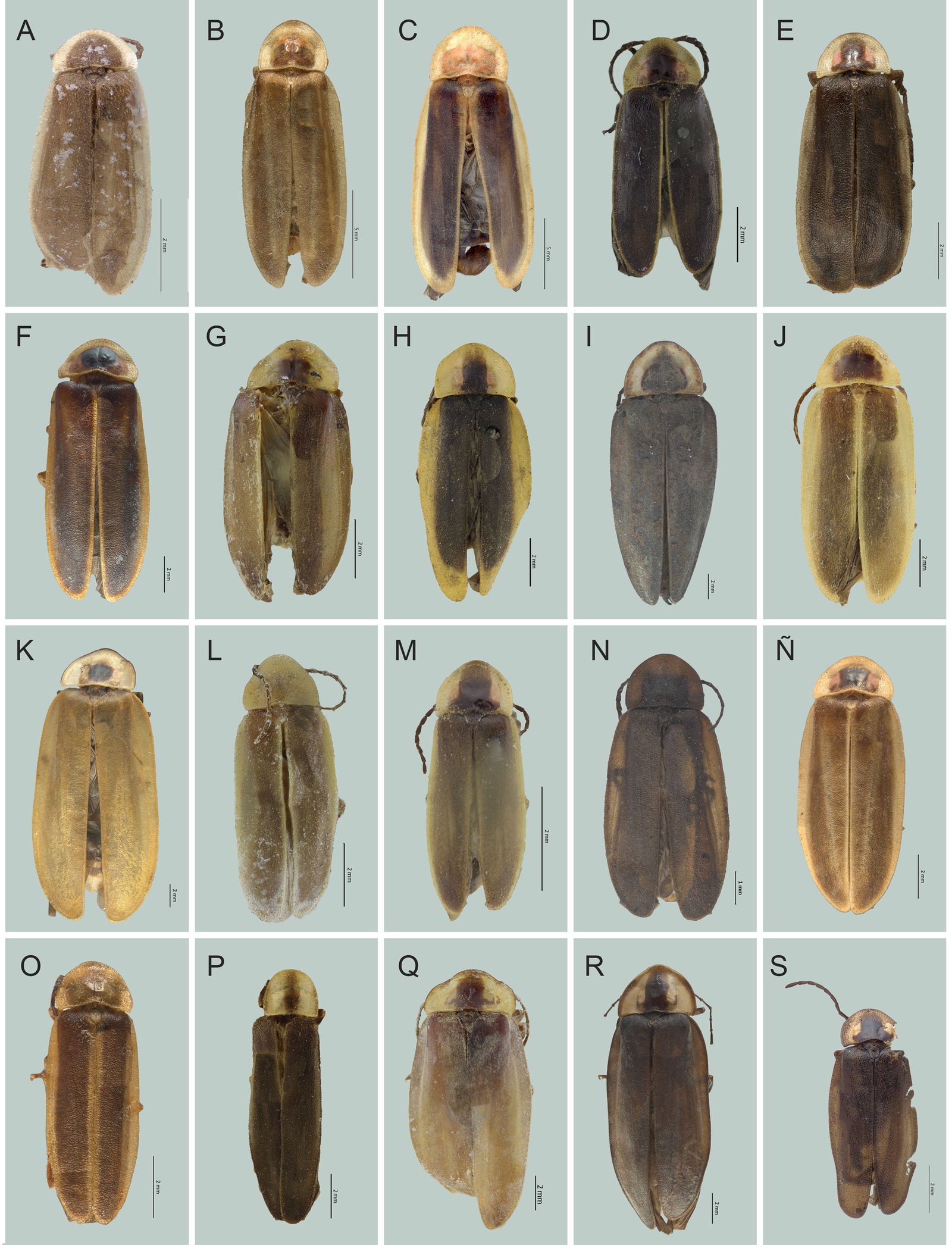
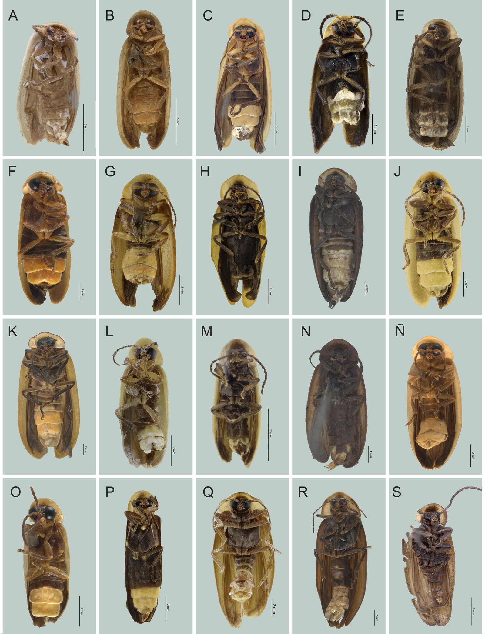
Figs. 5O, 6O, 7A, 8A
Macho. Largo 6.45 mm; ancho 2.72 mm; cuerpo semi paralelo; de color castaño claro, pronoto y élitros con márgenes amarillos, sin aparato luminoso. Cabeza: espacio interocular (0.55 mm) cóncavo, integumento brillante, chagrinado, con diminutas sedas; frente vertical, distancia interantenal (0.13 mm) más angosta que la fosita antenal (0.16 mm); ojos divergentes, finamente facetados, semiesféricos, un poco más largos (0.34 mm) que anchos (0.27 mm); antenas filiformes, comprimidas, 2 veces más largas que el pronoto, sin rebasar las coxas posteriores; escapo de 0.34 mm de longitud, casi cilíndrico, un poco más corto que los 2 siguientes antenitos juntos (0.41 mm), segundo corto (0.18 mm), tercero de 0.23 mm, cuarto-décimo de 0.25 a 0.27 mm, undécimo de 0.34 mm; sutura frontoclipeal membranosa, cóncava, clípeo trapezoidal, borde anterior recto, superficie espaciadamente punteada, con sedas largas y erectas; mandíbulas falcadas, con sedas basales; palpómero maxilar apical fusiforme, robusto, palpómero labial securiforme. Tórax: pronoto ojival, más ancho (1.81 mm) que largo (1.27 mm), borde posterior recto, ángulos posteriores obtusos, disco brillante, con mancha castaña más oscura en el tercio anterior, convexo, lados ampliamente explanados, con poros glandulares ordenados en el margen, más abundantes en la parte anterior del disco; escutelo casi triangular, con borde posterior romo, superficie brillante, diminutamente punteada y pilosidad decumbente, una más larga; epipleura angosta, espiráculos respiratorios no tubulares, transversos; patas cortas, pro y mesopatas semejantes entre sí, las posteriores un poco más largas, fémures comprimidos, casi fusiformes, tibias acanaladas, apicalmente dilatadas, borde externo no crenulado, tarsómeros lateralmente comprimidos, primer metatarsómero más corto (0.11 mm) que los 2 siguientes juntos (0.18 mm), cuarto cubriendo casi todo el quinto, uñas simples. Abdomen: penúltimo tergito con ángulos agudos, pigidio no visible; esternitos 5-6 más cortos que los precedentes, sin puntuaciones estigmatiformes, 7 con borde posterior cóncavo, 8 espatulado; edeago (figs. 4O, 5O, 6O) con pieza basal simétrica, casi tan larga (0.31 mm) como ancha (0.27 mm), más corta que los lóbulos laterales (0.38 mm), con el borde dorsal posterior cóncavo, el proximal convexo; lóbulo medio basalmente cilíndrico, 3 veces tan largo (0.27 mm) como ancho (0.09 mm), porción apical acuminada, casi alcanzando el ápice de los lóbulos laterales, parte dorsal membranosa, ventral quitinosa, ápice membranoso, orificio medio ventralmente abierto, excrecencias dorso basales cortas, robustas, simétricas, de ápice romo, basalmente juntas; lóbulos laterales robustos, un poco menos de 2 veces tan largos (0.37 mm) como anchos (0.20 mm), interiormente poco excavados, en vista dorsal con ápice redondeado, ventralmente dilatados en la parte media, lateralmente casi paralelos, sin modificaciones para la recepción de las excrecencias del lóbulo medio. Hembra desconocida.
Resumen taxonómico
Etimología: especie dedicada a Martha Isabel (Pati) Ruiz Corzo, activista reconocida por su labor a favor de la protección de la biodiversidad en la Sierra Gorda, en el estado de Querétaro, en el centro de México.
Material examinado: holotipo macho. México, Oaxaca, 1-12-2004, km 111 Carr. Oaxaca-Pto. Escondido, 1,443 m snm, 16º26’05” N, 97º01’58” O, E. Barrera. | COL.TIP-04017.
Comentarios taxonómicos
Especie semejante a Photinus elvirae sp. nov.; sin embargo, las excrecencias son más largas y dirigidas hacia arriba en P. isabelae, en P. elvirae se dirigen lateralmente. Los lóbulos laterales en P. isabelae se juntan en el ápice, en P. elvirae están juntos desde la mitad hacia el ápice. También cercana a P. stephanieae sp. nov., de la que se distingue por el tamaño y forma del edeago. Ver diagnosis Photinus juanrulfoi Zaragoza-Caballero y Domínguez-León, 2023
Diagnosis: en Zaragoza-Caballero et al. (2023).
Distribución: Durango. Nuevo registro: Chiapas (CNIN). Material examinado: México, Chiapas, III-13-53, R.C. Bechtel, E.I. Schinger collectors.
Photinus juliae Zaragoza-Caballero y González-Ramírez sp. nov.
http://zoobank.org/urn:lsid:zoobank.org:act:728CED0C-C87D-42AF-AEF7-F2BEAF902B39
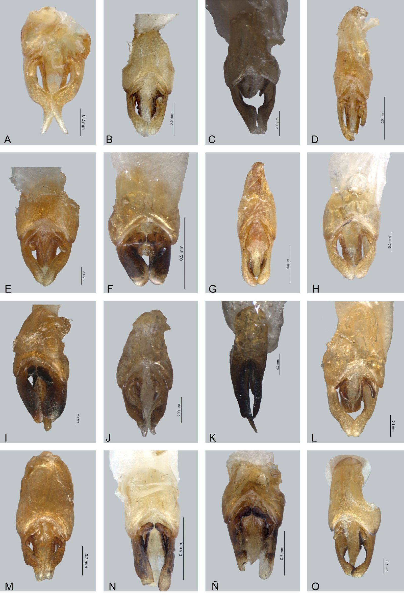
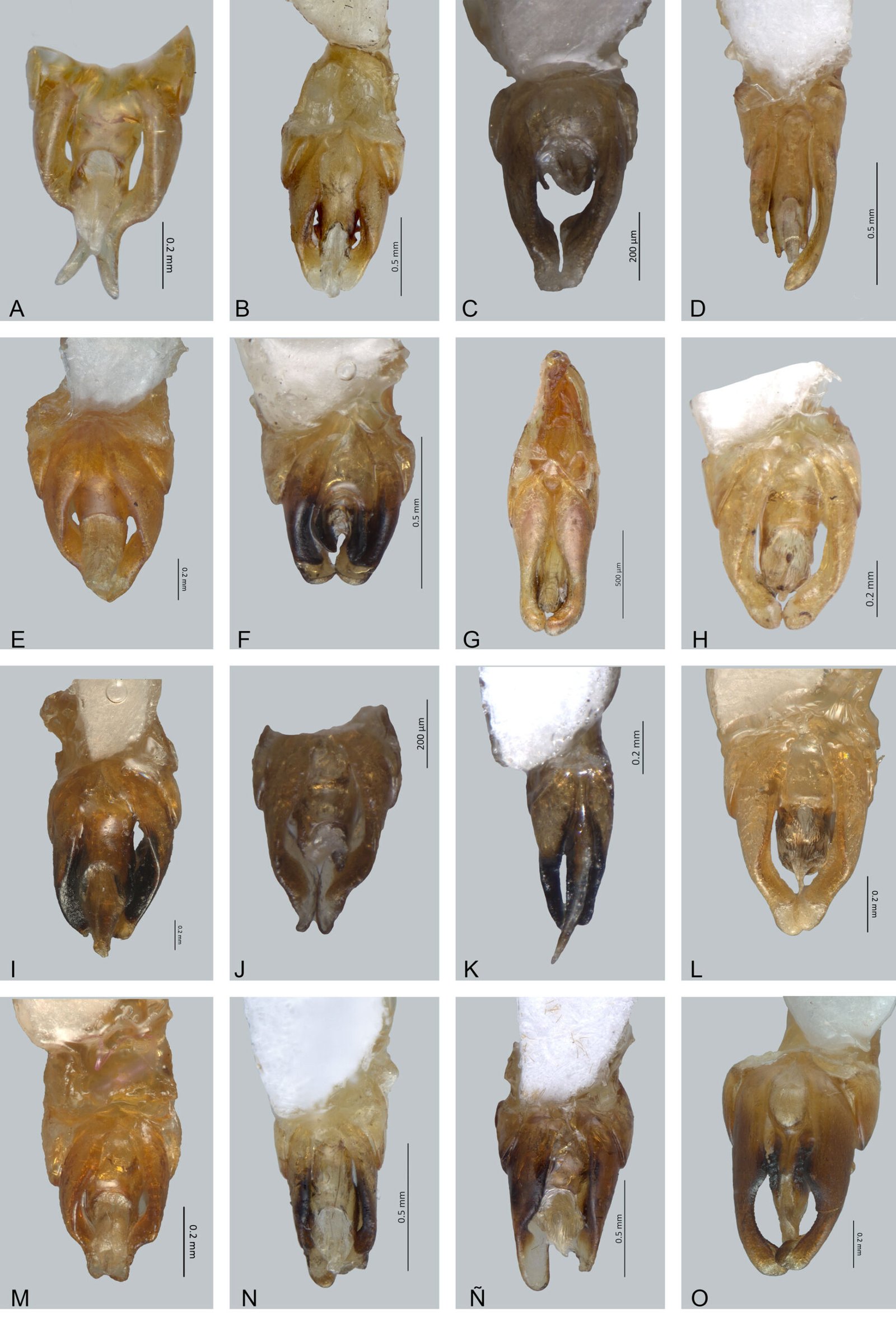
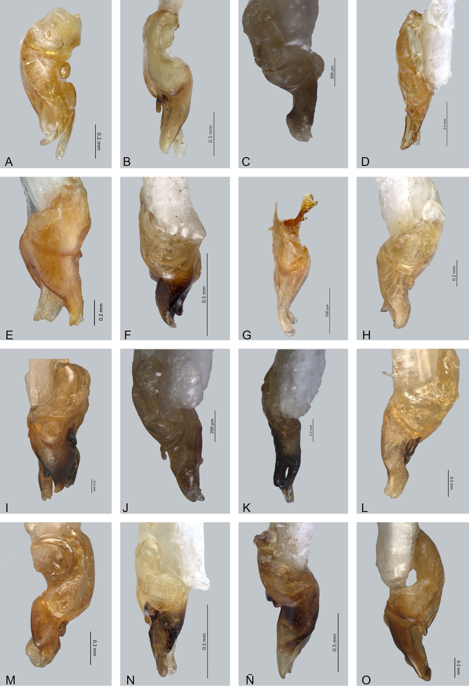
Figs. 7B, 8B, 9A, 10A, 11A
Macho. Largo 14.09 mm, ancho 4.54 mm; cuerpo casi paralelo; color castaño con excepción de: pronoto amarillo crema, con una banda longitudinal castaña en forma de rectángulo y esbozos de máculas laterales que posiblemente fueron rojizas; epipleura y élitros con una banda longitudinal amarillo crema; coxas, trocánteres y fémures ambarinos; aparato luminoso ocupando los esternitos 5-6. Cabeza: espacio interocular (0.88 mm), cóncavo, paralelo, integumento brillante, chagrinado, con sedas largas; frente vertical, distancia interantenal (0.29 mm), más angosta que fosita antenal (0.32 mm); ojos finamente facetados, semiesféricos, prominentes, tan largos (0.67 mm) como anchos (0.67 mm); antenas filiformes y largas (5.84 mm), casi 2 veces más largas que el pronoto, sobrepasando el borde posterior del metaesterno, escapo alcanzando 0.67 mm de longitud, claviforme, más corto que los 2 antenitos anteriores juntos (0.83 mm), segundo antenito corto (0.21 mm), tercero-décimo de 0.62 mm; sutura frontoclipeal membranosa, cóncava; clípeo diminuto y trapezoidal, con borde anterior recto, superficie espaciadamente punteada, con sedas largas escasas, uniformemente distribuidas; mandíbulas falcadas, esbeltas, angularmente arqueadas, con sedas en su base externa; palpómero maxilar apical securiforme, robusto; palpómero labial securiforme. Tórax: pronoto semicircular, más ancho (4.09 mm) que largo (3 mm), borde posterior recto, ángulos posteriores obtusos, disco convexo, explanaciones laterales anchas y acanaladas, con poros glandulares espaciadamente distribuidos con excepción de la superficie medio-posterior, superficie brillante con pilosidad abundante y decumbente; escutelo espatulado con borde posterior romo, superficie plana, lisa y con pilosidad escasa, decumbente; élitros paralelos, casi 5 veces más largos (11.09 mm) que anchos (2.27 mm), integumento chagrinado y rugosamente punteado, opaco, pilosidad decumbente de 2 tipos: una larga y otra corta; epipleura angosta, limitada por una costilla ancha en la base y que gradualmente disminuye al ápice; espiráculos respiratorios mesotorácicos tubulares; patas largas y esbeltas, pro y mesopatas semejantes entre sí, las posteriores un poco más largas, fémures comprimidos, fusiformes, tibias aplanadas y acanaladas, apicalmente dilatadas, borde externo crenulado, tarsómeros lateralmente comprimidos, primer metatarsómero más largo (0.55 mm) como los 2 siguientes juntos (0.51 mm), cuarto bífido, cubriendo casi todo el quinto, uñas simples. Abdomen: esternitos 5-6 más cortos que los precedentes, con puntuaciones estigmatiformes, borde posterior del sexto esternito escotado, séptimo con el borde posterior cóncavo, octavo espatulado, borde posterior del pigidio ligeramente bicóncavo; edeago (figs. 9A, 10A, 11A) con el borde dorsal posterior cóncavo de la pieza basal; lóbulo medio más de 3 veces más largo (0.61 mm) que ancho (0.20 mm), cónico, más corto que los lóbulos laterales (0.76 mm), con parte dorsal membranosa y ventral con base esclerosada y parte apical membranosa, orificio medio ventralmente abierto, excrecencias dorso basales simétricas, como lóbulos largos, divergentes; lóbulos laterales interiormente excavados y con ápice en vista dorsal agudos, ventralmente dilatados en la parte media, en vista lateral casi paralelos, no modificados para la recepción de las excrecencias del lóbulo medio. Hembra desconocida.
Resumen taxonómico
Etimología: especie dedicada a la Dra. Julia Carabias Lillo cuya carrera se ha centrado en la ecología, conservación de ecosistemas y el manejo de recursos naturales, especialmente en la selva Lacandona.
Material examinado: holotipo macho: México: Chiapas: Comitán, 14-VIII-1956. D. H. Janzen Collector. | Photinus sp. 63 (Laporte, 1833) | Colección del Instituto de Biología, UNAM, México D.F. | QR 51904 | COL. TIP-04018.
Comentarios taxonómicos
Especie morfológicamente similar a Photinus tepetzala Zaragoza-Caballero y Vega-Badillo, 2020 de la cual se distingue por la forma del espacio interocular y del borde posterior del clípeo, así como la longitud de los esternitos 5-6 y de las excrecencias dorso basales. En P. tepetzala el espacio interocular es convexo, en cambio en P. juliae sp. nov. es cóncavo; el borde anterior del clípeo en P. tepetzala es escotado, mientras que en P. juliae sp. nov., es recto; en P. tepetzala los esternitos 5-6 son más largos que los precedentes, en contraste en P. juliae sp. nov., son más cortos; las excrecencias dorso basales en P. tepetzala son lóbulos cortos, mientras que en P. juliae sp. nov., son lóbulos largos.
Photinus kaedii Zaragoza-Caballero y González-Ramírez, 2023 en Zaragoza-Caballero et al. (2023)
Diagnosis: en Zaragoza-Caballero et al. (2023).
Distribución: Sinaloa. Nuevo registro: Guerrero (CNIN). Material examinado: México, Guerrero, Olinala las Juntas, 26-VIII-2005, 1,485 m snm, 17°47’38” N, 98°46’17” O, L. Cervantes, R. Carranza.
Photinus kleriocaro Zaragoza-Caballero, 2005
Diagnosis: en Zaragoza-Caballero et al. (2023).
Distribución: Colima, Jalisco, Guerrero, Nayarit y Sinaloa.
Photinus lazcanoi Zaragoza-Caballero y González-Ramírez sp. nov.
http://zoobank.org/urn:lsid:zoobank.org:act:E1EF76C4-B4B4-4A92-A0C7-CCD852CB5859
Figs. 7C, 8C, 9B, 10B, 11B
Macho. Largo 20.45 mm, ancho 7.28 mm; cuerpo casi paralelo; color castaño con excepción de: pronoto ambarino y esbozos de máculas laterales rojizas; élitros el margen interno y externo ambarinos; coxas, trocánteres, fémures y parte media del metasterno amarillos; aparato luminoso ocupando los esternitos 5-6. Cabeza: espacio interocular (1.08 mm), plano, paralelo, integumento brillante, chagrinado, con largas sedas; frente vertical, distancia interantenal (0.26 mm), más angosta que fosita antenal (0.34 mm); ojos finamente facetados, semiesféricos, prominentes, más largos (1.13 mm) que anchos (0.62 mm); antenas filiformes y largas (6.24 mm), una vez y media más largas que el pronoto, sobrepasando el borde posterior del metaesterno, escapo alcanzando 0.62 mm de longitud, claviforme, más corto que los 2 antenitos anteriores juntos (0.78 mm), segundo antenito corto (0.26 mm), tercero más corto que los subsiguientes (0.52 mm), cuarto-décimo de 0.57 mm, undécimo 0.85 mm; sutura frontoclipeal membranosa, cóncava; clípeo trapezoidal, con borde anterior recto, superficie punteada, con sedas largas irregularmente distribuidas; mandíbulas falcadas, robustas, angularmente arqueadas, con sedas en su base externa; palpómero maxilar apical securiforme, robusto; palpómero labial securiforme. Tórax: pronoto semicircular, más ancho (5.64 mm) que largo (4.09 mm), borde posterior recto, ángulos posteriores obtusos, disco convexo, explanaciones laterales angostas y acanaladas, con poros glandulares espaciadamente distribuidos con excepción de la superficie medio-posterior, superficie brillante con pilosidad escasa y decumbente; escutelo espatulado con borde posterior romo, superficie plana, lisa y con pilosidad abundante, decumbente; élitros paralelos, 4 veces y media más largos (16.36 mm) que anchos (3.64 mm), integumento chagrinado y rugosamente punteado, opaca, pilosidad decumbente de 2 tipos: una larga y otra corta; epipleura angosta, limitada por una costilla ancha en la base y que gradualmente disminuye al ápice; espiráculos respiratorios mesotorácicos tubulares; patas cortas y esbeltas, pro y mesopatas semejantes entre sí, las posteriores un poco más largas, fémures ligeramente robustos, casi fusiformes, tibias aplanadas y acanaladas, apicalmente dilatadas, borde externo no crenulado, tarsómeros lateralmente comprimidos, primer metatarsómero más largo (0.79mm) como los 2 siguientes juntos (0.60 mm), cuarto bífido, cubriendo casi todo el quinto, uñas simples. Abdomen: esternitos 5-6 más cortos que los precedentes, con puntuaciones estigmatiformes, borde posterior del sexto esternito emarginado, borde posterior del pigidio cóncavo; edeago (figs. 9B, 10B, 11B) con pieza basal simétrica, tan larga (0.92 mm) como los lóbulos laterales (0.92 mm), con borde dorsal posterior cóncavo, anterior convexo; lóbulo medio 3 veces más largo (0.85 mm) que ancho (0.25), cónico, más corto que los lóbulos laterales, con parte dorsal membranosa y ventral con base esclerosada y parte apical membranosa, base con 2 estructuras de apariencia molar, orificio medio ventralmente abierto, excrecencias dorso basales simétricas, como lóbulos largos, divergentes; lóbulos laterales interiormente excavados y ápice en vista dorsal redondeada, ventralmente dilatados en la parte media, en vista lateral casi paralelos, con ápice romo, no modificados para la recepción de las excrecencias del lóbulo medio. Hembra desconocida.
Resumen taxonómico
Etimología: especie dedicada al Dr. Antonio Eusebio Lazcano Araujo Reyes, reconocido por su investigación en el origen y evolución temprana de la vida, así como por su destacada labor como divulgador científico.
Material examinado: holotipo macho: México: Guerrero: Petaquillas, 9 kms. O. Chilpancingo, 6-VII-1963. F. Pacheco, M | Photinus sp. 51 (Laporte, 1833) | Colección del Instituto de Biología, UNAM. México, D.F. | QR 51854 | COL.TIP-04019.
Comentarios taxonómicos
Especie morfológicamente similar a Photinus toledoi Zaragoza-Caballero, 2000 de la cual se distingue por su tamaño corporal, la forma del borde posterior del sexto esternito y del pigidio. En P. toledoi el tamaño corporal es menor con respecto a P. lazcanoi sp. nov.; el borde posterior del del sexto esternito en P. toledoi es escotado, mientras que en P. lazcanoi sp. nov., es emarginado; en P. toledoi el borde posterior del pigidio es trunco, en contraste en P. lazcanoi sp. nov. es cóncavo.
Photinus lekoalloios Zaragoza-Caballero, 1996
Diagnosis: en Zaragoza-Caballero et al. (2023).
Distribución: Chiapas y Veracruz.
Photinus lloydi Zaragoza-Caballero y Pérez-Flores sp. nov.
http://zoobank.org/urn:lsid:zoobank.org:act:64844775-876D-4FAF-88DF-854D0B7FCE3D
Figs. 7D, 8D, 9C, 10C, 11C
Macho. Largo 9.43 mm, ancho 3.94 mm; cuerpo paralelo; integumento mayormente de color castaño; pronoto con los márgenes laterales anchos, amarillo pálido, centralmente castaño rojizo, con las orillas ligeramente naranjas; élitros con sutura elitral y márgenes laterales amarillos, el resto de la superficie castaña; coxas, trocánteres y parte anterior de las patas, castaño amarillento; aparato luminoso ocupando los esternitos 5-7. Cabeza: espacio interocular (0.66 mm), ligeramente deprimido, integumento brillante, chagrinado, con sedas cortas; frente casi vertical, distancia interantenal (0.16 mm), más angosta que la fosita antenal (0.19 mm); ojos divergentes, finamente facetados, semiesféricos, más largos (0.68 mm) que anchos (0.49 mm); antenas comprimidas, largas, sobrepasando la mitad del metasterno, escapo de 0.34 mm de longitud, casi cilíndrico, ligeramente más ancho en el ápice, más corto que los 2 siguientes antenitos juntos (0.47 mm), el segundo corto (0.11 mm), el tercero de 0.24 mm, el cuarto de 0.26 mm, del quinto al décimo 0.19 mm, y el undécimo 0.29 mm de largo; sutura frontoclipeal membranosa, cóncava; clípeo trapezoidal, borde anterior ligeramente redondeado, superficie con abundantes puntos, poco separados, con sedas largas y erectas; mandíbulas falcadas, esbeltas, con sedas basales; palpómero maxilar apical fusiforme, robusto, labial securiforme. Tórax: pronoto más ancho (2.92 mm) que largo (1.97 mm), ojival, borde posterior ligeramente ondulado, ángulos posteriores casi rectos, disco brillante, convexo, lados ampliamente explanados, con poros glandulares ordenados en el margen, distribuidos homogéneamente; escutelo lingüiforme, con el borde posterior redondeado, superficie brillante, con puntuaciones toscas y pilosidad decumbente; élitros más de 3.5 veces más largos (7.48 mm) que anchos (2.07 mm), superficie rugosamente punteada, brillante, con 2 tipos de pilosidad decumbente, una más larga y menos abundante; epipleura reducida; espiráculos respiratorios mesotorácicos tubulares; patas largas, pro y meso-patas semejantes entre sí, las posteriores un poco más largas, fémures comprimidos, fusiformes, tibias acanaladas, apicalmente dilatadas, borde externo crenulado, tarsómeros comprimidos lateralmente, primer metatarsómero ligeramente más largo (0.24 mm) que los 2 siguientes juntos (0.21 mm), cuarto bífido, cubriendo la mitad del quinto, uñas simples. Abdomen: últimos tergitos con los ángulos posteriores agudos, pigidio convexo, con ángulos posteriores redondeados; esternitos 5-6 poco menos cortos que los precedentes, con puntuaciones estigmatiformes, 7 con el borde posterior ligeramente cóncavo, 8 espatulado; edeago (figs. 9C, 10C, 11C) con la pieza basal simétrica, más larga (0.58 mm) que ancha (0.44 mm), apenas más larga que los lóbulos laterales (0.56 mm), con borde dorsal posterior cóncavo, proximal convexo; lóbulo medio casi cilíndrico, con ápice no rebasando a los lóbulos laterales, parte dorsal membranosa y ventralmente quitinoso, excrecencias dorso basales cortas, simétricas, lobuladas, divergentes, basalmente separadas; lóbulos laterales esbeltos, casi 2.5 veces tan largos (0.56 mm) como altos (0.23 mm), interiormente excavados, en vista dorsal con ápice redondeado, en vista lateral más altos en su base, sin modificaciones para la recepción de excrecencias del lóbulo medio. Hembra desconocida.
Resumen taxonómico
Etimología: especie dedicada a James E. Lloyd, conocido como “Firefly Doc” por sus trabajos pioneros con luciérnagas y sus numerosas contribuciones al estudio de esta familia.
Material examinado: holotipo macho. México, Chiapas, Ocosingo, km 7 Ocosingo-San Cristóbal, 1,180 m snm, 16º50’33” N, 92º06’15” O, 27/VI/07, Bosque Pino-Encino, C. Mayorga, G. Ortega, L. Cervantes. | COL. TIP-04020.
Comentarios taxonómicos
Esta especie es morfológicamente similar a Photinus kleriocaro Zaragoza-Caballero, 2005 y a Photinus poncianoae Zaragoza-Caballero-Pérez-Flores, 2024, de las cuales se distingue por el tamaño y la forma del edeago. El tamaño de P. kleriocaro es menor que Photinus lloydi sp. nov., mientras que P. poncianoae es de mayor talla. Del edeago principalmente se diferencia P. lloydi sp. nov. de las otras 2 especies por las excrecencias dorso basales distintivamente cortas y el lóbulo medio acortado. La forma general también se asemeja a Photinus tuxtlaensis (Zaragoza-Caballero, 1995), del que se diferencia por la forma del edeago, notablemente más estrecho y sin excrecencias dorsobasales en P. tuxtlaensis, más ancho y con excrecencias dorsobasales cortas en P. lloydi sp. nov., además, el tamaño de esta última es mayor que en P. tuxtlaensis.
Photinus lunicollis Gorham, 1880
Diagnosis: en Zaragoza-Caballero, López-Pérez et al. (2020).
Distribución: Puebla y Oaxaca.
Photinus mariaeugeniae Zaragoza-Caballero y López-Pérez sp. nov.
http://zoobank.org/urn:lsid:zoobank.org:act:D14BE2C9-E2D5-4DFC-AE55-8B4C4BBC5241
Figs. 7E, 8E, 9D, 10D, 11D
Macho. Largo 11.2 mm, ancho 3.26 mm; cuerpo casi paralelo; cabeza, antenas y patas cafés; disco pronotal con 2 manchas rojizas a los lados, el centro y la base, castaño, el resto del pronoto amarillento; élitros cafés con excepción de los márgenes laterales y el margen elitral; mancha lútea ocupando esternitos 5-7. Cabeza: espacio interocular (0.5 mm), poco deprimido, paralelo, integumento opaco, finamente punteado, con sedas cortas esparcidas; frente vertical, distancia interantenal (0.1 mm), más angosta que fosita antenal (0.2 mm); ojos finamente facetados, esféricos, poco más largos (0.6 mm) que anchos (0.4 mm); escapo de 0.4 mm de longitud, casi cilíndrico, más corto que los 2 siguientes antenitos juntos (0.5 mm), segundo muy corto (0.1 mm), tercero 0.4 mm, cuarto 0.5 mm, quinto-séptimo 0.6, octavo 0.7 mm, noveno-décimo 0.6 mm, onceavo 0.8 mm; sutura frontoclipeal membranosa, sinuada; clípeo trapezoidal, borde anterior recto, superficie espaciadamente punteada, con sedas largas y erectas; mandíbulas falcadas, con sedas basales; palpómero maxilar y labial apical securiforme. Tórax: pronoto más ancho (2.3 mm) que largo (1.9 mm), semicircular, borde posterior recto, ángulos posteriores obtusos, disco brillante, con un surco en el medio, lados explanados, con poros glandulares ordenados en el margen, más abundantes en parte anterior del disco; escutelo espatulado, con el borde posterior redondo, superficie brillante, 3 puntos gruesamente marcados y con poca pilosidad; élitros más de 5 veces más largos (9.3 mm) que anchos (1.6 mm), superficie rugosamente punteada, opaca, con 2 pilosidades decumbentes; epipleura angosta; espiráculos respiratorios mesotorácicos tubulares; pro y meso-patas semejantes entre sí, posteriores un poco más largas, fémures comprimidos, fusiformes, tibias aplanadas, apicalmente dilatadas, borde externo no crenulado, tarsómeros lateralmente comprimidos, primer metatarsómero tan largo (0.3 mm) como los 2 siguientes juntos, cuarto bífido, cubriendo casi todo el quinto, uñas simples. Abdomen: penúltimo tergito con ángulos posteriores romos; esternitos 5-6 más cortos que los precedentes, sin puntuaciones estigmatiformes, 7 con borde posterior cóncavo, 8 espatulado; edeago (figs.9D, 10, D, 11D) con pieza basal simétrica, más corta (0.2 mm) que los lóbulos laterales (0.3 mm), con borde dorsal posterior cóncavo; lóbulo medio poco más de 2 veces más largo que ancho (0.3 mm), cilíndrico, misma longitud que lóbulos laterales (0.3 mm), membranoso en vista dorsal y ventralmente un tanto quitinoso, con excrecencias dorso basales, alargadas, dirigidas hacia los laterales; lóbulos laterales con el ápice redondeado, curvo en vista dorsal y ventral. Hembra desconocida.
Resumen taxonómico
Etimología: especie dedicada a la Biól. María Eugenia Díaz Batres por su larga trayectoria como curadora de la Colección Entomológica del Museo de Historia Natural y Cultura Ambiental de la Ciudad de México.
Material examinado: México: Chiapas: Reserva El
Triunfo, 6-V-1993. | COL.TIP-04021.
Comentarios taxonómicos
Es cercana a P. morronei Zaragoza-Caballero, 2020 en la forma del edeago; sin embargo, la longitud de este en P. morronei es más larga; el cuerpo de P. morronei es paralelo, sin líneas blanquecinas en los márgenes laterales de los élitros; el pronoto ojival en P. morronei y semicircular en P. mariaeugeniae sp. nov. La longitud total del cuerpo es mayor en P. morronei.
Photinus mariomolinai Zaragoza-Caballero y Cifuentes-Ruiz sp. nov.
http://zoobank.org/urn:lsid:zoobank.org:act:2D9229A2-E96A-41AA-8C3E-A379746EC457
Figs. 7F, 8F, 9E, 10E, 11E
Macho. Largo 17 mm, ancho 6 mm; cuerpo semi paralelo; de color negro con márgenes amarillos, pronoto amarillo, con una mancha negra en el disco y lados cremosos; aparato luminoso ocupando los esternitos 5-6. Cabeza: espacio interocular (1 mm), cóncavo, integumento brillante, chagrinado, con diminutas sedas; frente vertical, distancia interantenal (0.24 mm), más angosta que la fosita antenal (0.32 mm); ojos divergentes, finamente facetados, semiesféricos, más largos (1.2 mm) que anchos (0.76 mm); antenas filiformes, comprimidas, largas, 1.6 veces más largas que el pronoto, rebasando las coxas posteriores, escapo de 0.84 mm, casi cilíndrico, un poco más largo que los 2 siguientes antenitos juntos (0.76 mm), segundo corto (0.16 mm), tercero de 0.6 mm, cuarto-décimo 0.52, undécimo 0.64 mm; sutura frontoclipeal membranosa, cóncava; clípeo trapezoidal, borde anterior recto, superficie espaciadamente punteada, con sedas largas y erectas; mandíbulas falcadas, con sedas basales; palpómero maxilar apical securiforme, robusto; palpómero labial securiforme. Tórax: pronoto semicircular, más ancho (5 mm) que largo (3.6 mm), borde posterior recto, ángulos posteriores obtusos, disco brillante, convexo, lados ampliamente explanados, con poros glandulares ordenados en el margen, más abundantes en la parte anterior del disco; escutelo casi triangular, con el borde posterior romo, superficie brillante, diminutamente punteada y pilosidad decumbente; élitros casi 5 veces más largos (13.4 mm) que anchos (3 mm), superficie rugosamente punteada, brillante, con 2 tipos de pilosidad decumbente, una más larga; epipleura angosta; espiráculos respiratorios mesotorácicos tubulares; patas cortas, pro y mesopatas semejantes entre sí, las posteriores un poco más largas, fémures comprimidos, casi fusiformes, tibias acanaladas, apicalmente dilatadas, borde externo no crenulado, tarsómeros lateralmente comprimidos, primer metatarsómero más largo (0.6 mm) que los 2 siguientes juntos (0.44 mm), cuarto bífido, cubriendo casi todo el quinto, uñas simples. Abdomen: penúltimo tergito con ángulos posteriores agudos, pigidio convexo; esternitos 5-6 casi tan largos como precedentes, con puntuaciones estigmatiformes, 7 con el borde posterior cóncavo, 8 espatulado; edeago (figs. 9E, 10E, 11E) con la pieza basal simétrica, un poco más ancha (0.64 mm) que larga (0.54 mm), más corta que los lóbulos laterales (0.78 mm), con el borde dorsal posterior cóncavo, el proximal convexo; lóbulo medio basalmente cilíndrico, una vez y media tan largo (0.38 mm) como ancho (0.24 mm), porción apical acuminada, casi alcanzando el ápice de los lóbulos laterales, parte dorsal membranosa, la ventral quitinosa, ápice membranoso, orificio medio ventralmente abierto, excrecencias dorso basales robustas, simétricas, lobuladas, divergentes, basalmente separadas; lóbulos laterales esbeltos, casi 2 veces y media tan largos (0.78 mm) como altos (0.32 mm), interiormente excavados, en vista dorsal con el ápice redondeado, lúteo, ventralmente dilatados en la parte media, lateralmente casi paralelos, sin modificaciones para la recepción de las excrecencias del lóbulo medio. Hembra desconocida.
Resumen taxonómico
Etimología: especie dedicada a Mario Molina, científico mexicano, premio Nobel de Química en 1995. También se dedicó a la promoción de políticas públicas para la calidad del aire y la protección ambiental en México y el mundo.
Material examinado: holotipo macho. México, Simojovel, Chiapas, VIII-1-16-58. J.A. Chemsak. | COL. TIP-04022.
Comentarios taxonómicos
Especie similar a Photinus avendanoae sp. nov., pero difiere en las excrecencias dorsobasales del edeago; además, los lóbulos laterales en P. mariomolinae en vista ventral rodean al lóbulo medio, en P. avendanoae quedan juntos en el ápice y no rodean el lóbulo medio. También se asemeja a P. franckei; sin embargo, en esta última especie los lóbulos laterales en vista dorsal se juntan desde la mitad hacia el ápice formando una curva pronunciada, en P. mariomolinai se juntan solo en el ápice; la forma de las excrecencias es distinta entre las 2 especies.
Photinus margaritae Zaragoza-Caballero y Domínguez-León sp. nov.
http://zoobank.org/urn:lsid:zoobank.org:act:500FA875-9606-4291-9A98-33E629387D60
Figs. 7G, 8G, 9F, 10F, 11F
Macho. Largo 9.36 mm, ancho 4.43 mm; cuerpo semi paralelo; color castaño claro, pronoto con márgenes laterales ambarinos y el centro con mancha castaña que se extiende cerca del margen anterior; aparato luminoso ocupando esternitos 5-7. Cabeza: espacio interocular (0.62 mm), cóncavo, integumento brillante, chagrinado, con diminutas sedas; frente vertical, distancia interantenal (0.16 mm), más angosta que fosita antenal (0.21 mm); ojos divergentes, finamente facetados, semiesféricos, más largos (1.01 mm) que anchos (0.57 mm); antenas comprimidas, largas, rebasando coxas posteriores, escapo 0.72 mm de longitud, semitriangular, un poco más largo que los 2 siguientes antenitos juntos (0.62 mm), segundo corto (0.23 mm), tercero de 0.39 mm, del cuarto-décimo 0.42 mm y, undécimo 0.96 mm de largo; sutura frontoclipeal membranosa, cóncava; clípeo trapezoidal, borde anterior recto, superficie espaciadamente punteada, con sedas largas y erectas; mandíbulas falcadas, con sedas basales; palpómero maxilar apical fusiforme, robusto; labial securiforme. Tórax: pronoto más ancho (3.33 mm) que largo (2.41 mm), ojival, borde posterior recto, ángulos posteriores obtusos, disco brillante, convexo, lados ampliamente explanados, poros glandulares ordenados en el margen, más abundantes en parte anterior del disco; escutelo casi triangular, con el borde posterior romo, superficie brillante, diminutamente punteada y pilosidad decumbente; élitros un poco más de 5 veces más largos (7.72 mm) que anchos (1.71 mm), superficie rugosamente punteada, opaca, con 2 tipos de pilosidad decumbente, una un poco más larga; epipleura angosta; espiráculos respiratorios mesotorácicos tubulares; patas cortas, pro y meso-patas semejantes entre sí, posteriores un poco más largas, fémures comprimidos, casi fusiformes, tibias acanaladas, apicalmente dilatadas, borde externo no crenulado, tarsómeros lateralmente comprimidos, primer metatarsómero más largo (0.5 mm) que 2 siguientes juntos (0.41 mm), cuarto bífido, cubriendo casi todo el quinto, uñas simples. Abdomen: pigidio convexo; esternitos 5-6 tan largos como precedentes, con puntuaciones estigmatiformes, 7 con borde posterior cóncavo, 8 no se encuentra en el ejemplar; edeago (figs. 9F, 10F, 11F) con pieza basal simétrica, tan larga (0.51 mm) como ancha (0.5 mm), más larga que lóbulos laterales (0.36 mm), con borde dorsal posterior cóncavo, proximal convexo; lóbulo medio basalmente cilíndrico, un poco más de 5 veces tan largo (0.32 mm) como ancho (0.06 mm), porción apical acuminada, no alcanzando ápice de lóbulos laterales, parte dorsal y ventral membranosa, orificio medio ventralmente abierto, excrecencias dorso basales más anchas en base, de forma ovalada; lóbulos laterales anchos, un poco más de 8 veces tan largos (0.36 mm) como anchos (0.17 mm), vista lateral con ápice con un dentículo, ventralmente con ápice no esclerotizado. Hembra desconocida.
Resumen taxonómico
Etimología: especie dedicada con cariño a la Dra. Margarita Canales de la Facultad de Estudios Superiores Iztacala, que confió, apoyó y motivó incondicionalmente durante su licenciatura al segundo autor de la especie para seguir en su desarrollo académico y personal.
Material examinado: holotipo macho. México: Oaxaca:
La Ventosa, 48 millas, VII/21/63 | COL.TIP-04023.
Comentarios taxonómicos
Especie morfológicamente similar a Photinus congruus Chevrolat, 1834 de la cual se distingue por su tamaño corporal y por la forma particular del edeago. El tamaño de P. congruus es mayor con respecto a P. margaritae sp. nov., además la primera especie mencionada presenta 2 líneas características en los élitros; el edeago en vista dorsal es muy parecido, sin embargo, las excrecencias dorsobales en P. margaritae sp. nov. son más pequeñas y más anchas en la base y sus lóbulos laterales son igual de anchos en la porción basal y distal, convergiendo en el ápice.
Photinus marthae Zaragoza-Caballero y Cifuentes-Ruiz, 2023 en Zaragoza-Caballero et al. (2023)
Diagnosis: en Zaragoza-Caballero et al. (2023)
Distribución: Oaxaca y Michoacán.
Photinus magdalenae Zaragoza-Caballero, 2015
Diagnosis: en Zaragoza-Caballero (2015).
Distribución: Guerrero y Oaxaca.
Photinus medellini Zaragoza-Caballero y Gutiérrez-Carranza sp. nov.
http://zoobank.org/urn:lsid:zoobank.org:act:B4CDBA69-890D-4EF9-8885-1B9E9812B0A6
Figs. 7H, 8H, 9G, 10G, 11G
Macho. Largo 12.55 mm, ancho 5.64 mm; cuerpo ovoide; color castaño oscuro con excepción de: pronoto amarillento con una mancha paralela negra, borde lateral y apical de los élitros y trocánteres amarillo pajizo; sin aparato luminoso. Cabeza: espacio interocular (0.72 mm), plano, paralelo, integumento chagrinado con pocas sedas; frente vertical; distancia interantenal más angosta (0.14 mm) que fosita antenal (0.28 mm); ojos finamente facetados, ovoides, más largos (0.62 mm) que anchos (0.48 mm); antenas comprimidas, largas (6.69 mm), sobrepasan las coxas posteriores, escapo (0.69 mm), cilíndrico, más corto que los 2 siguientes antenitos juntos (0.83), segundo corto (0.28 mm), tercero (0.55 mm), cuarto al décimo de 0.62 mm, undécimo de 0.83 mm; sutura frontoclipeal membranosa, cóncava; clípeo trapezoidal, superficie punteada, con sedas largas irregularmente distribuidas; mandíbulas falcadas; palpómero maxilar apical fusiforme, robusto; labial triangular. Tórax: pronoto semicircular, más ancho (4.18 mm) que largo (2.73 mm), borde posterior redondeado, ángulos posteriores ligeramente obtusos, disco convexo, lados ampliamente explanados, superficie chagrinada y opaca, poros glandulares distribuidos irregularmente con excepción del centro del disco; escutelo espatulado, con borde posterior truncado, superficie brillante, punteada y con poros; élitros 4 veces más largos (10.91 mm) que anchos (2.73 mm), superficie rugosamente punteada, opaca, con 2 tipos de pilosidad decumbente, una más larga; epipleura amplia en la base y reducida hacia el ápice; espiráculos respiratorios mesotorácicos tubulares; patas cortas y semejantes entre sí (4.14 mm), fémures comprimidos, casi fusiformes, tibias acanaladas, levemente dilatadas hacia el ápice, borde externo crenulado, tarsómeros lateralmente comprimidos, primer metatarsómero más largo (0.69 mm) que los 2 siguientes juntos (0.38 mm), cuarto bífido, casi cubriendo al quinto, uñas simples. Abdomen: tergitos 4-6 con ángulos posteriores agudos y proyectados, ventritos 5-6 más cortos que los precedentes, sin puntuaciones estigmatiformes, 7 con borde posterior cóncavo, pigidio truncado con borde posterior bisinuado, 8 ojival; edeago (figs. 9G, 10G, 11G): pieza basal simétrica, casi tan larga (0.97 mm) como los lóbulos laterales (1.00 mm), con borde dorsal posterior cóncavo con forma de “V”, lóbulo medio 4 veces más largo (0.83 mm) que ancho (0.21 mm), cilíndrico, ápice no rebasando los lóbulos laterales, dorso-lateralmente esclerosado, superficie glabra con excepción de la región latero-apical que es rugosa, membranoso hacia el ápice, orificio medio ventralmente abierto, excrecencias dorso basales robustas, en forma de protuberancias connadas al lóbulo medio, lóbulos laterales dilatados en la base, sin modificaciones para la recepción de las excrecencias del lóbulo medio. Hembra desconocida.
Resumen taxonómico
Etimología: especie dedicada al Dr. Rodrigo A. Medellín Legorreta, en reconocimiento a su trayectoria centrada en la ecología y conservación de murciélagos. El epíteto específico corresponde al primer apellido.
Material examinado: holotipo macho. México, Guerrero, Atoyac de Álvarez, La Golondrina, 1,800 m snm, 06-VIII-1983, B. M. M. (bosque mesófilo de montaña), Col. Luis A. | QR 51990 | COL.TIP-04023.
Comentarios taxonómicos
Especie cercana a Photinus davilae sp. nov. de la que se distingue por el tamaño, patrón de coloración y forma del edeago. P. medellini sp. nov. tiene una longitud menor de 12.55 mm, no obstante, presenta un cuerpo más ovoide con un ancho total de 5.64 mm, la mancha del pronoto es paralela, el margen amarillento de los élitros es ancho, la pieza basal es más larga que ancha y el lóbulo medio no rebasa los lóbulos laterales. P. davilae presenta una longitud de 14.18 mm y 4.18 mm de ancho, la mancha del pronoto cubre casi en su totalidad el disco, el margen amarillento de los élitros es esbelto, la pieza basal es tan larga como ancha y el lóbulo medio casi alcanza el ápice de los lóbulos laterales.
Photinus meteoralis Gorham, 1881
Fig. 7I, 8I
Longitud 19-21 mm largo, ancho. Color castaño oscuro con excepción del pronoto que es amarillento con una mancha oscura en el disco. Pronoto ojival, margen basal casi recto, margen apical redondeado, ángulos poco agudos. Élitros más anchos en la base, angostos hacia el ápice, con una costilla en el medio poco visible. Aparato luminoso ocupando parte del esternito 4 y 5-6. Edeago: pieza basal tan larga como los lóbulos laterales que dorsalmente cubren al lóbulo medio en su base, excrecencias dorso-basales más menos de forma renada y estrechamente relacionadas, lóbulos laterales casi triangulares en vista lateral y modificados para la recepción de las excrecencias.
Distribución: Chiapas.
Photinus mireyae Zaragoza-Caballero, 2015
Diagnosis: en Zaragoza-Caballero (2015).
Distribución: Guerrero.
Photinus moestus (Perty, 1830)
Diagnosis: coloración oscura, cuerpo paralelo; antenas serradas; pronoto semicircular con el disco negro; élitros paralelos.
Distribución: Oaxaca.
Photinus morelosensis Zaragoza-Caballero, 2000
Diagnosis: en Zaragoza-Caballero, López-Pérez et al. (2020).
Distribución: Ciudad de México, Chiapas, Estado de México, Hidalgo, Morelos y Oaxaca.
Photinus navarretei Zaragoza-Caballero, 2015
Diagnosis: en Zaragoza-Caballero (2015).
Distribución: Guerrero.
Photinus nerivelai Zaragoza-Caballero y Rodríguez-Mirón sp. nov.
http://zoobank.org/urn:lsid:zoobank.org:act:DFE91B74-9E56-4D15-9AC2-1363DE8DB987
Figs. 7J, 8J, 9H, 10H, 11H
Macho. Largo 12.19 mm, ancho 2.29 mm. Cuerpo casi paralelo; de color castaño; pronoto con lados amarillos, disco castaño; élitros cafés, lados amarillentos; aparato luminoso ocupando esternitos 5-7. Cabeza: espacio interocular (0.79 mm), deprimido, paralelo, integumento lustroso, finamente punteado, con sedas conspicuas; frente vertical, distancia interantenal (0.15 mm), más angosta que la fosita antenal (0.22 mm); ojos finamente facetados, semiesféricos, más largos (0.87 mm) que anchos (0.93 mm); antenas comprimidas, escapo de 0.6 mm de longitud, cilíndrico, más corto que los 2 siguientes antenitos juntos (0.66 mm), segundo corto (0.22 mm), tercero de 0.48 mm, cuarto 0.43 mm, quinto 0.43 mm; sutura frontoclipeal membranosa, cóncava; clípeo trapezoidal, borde anterior recto, superficie espaciadamente punteada, con sedas largas y erectas; mandíbulas falcadas, con sedas basales; palpómero maxilar apical securiforme, robusto; palpómero labial triangular. Tórax: pronoto semicircular, más ancho (3.91 mm) que largo (2.64 mm), parte media del borde posterior redondeado, ángulos posteriores con una muesca, disco brillante, diminutamente punteado, lados explanados, con poros glandulares ordenados en el margen, más abundantes en la parte anterior del disco; escutelo subtriangular, con el borde posterior obtuso, diminutamente punteada y pilosidad decumbente; élitros 4.2 veces más largos (9.55 mm) que anchos (2.27 mm), superficie rugosamente punteada, opaca, con 2 tipos de pilosidad decumbente, una de blanquecina muy pequeña y la otra amarilla; epipleura amplia en la base y reducida hacia el ápice; espiráculos respiratorios mesotorácicos tubulares; pro y mesopatas semejantes entre sí, posteriores un poco más largas, fémures comprimidos, casi fusiformes, tibias aplanadas, apicalmente dilatadas, borde externo no crenulado, tarsómeros lateralmente comprimidos, primer metatarsómero más largo (0.44 mm) que los 2 siguientes juntos (0.29 mm), cuarto bífido, cubriendo casi todo el quinto, uñas simples. Abdomen: penúltimo tergito con ángulos posteriores romos; esternitos 5-6 más cortos que los precedentes, cóncavo, sin puntuaciones estigmatiformes, del 5-7 con borde posterior cóncavo, 8 ojival; edeago (figs. 9H, 10H, 11H) con pieza basal simétrica, más corta (0.44 mm) que los lóbulos laterales (0.7 mm), con borde dorsal posterior cóncavo; lóbulo medio 2 veces tan largo como ancho (0.20 mm), cilíndrico, longitud similar a lóbulos laterales (0.40 mm), membranoso en vista dorsal y ventralmente quitinoso, ápice membranoso, orificio medio ventralmente abierto, excrecencias dorso basales con forma de clava; lóbulos laterales con ápice en vista dorsal ligeramente dilatados, tocándose, cubriendo parte del lóbulo medio, ventralmente dilatados en el ápice, dejando expuesto el lóbulo medio, en vista lateral curvos, con ápice redondo, exponiendo el ápice del lóbulo medio. Hembra desconocida.
Resumen taxonómico
Etimología: dedicada al Dr. Rodolfo Neri Vela, astronauta, académico y promotor de la ciencia en México. El epíteto específico refiere a la conjugación de los 2 apellidos.
Material examinado: holotipo macho. México, Oaxaca, 63 min O Tehuantepec, 21-VIII-1952 | E. E. Gilbert, C. D. MacNell, cols. | COL.TIP-04024 | Paratipo. Un macho. México, Oaxaca, Puerto Angel, VII-20-1963, A. B. Lau, Col. | COL.TIP-04025.
Comentarios taxonómicos
Especie morfológicamente similar a Photinus abrilae Zaragoza-Caballero y González-Ramírez, 2020 de la que se distingue por diferencias en los esternito 5 y 6. En P. nerivelai sp. nov. carece de puntuaciones estigmatiformes y los ángulos posteriores son truncados. En P. abrilae, presentan poros estigmatiformes y los ángulos posteriores son redondeado, de manera adicional el tono de color de P. abrilae es más oscuro que el de P. nerivelai sp. nov. También similar a Photinus euguii quien es mayor en longitud y presenta puntuaciones estigmatiformes.
Photinus nigridorsis Gorham, 1881
Diagnosis: parte ventral del cuerpo, patas y antenas blancas, élitros opacos con la parte explanada amarillenta, pronoto con el disco negro, márgenes amarillentos. Pronoto semicircular, más ancho que largo, márgenes laterales sinuados, poco reflexos. Élitros ovalados, anchos en el medio, estrechándose al ápice.
Distribución: Guerrero y Oaxaca.
Photinus ovatus Gorham, 1881
Diagnosis: longitud 13 mm. Pronoto con una franja media oscura, el resto de color amarillento, élitros amarillentos en la parte media basal, el resto de coloración oscura. Pronoto semicircular, margen basal casi recto, élitros ovalados, más anchos en la parte media.
Distribución: Oaxaca.
Photinus paracongruus Zaragoza-Caballero, 1996
Diagnosis: en Zaragoza-Caballero et al. (2023).
Distribución: Veracruz. Nuevo registro: Chiapas (CNIN). Material examinado: México, Chiapas, 5 km N Par. Nac. Laguna Bélgica. México, Chiapas, Ocozocoautla de Espinosa, 13/04/2002, E.D. Rincón N. México, Chiapas, Mal Paso, 15-04-1974, 542, A. Riestra. México, Chiapas, Cintalapa. 19 km N. Rizo de Oro.
Photinus pachecoi Zaragoza-Caballero y González-Ramírez sp. nov.
http://zoobank.org/urn:lsid:zoobank.org:act:15BF1E64-83E1-40B8-8DBC-CB3C8285F6FC
Figs. 7K, 8K, 9I, 10I, 11I
Macho. Largo 24.72 mm, ancho 9.64 mm; cuerpo casi paralelo; color castaño con excepción de: pronoto amarillo crema, con una banda longitudinal castaño en forma de hexágono y esbozos de máculas laterales que posiblemente fueron rojizas; élitros ambarinos; coxas, trocánteres, márgenes internos y externos de los fémures ambarinos; márgenes laterales y parte central de los esternitos abdominales 1-4 y esternitos 7 y 8 que son ambarinos; aparato luminoso ocupando los esternitos 5-6. Cabeza: espacio interocular (1.05 mm), cóncavo, paralelo, integumento brillante, chagrinado, con diminutas sedas; frente vertical, distancia interantenal (0.21 mm), más angosta que fosita antenal (0.40 mm); ojos finamente facetados, semiesféricos, prominentes, más largos (1.74 mm) que anchos (0.98 mm); antenas filiformes y largas (10.01 mm), 2.2 veces más largas que el pronoto, sobrepasando el borde posterior del metaesterno, escapo alcanzando 0.98 mm de longitud, claviforme, un poco más corto que los 2 antenitos anteriores juntos (1.08 mm), segundo antenito corto (0.31 mm), tercero más corto que los subsiguientes (0.77 mm), cuarto 0.93 mm, quinto-décimo 0.97 mm, undécimo 1.20 mm; sutura frontoclipeal membranosa, cóncava; clípeo trapezoidal, con borde anterior recto, superficie espaciadamente punteada, con sedas largas irregularmente distribuidas; mandíbulas falcadas, esbeltas, angularmente arqueadas, con sedas en su base externa; palpómero maxilar apical securiforme, robusto; palpómero labial securiforme. Tórax: pronoto semicircular, más ancho (6.36 mm) que largo (4.45 mm), borde posterior biemarginado, ángulos posteriores obtusos, disco convexo, explanaciones laterales anchas y acanaladas, con poros glandulares espaciadamente distribuidos con excepción de la superficie medio-posterior, superficie brillante con pilosidad escasa y decumbente; escutelo espatulado con borde posterior emarginado, superficie plana, lisa y con pilosidad escasa, decumbente; élitros paralelos, 4 veces más largos (20.27 mm) que anchos (4.82 mm), integumento chagrinado y rugosamente punteado, opaca, pilosidad decumbente; epipleura angosta; espiráculos respiratorios mesotorácicos tubulares; patas largas y esbeltas, pro y mesopatas semejantes entre sí, las posteriores un poco más largas, fémures comprimidos, casi fusiformes, tibias aplanadas y acanaladas, apicalmente dilatadas, borde externo no crenulado, tarsómeros lateralmente comprimidos, primer metatarsómero más largo (1.16 mm) como los 2 siguientes juntos (0.93 mm), cuarto bífido, cubriendo casi todo el quinto, uñas simples. Abdomen: esternitos 5-6 más cortos que los precedentes, con puntuaciones estigmatiformes, borde posterior del sexto esternito emarginado, séptimo esternito con el borde posterior cóncavo, el octavo ojival; borde posterior del pigidio biemarginado; edeago (figs. 9I, 10I, 11I) con pieza basal simétrica, más cortos (0.97 mm) que los lóbulos laterales (1.20 mm), con borde dorsal posterior cóncavo, anterior convexo; lóbulo medio casi 3 veces más largo (1.34 mm) que ancho (0.46), cónico, más largo que los lóbulos laterales, con parte dorsal membranosa y ventral con base esclerosada y parte apical membranosa, orificio medio ventralmente abierto, excrecencias dorso basales simétricas, como lóbulos largos, robustos, y juntas; lóbulos laterales interiormente excavados, ventralmente dilatados en la parte media, en vista lateral casi paralelos, con ápice romo, no modificados para la recepción de las excrecencias del lóbulo medio. Hembra desconocida.
Resumen taxonómico
Etimología: especie dedicada al destacado escritor mexicano José Emilio Pacheco Berny, reconocido por su versatilidad en géneros como la poesía, narrativa, ensayo y traducción.
Material examinado: holotipo macho: México: Chiapas: Tapalapa 6 km NO Tapalapa, 18-III-94. B. Gómez. Trampa de luz U.V. | ♂ | Colección del Instituto de
Biología, UNAM. México, D.F. | COL.TIP-04026.
Comentarios taxonómicos
Especie morfológicamente similar a Photinus guatemalae Gorham, 1880 de la cual se distingue por su tamaño corporal, cercanía de las excrecencias dorso basales y longitud del lóbulo medio. En P. guatemalae el tamaño corporal es menor con respecto a P. pachecoi sp. nov.; las excrecencias dorso basales en P. guatemalae están juntas, mientras que en P. pachecoi sp. nov., las excrecencias dorso basales están separadas; en P. guatemalae el lóbulo medio es tan largo como los lóbulos laterales, en contraste en P. pachecoi sp. nov., el lóbulo medio es más largo que los lóbulos laterales.
Photinus pelegae Zaragoza-Caballero y Pérez-Flores sp. nov.
http://zoobank.org/urn:lsid:zoobank.org:act:7C27C85B-83A2-420D-A428-0E5E84E0FFDE
Figs. 7L, 8L, 9J, 10J, 11J
Macho. Largo 9.08 mm, ancho 3.36 mm; cuerpo paralelo; integumento mayormente castaño claro, más oscuro ventralmente; pronoto con los márgenes laterales amarillo pálido, centralmente amarillo; élitros con sutura elitral y márgenes laterales amarillo pálido, el resto de la superficie castaña; aparato luminoso ocupando los esternitos 5-6. Cabeza: espacio interocular (0.52 mm), cóncavo, integumento brillante, chagrinado, con diminutas sedas; frente vertical, distancia interantenal (0.11 mm), más angosta que la fosita antenal (0.22 mm); ojos divergentes, finamente facetados, semiesféricos, más largos (0.63 mm) que anchos (0.52 mm); antenas comprimidas, largas, alcanzando el borde posterior del metasterno, escapo de 0.38 mm de longitud, casi cilíndrico, más corto que los 2 siguientes antenitos juntos (0.55 mm), segundo corto (0.17 mm), tercero de 0.38 mm, cuarto al décimo 0.42 mm y undécimo 0.44 mm de largo; sutura frontoclipeal membranosa, cóncava; clípeo trapezoidal, borde anterior ligeramente redondeado, superficie espaciadamente punteada, con sedas largas y erectas; mandíbulas falcadas, robustas, con sedas basales; palpómero maxilar apical securiforme, robusto, labial securiforme. Tórax: pronoto más ancho (2.52 mm) que largo (1.88 mm), ojival, borde posterior recto, ángulos posteriores ligeramente obtusos, disco brillante, convexo, lados ampliamente explanados, con poros glandulares ordenados en el margen, distribuidos homogéneamente; escutelo triangular, con el borde posterior romo, superficie brillante, diminutamente punteada y pilosidad decumbente abundante; élitros casi 4 veces más largos (7.21 mm) que anchos (1.84 mm), superficie rugosamente punteada, brillante, con 2 tipos de pilosidad decumbente, una más larga; epipleura angosta; espiráculos respiratorios mesotorácicos tubulares; patas largas, pro y meso-patas semejantes entre sí, posteriores un poco más largas, fémures comprimidos, algo fusiformes, tibias acanaladas, apicalmente dilatadas, borde externo crenulado, tarsómeros comprimidos lateralmente, primer metatarsómero más largo (0.27 mm) que los 2 siguientes juntos (0.16 mm), cuarto bífido, cubriendo casi todo el quinto, uñas simples. Abdomen: últimos tergitos con ángulos posteriores agudos, pigidio convexo, con ángulos posteriores redondeados; esternitos 5-6 tan largos que los precedentes, con puntuaciones estigmatiformes, 7 con el borde posterior ligeramente cóncavo, 8 espatulado; edeago (figs. 9J, 10J, 11J) con pieza basal simétrica, más larga (0.57 mm) que ancha (0.42 mm), más larga que lóbulos laterales (0.49 mm), con borde dorsal posterior cóncavo, el proximal convexo; lóbulo medio casi cilíndrico, más de 3 veces tan largo (0.29 mm) como ancho (0.08 mm), con ápice no rebasando lóbulos laterales, parte dorsal membranosa y ventralmente quitinoso, ápice membranoso, orificio medio apicalmente abierto, excrecencias dorso basales largas, simétricas, lobuladas, divergentes, basalmente separadas; lóbulos laterales esbeltos, casi 3 veces tan largos (0.49 mm) como altos (0.17 mm), interiormente excavados, en vista dorsal con ápice redondeado, ventralmente dilatados en parte media, en vista lateral más altos en su base, sin modificaciones para la recepción de las excrecencias del lóbulo medio. Hembra desconocida.
Resumen taxonómico
Etimología: especie dedicada a Orit Peleg por sus investigaciones en el campo de la comunicación sincrónica de las luciérnagas.
Material examinado: holotipo macho. México, Guerrero, Zumpango del Río, km 27 desv. a Filo de Caballo, 1,739 m snm, 25/VIII/2005, 17º46’33” N, 99º42’16” O.
Cols. L. Cervantes, R. Carranza. | COL.TIP-04027.
Comentarios taxonómicos
Especie morfológicamente similar a Photinus pulchellus Gorham, 1880, de la cual se distingue por la forma del pronoto y del edeago. El pronoto en P. pulchellus es semicircular apicalmente con respecto a P. pelegae sp. nov. que es más conspicuo; adicionalmente, la forma del escutelo en esta última es triangular. La forma del edeago en P. pulchellus presenta la pieza basal y el lóbulo medio más anchos, mientras que en P. pelegae sp. nov. son menos anchos, con el borde dorsal posterior de la pieza basal notoriamente más cóncavo, además los lóbulos laterales en P. pulchellus están curvados en vista lateral, mientras que en P. pelegae sp. nov. son más rectos lateralmente.
Photinus pitstiki Zaragoza-Caballero y Vega-Badillo sp. nov.
http://zoobank.org/urn:lsid:zoobank.org:act:F5681E89-31D4-416A-ABCA-FE506B606E2D
Figs. 7M, 8M, 9K, 10K, 11K
Macho. Largo 6.50 mm, ancho 2.25 mm; cuerpo semi paralelo; color castaño, con excepción de partes laterales del pronoto, márgenes laterales de los élitros y la parte apical del esternito 7 y del pigidio que son ambarinos; pronoto con una mancha negra más menos paralela basalmente ensanchada; sin aparato luminoso. Cabeza: espacio interocular (0.59 mm), plano, paralelo, integumento brillante, liso, con diminutas sedas; frente vertical, distancia interantenal (0.09 mm), 2 veces más angosta que la fosita antenal (0.19 mm); ojos finamente facetados, semiesféricos, más largos (0.37 mm) que anchos (0.22 mm); antenas comprimidas, largas, apenas rebasando las coxas posteriores, escapo de 0.41 mm de longitud, en forma de clava, tan largo como los 2 siguientes antenitos juntos, segundo corto (0.15 mm), tercero-noveno de 0.26mm, décimo 0.21 mm y undécimo de 0.26 mm; sutura frontoclipeal membranosa, cóncava; clípeo trapezoidal, borde anterior cóncavo, superficie densamente punteada, con sedas largas y erectas; mandíbulas falcadas, con sedas basales; palpómero maxilar apical securiforme, robusto; labial fusiforme. Tórax: pronoto más ancho (1.92 mm) que largo (1.38 mm), ojival, borde posterior recto, ángulos posteriores obtusos, disco brillante, convexo, lados ampliamente explanados, con poros glandulares ordenados en el margen, más abundantes en la parte anterior del disco; escutelo casi triangular, con el borde posterior romo, superficie brillante, gruesamente punteada y pilosidad decumbente; élitros 4.2 veces más largos (5.05 mm) que anchos (1.20 mm), superficie rugosamente punteada, opaca, con 2 tipos de pilosidad decumbente, una más larga; epipleura ancha; espiráculos respiratorios mesotorácicos tubulares; patas cortas, pro y mesopatas semejantes entre sí, las posteriores un poco más largas, fémures comprimidos, casi fusiformes, tibias acanaladas, apicalmente dilatadas, borde externo crenulado, tarsómeros lateralmente no comprimidos, primer metatarsómero un poco más largo (0.21 mm) como los 2 siguientes juntos (0.19 mm), el cuarto bífido, cubriendo la mitad del quinto, uñas simples. Abdomen: penúltimo tergito con los ángulos posteriores agudos, pigidio con borde posterior obcordado; esternitos del 1-6 con longitud semejante, sin puntuaciones estigmatiformes, séptimo esternito con el borde posterior escotado, el octavo ojival; edeago (figs. 9K, 10K, 11K) con la pieza basal simétrica, más larga (0.62 mm) que los lóbulos laterales (0.45 mm), con el borde dorsal posterior cóncavo, el proximal convexo; lóbulo medio cónico, 6 veces tan largo como ancho (0.68 mm), porción apical acuminada, rebasando el ápice de los lóbulos laterales, parte dorsal membranosa, la ventral con la base esclerosada y el ápice membranoso, orificio medio apical, excrecencias dorsobasales largas, simétricas, con apariencia lobular, proyectadas hacia el ápice formando una apariencia de corazón, lóbulos laterales interiormente paralelos con el borde interno crenulado, en vista dorsal con el ápice redondeado, ventralmente dilatados en la parte basal protegiendo el lóbulo medio, en vista lateral estrechados bruscamente hacia el ápice desde su parte medía, con una pequeña modificación para la recepción de las excrecencias del lóbulo medio. Hembra desconocida.
Resumen taxonómico
Etimología: el epíteto específico proviene de la palabra pitstik, del náhuatl, que significa estrecho y alude a la forma que presentan los lóbulos laterales.
Material examinado: holotipo macho. México, Guerrero, Los Ocotes km 6 carr. Taxco-Ixcateapan. 23-XI-2005. H. Brailovsky, E. Barrera cols. | COL.TIP-04028.
Comentarios taxonómicos
Especie morfológicamente similar a Photinus tepeyollotli Zaragoza-Caballero y Vega-Badillo, 2020 y Photinus xipei Zaragoza-Caballero y Vega-Badillo. En ambas especies, el edeago presenta lóbulos laterales que se estrechan bruscamente en vista lateral, de manera similar a lo que ocurre en P. pitstiki sp. nov. Sin embargo, el inicio de este estrechamiento varía según la especie. Por otro lado, se puede distinguir de P. tepeyollotli por la distancia interantenal. En P. pitstiki sp. nov. la distancia interantenal es 2 veces más angosta que la fosita antenal, en cambio en P. tepeyollotli es tan amplia como lo ancho de la fosita antenal.
Photinus platyphallos Zaragoza-Caballero, 1995
Figs. 7N, 8N
Diagnosis: largo 8.39 mm; ancho 3.21. Color negro con excepción de 2 pequeñas manchas laterales sobre el séptimo esternito que representa el aparato luminoso. Pronoto semicircular, élitros casi paralelos. Edeago ancho y aplanado, pieza basal simétrica; lóbulos laterales en vista dorso-ventral angostos con forma de gancho; lóbulo medio aplanado; excrecencias dorsobasales simétricas poco prominentes, dirigidas hacia atrás.
Distribución: Oaxaca.
Photinus productus Gorham, 1881
Diagnosis: en Zaragoza-Caballero, López-Pérez et al. (2020).
Distribución: Chiapas, Oaxaca, Puebla y Veracruz.
Photinus pseudogliscens Zaragoza-Caballero y Vega-Badillo sp. nov.
http://zoobank.org/urn:lsid:zoobank.org:act:C794E7BC-2766-4724-802D-58925BAE5B51
Figs. 7Ñ, 8Ñ, 9L, 10L, 11L
Macho. Largo 13.42 mm, ancho 5.00 mm; cuerpo semi paralelo; color castaño, pronoto con una mancha castaña casi rectangular basalmente ensanchada, lados cremosos; élitros con bandas amarillas marginales y sobre el disco; aparato luminoso ocupando esternitos 5-6. Cabeza: espacio interocular (0.79 mm), cóncavo, paralelo, integumento brillante, liso, con diminutas sedas; frente vertical, distancia interantenal (0.16 mm), más angosta que la fosita antenal (0.26 mm); ojos finamente facetados, semiesféricos, más largos (1.17 mm) que anchos (0.73 mm); antenas comprimidas, largas, rebasando las coxas posteriores, escapo de 0.82 mm de longitud, en forma de clava, un poco más largo que los 2 siguientes antenitos juntos (0.76 mm), segundo corto (0.19 mm), tercero-décimo 0.57mm, undécimo 0.67 mm; sutura frontoclipeal membranosa, cóncava; clípeo trapezoidal, borde anterior recto, superficie densamente punteada, con sedas largas y erectas; mandíbulas falcadas, con sedas basales; palpómero maxilar apical securiforme, robusto; labial securiforme. Tórax: pronoto más ancho (3.65 mm) que largo (2.77 mm), semicircular, borde posterior recto, ángulos posteriores obtusos, disco brillante, convexo, lados ampliamente explanados, con poros glandulares ordenados en el margen; escutelo casi triangular, con borde posterior romo, superficie brillante, gruesamente punteada y pilosidad decumbente; élitros 3.4 veces más largos (10.00 mm) que anchos (2.91 mm), superficie rugosamente punteada, brillante, con 2 tipos de pilosidad decumbente, una más larga; epipleura ancha; espiráculos respiratorios mesotorácicos tubulares; patas cortas, pro y mesopatas semejantes entre sí, las posteriores un poco más largas, fémures comprimidos, casi fusiformes, tibias acanaladas, apicalmente dilatadas, borde externo crenulado, tarsómeros lateralmente no comprimidos, primer metatarsómero más largo (0.53 mm) como los 2 siguientes juntos (0.48 mm), cuarto bífido, cubriendo casi todo el quinto, uñas simples. Abdomen: penúltimo tergito con los ángulos posteriores agudos, pigidio obcordado; esternitos 5-6 más cortos que los precedentes, con puntuaciones estigmatiformes, séptimo esternito con borde posterior escotado, octavo ojival; edeago (figs. 9L, 10L, 11L ) con pieza basal simétrica, más larga (0.78 mm) que los lóbulos laterales (0.52 mm), con el borde dorsal posterior cóncavo, el proximal convexo; lóbulo medio cilíndrico, 1.5 veces tan largo (0.37 mm) como ancho, porción apical trunca, sin alcanzando el ápice de los lóbulos laterales, dorsalmente membranoso en la base y quitinoso de la parte media hacia el ápice, la ventral quitinosa, orificio medio terminal, excrecencias dorsobasales largas, simétricas, lobuladas, divergentes, aplanadas lateralmente; lóbulos laterales interiormente excavados y, en vista dorsal con el ápice redondeado, ventralmente dilatados en la parte media, lateralmente en forma de S con el ápice transparente, sin modificaciones para la recepción de las excrecencias del lóbulo medio. Hembra desconocida.
Resumen taxonómico
Etimología: el epíteto específico pseudogliscens hace referencia al parecido de esta especie con Photinus gliscens Gorham, 1881.
Material examinado: holotipo macho. México, Chiapas, km 84, Tuxtla Gutiérrez – Villa Flores. 530 m snm. 12-VII-1988. A. Cadena, L. Cervantes cols. | QR 51907 | COL.TIP-04029.
Comentarios taxonómicos
Especie morfológicamente similar a Photinus gliscens Gorham, 1881 de la cual se distingue por su tamaño corporal y por la forma particular del lóbulo medio del edeago. En P. gliscens el tamaño corporal es mayor con respecto a P. pseudogliscens sp. nov.; el lóbulo medio en P. gliscens es totalmente membranoso en vista dorsal, en contraste con P. pseudogliscens sp. nov. que dorsalmente es membranoso en la base y quitinoso de la parte media hacia el ápice.
Photinus pyralis (Linnaeus, 1767)
Diagnosis: en Zaragoza-Caballero et al. (2023).
Distribución: Colima, Estado de México, Guanajuato, Guerrero, Jalisco, Michoacán, Morelos, Nuevo León, San Luis Potosí y Sinaloa.
Photinus reichei Gorham, 1881
Diagnosis: en Zaragoza-Caballero et al. (2024).
Distribución: Oaxaca y Veracruz.
Photinus reynosoi Zaragoza-Caballero y Vega-Badillo sp. nov.
http://zoobank.org/urn:lsid:zoobank.org:act:C8FCFE43-0ABD-40D0-A786-6CFEC9DAB582
Figs. 7O, 8O, 9M, 10M, 11M
Macho. Largo 9.27 mm, ancho 3.00 mm; cuerpo semi paralelo; color castaño, con excepción del pronoto, sutura elitral, márgenes laterales de los élitros, escutelo, coxas, trocánter, fémur que son ambarinos, aparato luminoso ocupando los esternitos 5-6. Cabeza: espacio interocular (0.95 mm), cóncavo, paralelo, integumento brillante, liso, con diminutas sedas; frente vertical, distancia interantenal (0.16 mm), 2 veces más angosta que la fosita antenal (0.32 mm); ojos finamente facetados, semiesféricos, más largos (0.98 mm) que anchos (0.69 mm); antenas comprimidas, largas, casi alcanzando las coxas posteriores, escapo de 0.53 mm de longitud, en forma de clava, tan largo como los 2 siguientes antenitos juntos (0.52 mm), segundo corto (0.15 mm), tercero y cuarto 0.37 mm, quinto-décimo (0.32 mm), undécimo 0.43 mm; sutura frontoclipeal membranosa, cóncava; clípeo trapezoidal, borde anterior recto, superficie espaciadamente punteada, con sedas largas y erectas; mandíbulas falcadas, con sedas basales; palpómero maxilar apical securiforme, robusto; labial fusiforme. Tórax: pronoto más ancho (2.57 mm) que largo (1.96 mm), semicircular, en su mitad basal, con una quilla, ángulos posteriores agudos, disco brillante, convexo, lados ampliamente explanados, con poros glandulares ordenados en el margen, más abundantes en la parte anterior del disco; escutelo casi triangular, con el borde posterior romo, superficie brillante, diminutamente punteada y pilosidad decumbente; élitros 4.4 veces más largos (6.76 mm) que anchos (1.53 mm), superficie rugosamente punteada, brillante, con pilosidad decumbente; epipleura reducida; espiráculos respiratorios mesotorácicos tubulares; patas cortas, pro y mesopatas semejantes entre sí, las posteriores un poco más largas, fémures comprimidos, casi fusiformes, tibias acanaladas, apicalmente dilatadas, borde externo crenulado, tarsómeros lateralmente no comprimidos, primer metatarsómero más largo (0.38 mm) que los 2 siguientes juntos (0.30 mm), el cuarto escotado, cubriendo casi todo el quinto, uñas simples. Abdomen: esternitos 5-6 más cortos que los precedentes, con puntuaciones estigmatiformes; edeago (figs. 9M, 10M, 11M) con la pieza basal simétrica, más larga (0.53 mm) que los lóbulos laterales (0.30 mm), con el borde dorsal posterior cóncavo, el proximal convexo; lóbulo medio cónico, 3 veces tan largo (0.38 mm) como ancho (0.11 mm), alcanzando el ápice de los lóbulos laterales, parte dorsal membranosa y la ventral con la base esclerosada, ápice membranoso, orificio medio terminal, excrecencias dorsobasales cortas, simétricas, aplanadas, divergentes y separadas; lóbulos laterales interiormente excavados y, en vista dorsal con el ápice redondeado, ventralmente casi paralelos, lateralmente en forma de S con el ápice transparente, con modificaciones para la recepción de las excrecencias del lóbulo medio. Hembra desconocida.
Resumen taxonómico
Etimología: especie dedicada a Daniel Reynoso Velasco, destacado entomólogo dedicado al estudio de los hemípteros acuáticos.
Material examinado: holotipo macho. México, Oaxaca, La Ventosa, 72 min E Oaxaca. 21-VII-1963. J.
Doyen col. | COL.TIP-0430.
Comentarios taxonómicos
Especie morfológicamente similar a Photinus marthae Zaragoza-Caballero y Cifuentes-Ruiz, 2023 y a Photinus alanisae sp. nov. de las cuales se distingue por la forma y tamaño de la pieza basal del edeago.
Photinus romeronapolesi Zaragoza-Caballero y López-Pérez sp. nov.
http://zoobank.org/urn:lsid:zoobank.org:act:ECC97A9B-2AE3-4F6E-9859-33DA596F0F2D
Figs. 7P, 8P, 9N, 10N, 11N
Macho. Largo 11.45 mm, ancho 3.28 mm; cuerpo paralelo; de color negro, pronoto alargado, con mancha negra difusa, más menos paralela, lados cremosos; aparato luminoso ocupando esternitos 5-6. Cabeza: espacio interocular (0.88 mm), plano, integumento brillante, chagrinado, con diminutas sedas; frente vertical, distancia interantenal (0.36 mm), más angosta que fosita antenal (0.16 mm); ojos paralelos, finamente facetados, semiesféricos, más largos (0.62 mm) que anchos (0.42 mm); antenas comprimidas, cortas, no alcanzando las coxas posteriores, escapo de 0.50 mm de longitud, claviforme, un poco más largo que los 2 siguientes antenitos juntos (0.53 mm), segundo corto (0.17 mm), tercero de 0.36 mm, cuarto-décimo 0.38 mm, undécimo 0.39 mm; sutura frontoclipeal membranosa, cóncava; clípeo trapezoidal, borde anterior cóncavo, superficie, con sedas largas y erectas; mandíbulas falcadas, con sedas basales; palpómero maxilar apical fusiforme, robusto; labial securiforme. Tórax: pronoto un poco más ancho (2.54 mm) que largo (2.27 mm), borde posterior casi recto, ángulos posteriores obtusos, disco opaco, un poco convexo, lados explanados, con poros glandulares ordenados en el margen, más abundantes en parte anterior del disco; escutelo espatulado, con borde posterior romo, diminutamente punteada con pilosidad decumbente; élitros poco más de 5 veces más largos (9.18 mm) que anchos (1.64 mm), superficie rugosamente punteada, opaca, con 2 tipos de pilosidad decumbente, una más larga; epipleura angosta; espiráculos respiratorios mesotorácicos membranosos; patas cortas, pro y mesopatas semejantes entre sí, las posteriores un poco más largas, fémures comprimidos, casi fusiformes, tibias acanaladas, apicalmente dilatadas, borde externo no crenulado, tarsómeros lateralmente comprimidos, primer metatarsómero tan largo (0.50 mm) como los 2 siguiente juntos, cuarto bífido, cubriendo casi todo el quinto, uñas simples. Abdomen: penúltimo tergito con los ángulos posteriores romos, pigidio cóncavo; esternitos 5-6 tan largos que los precedentes, sin puntuaciones estigmatiformes, sexto y séptimo esternito con borde posterior cóncavo, octavo ojival; edeago (figs. 9N, 10N, 11N) con pieza basal simétrica, casi tan larga (0.50 mm) como ancha (0.45 mm), un poco más corta (0.53 mm) que los lóbulos laterales, con borde dorsal posterior angulado, proximal convexo; lóbulo medio cilíndrico, más de 3 veces y media tan largo (0.60 mm) como ancho (0.16 mm), sobrepasando un poco los lóbulos laterales, parte dorsal membranosa, ventralmente quitinosa, ápice membranoso, orificio medio ventralmente abierto, excrecencias dorso basales largas, lobuladas medianas, simétricas, lobuladas, divergentes, basalmente muy cercanas; lóbulos laterales casi triangulares, un poco más de 2 veces tan largos (0.53 mm) como altos (0.26 mm), en vista dorsal con el ápice redondeado, en vista ventral más o menos paralelos, en vista lateral más altos en su base, con modificaciones para la recepción de las excrecencias del lóbulo medio. Hembra desconocida.
Resumen taxonómico
Etimología: especie dedicada a Jesús Romero Nápoles, entomólogo especialista en el estudio de escarabajos de la subfamilia Bruchinae.
Material examinado: holotipo macho. México, Oaxaca, 4 km Teotitlán del Valle, 10-VIII-1992, D. S.
Verity Col. | COL.TIP-04031.
Comentarios taxonómicos
Esta especie es de talla y forma similar a P. diegoriverai, Zaragoza-Caballero y Domínguez-León, 2023 que tiene el pronoto rojizo y paralelo en la base, el espacio interocular excavado, los esternitos 5-6 con puntuaciones estigmatiformes, el edeago con las excrecencias dorso basales, separada en la base, curvadas y distalmente dilatadas. En Photinus romeronapolesi sp. nov., el pronoto es cremoso, semicircular, con una banda longitudinal oscura, espacio interocular plano, los esternitos 5-6 sin puntuaciones estigmatiformes, el edeago con las excrecencias dorso basales cercanas en la base más menos paralelas.
Photinus ruficollis Gorham, 1880
Diagnosis: en Zaragoza-Caballero, López-Pérez et al. (2020).
Distribución: Ciudad de México, Guerrero y Morelos.
Photinus salvini Gorham, 1881
Longitud 15 mm. Pronoto rojizo con una banda ancha longitudinal en el medio, élitros negros. Pronoto semicircular, más ancho que largo, margen anterior pronunciado al frente. Sin aparato luminoso.
Distribución: Oaxaca.
Photinus samirfloresi Zaragoza-Caballero y Cifuentes-Ruiz sp. nov.
http://zoobank.org/urn:lsid:zoobank.org:act:B2B71CBC-D706-4B07-91C7-CBDB0B1F6896
Figs. 7Q, 8Q, 9Ñ, 10Ñ, 11Ñ
Macho. Largo 17.2 mm, ancho 7.4 mm; cuerpo semi paralelo; de color castaño; pronoto amarillo con una mancha castaño oscuro en el centro, extendiéndose del margen anterior al margen posterior; aparato luminoso ocupando los esternitos 5-6. Cabeza: espacio interocular (0.8 mm) cóncavo, integumento brillante, chagrinado, con diminutas sedas; frente vertical, distancia interantenal (0.3 mm); más ancha que la fosita antenal (0.22 mm); ojos divergentes finamente facetados, semiesféricos, más largos (1.2 mm) que anchos (0.9 mm); antenas filiformes, comprimidas, incompletas, escapo de 0.93 mm, casi cilíndrico, igual de largo que los 2 siguientes antenitos juntos (0.93 mm), segundo corto (0.32 mm), tercero de 0.60 mm, cuarto-décimo 0.79 mm, undécimo ausente, sutura frontoclipeal membranosa, cóncava; clípeo trapezoidal, borde anterior recto, superficie espaciadamente punteada, con sedas largas y erectas; mandíbulas falcadas, con sedas basales; palpómero maxilar apical securiforme, robusto; palpómero labial securiforme. Tórax: pronoto semicircular, más ancho (5.54 mm) que largo (3.45 mm), borde posterior recto, ángulos posteriores obtusos, disco brillante, convexo, lados ampliamente explanados, con poros glandulares en el margen; escutelo casi triangular, con borde posterior romo, superficie brillante, diminutamente punteada y pilosidad decumbente; élitros casi 4 veces más largos (13.72 mm) que anchos (3.54 mm), superficie rugosamente punteada, opaca, con 2 tipos de pilosidad decumbente, una más larga; epipleura angosta; espiráculos respiratorios mesotorácicos tubulares; patas cortas, pro y mesopatas semejantes entre sí, posteriores un poco más largas, fémures comprimidos, casi fusiformes, tibias acanaladas, apicalmente dilatadas, borde externo no crenulado, tarsómeros lateralmente comprimidos, primer metatarsómero un poco más largo (0.79 mm) que los 2 siguientes juntos (0.69 mm), cuarto bífido, cubriendo casi todo el quinto, uñas simples. Abdomen: penúltimo tergito con ángulos posteriores agudos, pigidio convexo; esternitos 5-6 un poco menos largos que los precedentes, sin puntuaciones estigmatiformes, 7 con borde posterior cóncavo, 8 espatulado; edeago (figs. 9Ñ, 10Ñ, 11Ñ) con pieza basal simétrica, un poco más larga (0.74 mm) que ancha (0.65 mm), más corta que lóbulos laterales (0.93 mm), con borde dorsal posterior cóncavo, proximal convexo; lóbulo medio basalmente cilíndrico, 3 veces tan largo (0.69 mm) como ancho (0.23 mm), porción apical acuminada, sin alcanzar el ápice de lóbulos laterales, parte dorsal membranosa, ventral quitinosa, ápice membranoso, orificio medio ventralmente abierto, excrecencias dorso basales simétricas, lobuladas, base más ancha que el ápice, divergentes, basalmente juntas; lóbulos laterales esbeltos, 2 veces y media tan largos (0.93 mm) como anchos (0.37 mm), interiormente excavados, en vista dorsal con ápice redondeado, lúteo, ventralmente dilatados en la parte media, lateralmente casi paralelos, sin modificaciones para la recepción de las excrecencias del lóbulo medio. Hembra desconocida.
Resumen taxonómico
Etimología: la especie está dedicada a Samir Flores Soberanes, defensor de la naturaleza y promotor de la justicia socioambiental. Se opuso a la construcción de la termoeléctrica en Huexca y de un gasoducto asociado en territorio de Amilcingo, Morelos.
Material examinado: holotipo macho. México, Guerrero, P. Roveglia Omilteme, 27-V-37. | COL. TIP-04032.
Comentarios taxonómicos
Especie similar a Photinus margaritae sp. nov.; sin embargo, los lóbulos laterales son más pequeños y las excrecencias dorsobasales son más largas y delgadas en
Photinus saniphallos Zaragoza-Caballero, 2000
Diagnosis: en Zaragoza-Caballero, López-Pérez et al. (2020).
Distribución: Guerrero, Hidalgo y Morelos.
Photinus sarae Zaragoza-Caballero, 2015
Diagnosis: en Zaragoza-Caballero (2015).
Distribución: Guerrero.
Photinus sarukhani Zaragoza-Caballero y Núñez-Monroy sp. nov.
http://zoobank.org/urn:lsid:zoobank.org:act:C3F67BCC-83B3-4A7B-90DB-DB7652B90979
Figs, 7R, 8R, 9O, 10O, 11O
Macho. Largo 17.27 mm, ancho 5.82 mm; cuerpo semi paralelo; de color negro, pronoto con márgenes laterales negros y mancha negra casi paralela basalmente ensanchada, lados cremosos; aparato luminoso ocupando los esternitos 5-6. Cabeza: espacio interocular (0.88 mm), cóncavo, integumento brillante, chagrinado, con diminutas sedas; frente vertical, distancia interantenal (0.18 mm), más angosta que fosita antenal (0.46 mm); ojos divergentes, finamente facetados, semiesféricos, más largos (1.13 mm) que anchos (0.52 mm); antenas comprimidas, largas, rebasando las coxas posteriores, escapo de 0.90 mm de longitud, casi cilíndrico, un poco más corto que los 2 siguientes antenitos juntos (1.03 mm), el segundo corto (0.35 mm), el tercero de 0.68 mm, del cuarto al décimo 0.90 mm y, el undécimo 0.77 mm de largo; sutura frontoclipeal membranosa, cóncava; clípeo trapezoidal, borde anterior recto, superficie espaciadamente punteada, con sedas largas y erectas; mandíbulas falcadas, con sedas basales; palpómero maxilar apical fusiforme, robusto; labial securiforme. Tórax: pronoto más ancho (5.27 mm) que largo (2.91 mm), ojival, borde posterior recto, ángulos posteriores obtusos, disco brillante, convexo, lados ampliamente explanados, con poros glandulares ordenados en el margen, más abundantes en la parte anterior del disco; escutelo casi triangular, con borde posterior romo, superficie brillante, diminutamente punteada y pilosidad decumbente; élitros casi 5 veces más largos (14.36 mm) que anchos (2.91 mm), superficie rugosamente punteada, opaca, con 2 tipos de pilosidad decumbente, una más larga; epipleura angosta; espiráculos respiratorios mesotorácicos tubulares; patas cortas, pro y meso-patas semejantes entre sí, las posteriores un poco más largas, fémures comprimidos, casi fusiformes, tibias acanaladas, apicalmente dilatadas, borde externo no crenulado, tarsómeros lateralmente comprimidos, primer metatarsómero más largo (1.0 mm) que los 2 siguientes juntos (0.72 mm), cuarto bífido, cubriendo casi todo el quinto, uñas simples. Abdomen: penúltimo tergito con ángulos posteriores agudos, pigidio convexo, con ángulos posteriores agudos; esternitos 5-6 tan largos que los precedentes, sin puntuaciones estigmatiformes, séptimo esternito con borde posterior cóncavo, octavo espatulado; edeago (figs. 9O, 10O, 11O) con pieza basal simétrica, casi tan larga (0.86 mm) como ancha (0.80 mm), más larga que lóbulos laterales (0.67 mm), con borde dorsal posterior cóncavo, proximal convexo; lóbulo medio basalmente cilíndrico, un poco más de 5 veces tan largo (0.89 mm) como ancho (0.16 mm), porción apical acuminada, casi alcanzando el ápice de los lóbulos laterales, parte dorsal membranosa, ventral quitinosa y base con 10 estructuras de apariencia molar, ápice membranoso, orificio medio ventralmente abierto, excrecencias dorsobasales largas, simétricas, lobuladas, divergentes, basalmente separadas; lóbulos laterales esbeltos, un poco más de 2 veces y media tan largos (0.67 mm) como altos (0.25 mm), interiormente excavados, en vista dorsal con ápice redondeado, ventralmente dilatados en parte media, casi paralelos lateralmente, sin modificaciones para la recepción de las excrecencias del lóbulo medio. Hembra desconocida.
Resumen taxonómico
Etimología: mucho nos satisface dedicar esta especie al exrector de la Universidad Nacional Autónoma de México Dr. José Sarukhán Kermez por sus esfuerzos en beneficio de la biodiversidad. Pero, sobre todo, por ser fundador y promotor de la Fundación UNAM, instancia que, entre otras cosas, apoya a estudiantes universitarios de bajos recursos.
Material examinado: holotipo macho. México: Chiapas, Finca Prusia, 8-V-85, Cols. F. Arias, M. Vértiz, M. Velasco. | COL.TIP-04033.
Comentarios taxonómicos
Especie morfológica y morfométricamente similar a Photinus tiferethae sp. nov. de la cual se distingue por ser un poco más esbelta con una relación largo/ancho de los élitros 4.93 por 4.43. Pero sobre todo difieren por la apariencia dorsal membranosa del lóbulo medio y la forma alargada de las excrecencias dorso-basales del aparato reproductor.
Photinus simplex Gorham, 1881
Diagnosis: en Zaragoza-Caballero, López-Pérez et al. (2020).
Distribución: Morelos y Oaxaca.
Photinus silveirai Zaragoza-Caballero y González-Ramírez, 2023
Diagnosis: en Zaragoza-Caballero et al. (2023)
Distribución: Jalisco. Nuevo registro: Guerrero (CNIN). Material examinado: México, Guerrero, 32 km O Iguala, IX-15-1982, elev. 1,350 m. | J. A. Powell, J. A. Chemsak, at light.
Photinus souzae Zaragoza-Caballero y Cifuentes-Ruiz sp. nov.
http://zoobank.org/urn:lsid:zoobank.org:act:28463740-548B-4582-B722-8315E2BBF957
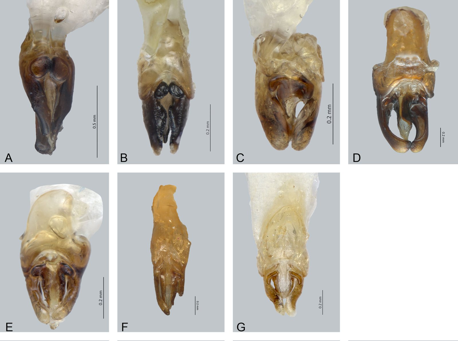
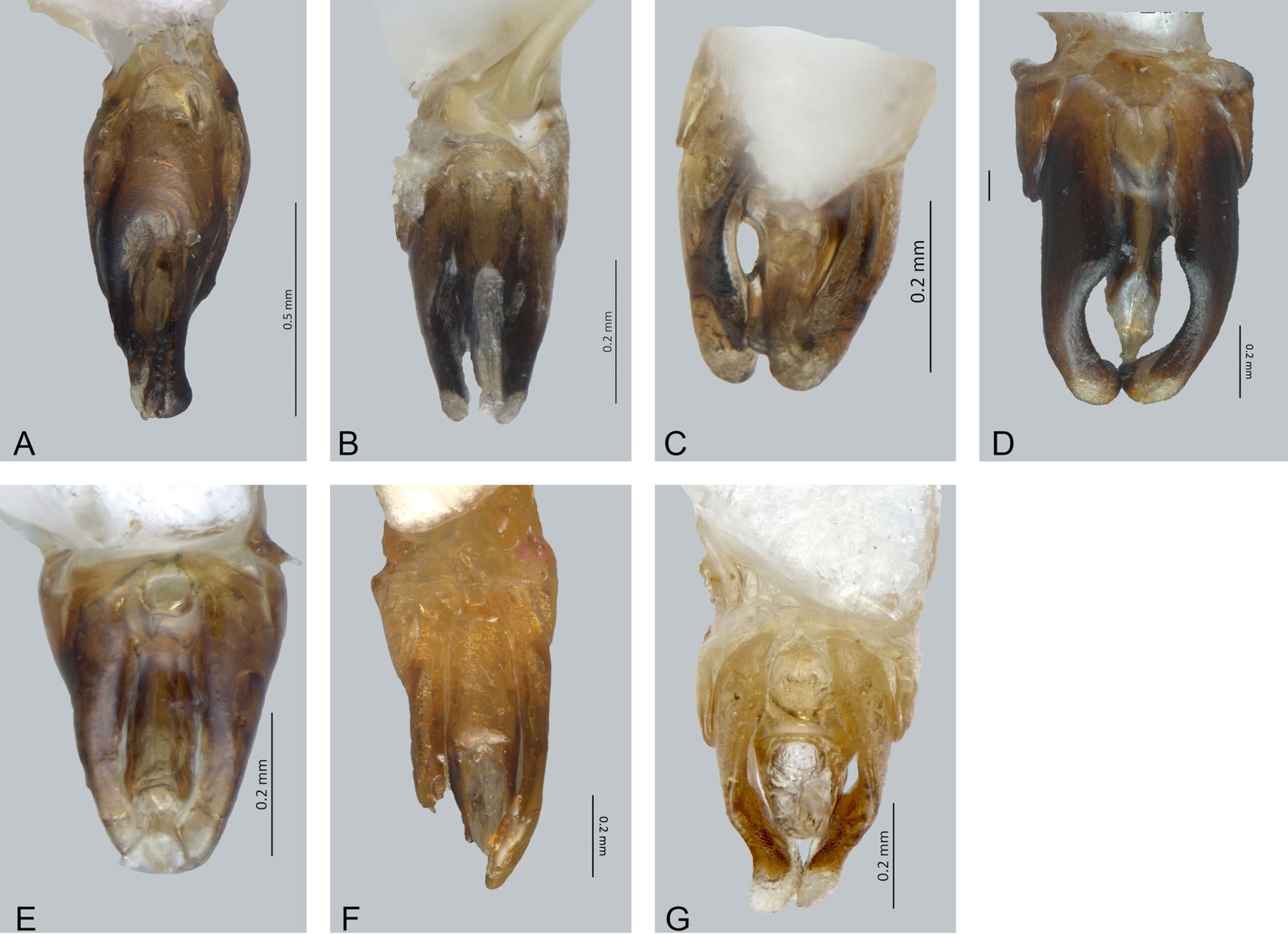
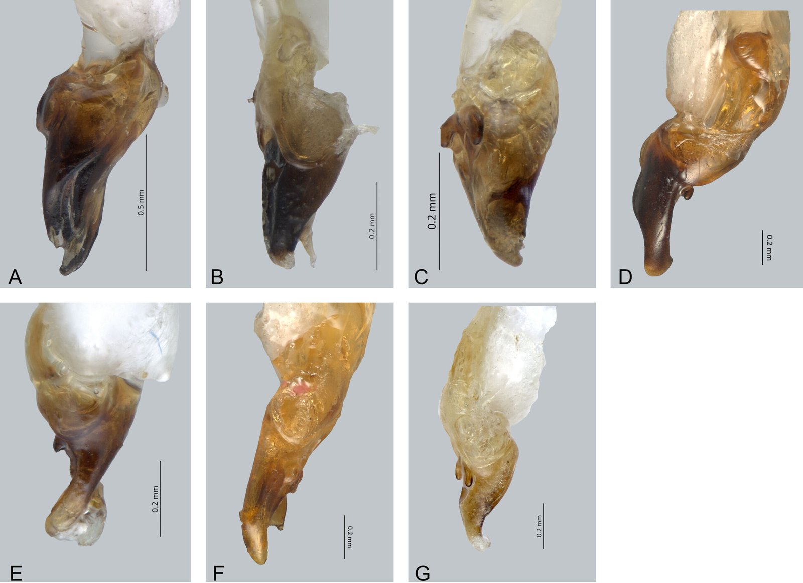
Figs. 7S, 8S, 12A, 13A, 14A
Macho. Largo 9.17 mm, ancho 4 mm; Cuerpo semi paralelo, de color negro, pronoto con los márgenes laterales amarillos con negro; abdomen incompleto. Cabeza: espacio interocular (0.69 mm), cóncavo, integumento brillante, chagrinado, con diminutas sedas; frente vertical, distancia interantenal (0.16 mm), más angosta que fosita antenal (0.24 mm); ojos divergentes finamente facetados, semiesféricos, más largos (0.66 mm) que anchos (0.22 mm); antenas filiformes, comprimidas, largas, 2 veces más largas que el pronoto, rebasando las coxas posteriores, escapo de 0.46 mm de longitud, casi cilíndrico, un poco más corto que los 2 siguientes antenitos juntos (0.48 mm), segundo corto (0.18 mm), tercero de 0.30 mm, cuarto-noveno 0.35 mm, décimo de 0.39 mm, undécimo de 0.58 mm; sutura frontoclipeal membranosa, cóncava, clípeo trapezoidal, borde anterior recto, superficie espaciadamente punteada, con sedas largas y erectas; mandíbulas falcadas, con sedas basales; palpómero maxilar fusiforme, robusto, palpómero labial securiforme. Tórax: pronoto ojival, más ancho (2.77 mm) que largo (2 mm), borde posterior recto, ángulos posteriores obtusos, disco con una mancha negra en el centro, basalmente ensanchada; convexo, lados ampliamente explanados, con poros glandulares ordenados en el margen, más abundantes en la parte anterior del disco; escutelo casi triangular, con borde posterior romo, superficie brillante, diminutamente punteada y pilosidad decumbente, una más larga; élitros poco más de 3.5 veces más largos (7.17 mm) que anchos (2.00 mm); epipleura angosta, espiráculos respiratorios no tubulares, transversos; patas cortas, pro y mesopatas semejantes entre sí, las posteriores un poco más largas, fémures comprimidos, casi fusiformes, tibias acanaladas, apicalmente dilatadas, borde externo no crenulado, tarsómeros lateralmente comprimidos, primer metatarsómero casi de igual longitud (0.23 mm) que los 2 siguientes juntos (0.22 mm), cuarto cubriendo casi todo el quinto, uñas simples. Abdomen incompleto; edeago (figs. 12A, 13A, 14A) con pieza basal no visible, lóbulo medio basalmente cilíndrico, 4 veces tan largo (0.40 mm) como ancho (0.09 mm), porción apical ensanchada, rebasando el ápice de los lóbulos laterales, parte dorsal membranosa, ventral quitinosa, ápice membranoso, orificio medio ventralmente abierto, excrecencias dorso basales ovaladas, aplanadas, grandes, simétricas, basalmente juntas; lóbulos laterales robustos, un poco más de 2 veces tan largos (0.74 mm) como anchos (0.31 mm), interiormente poco excavados, en vista dorsal con ápice acuminado, ventralmente dilatados en la parte media, lateralmente casi paralelos, incluye cavidades para la recepción de las excrecencias del lóbulo medio. Hembra desconocida.
Resumen taxonómico
Etimología: la especie está dedicada a Valeria Souza Saldívar, notable ecóloga microbiana que durante parte de su trayectoria profesional ha estudiado y defendido la cuenca de Cuatro Ciénegas, en el desierto de Coahuila,
Material examinado: holotipo macho. México, Chiapas, San Cristóbal, 16 min O, VII-16-57. J. A. Chemsak, P.D. Hurd, B.J. Rannells Collectors. | COL. TIP-04034.
Comentarios taxonómicos
Especie similar a Photinus kardiophallus Zaragoza-Caballero y López-Pérez, 2023; sin embargo, las excrecencias dorsobasales en P. souzae son más redondas y los lóbulos laterales son curvos hacia el ápice.
Photinus stephanieae Zaragoza-Caballero y Gutiérrez-Carranza sp. nov.
http://zoobank.org/urn:lsid:zoobank.org:act:2EB982F7-5EE6-427D-8726-E4AA57ED122B
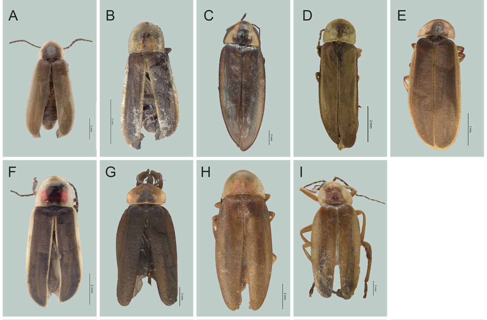
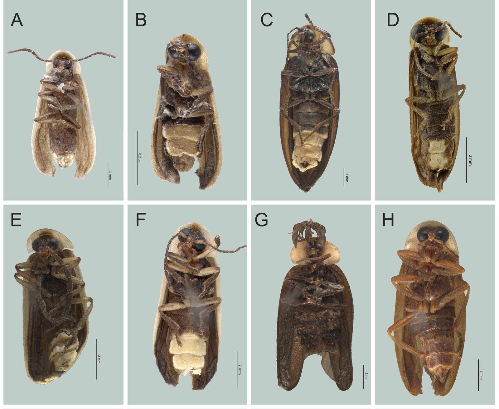
Figs. 12B, 13B, 14B, 15A, 16A
Macho. Largo 4.91 mm, ancho 2.00 mm; cuerpo casi paralelo; color castaño claro con excepción de: margen del pronoto y ventrito VII cremoso; sin aparato luminoso. Cabeza: espacio interocular (0.43 mm), plano, paralelo, integumento diminutamente punteado, lustroso y con pocas sedas decumbentes; frente vertical; distancia interantenal más angosta (0.09 mm) que la fosita antenal (0.13 mm); ojos finamente facetados, semiesféricos, casi tan largos (0.31 mm) como anchos (0.29 mm); antenas filiformes, cortas (2.04 mm), no sobrepasan las coxas posteriores, escapo (0.27 mm), cilíndrico, tan largo como los 2 siguientes antenitos juntos (0.27), segundo corto (0.09 mm), tercero-noveno de 0.18 mm, décimo y undécimo de 0.22 mm; sutura frontoclipeal membranosa, cóncava; clípeo trapezoidal, superficie brillante, con sedas largas irregularmente distribuidas; mandíbulas falcadas; palpómero maxilar apical fusiforme, robusto; el labial triangular. Tórax: pronoto semicircular, más ancho (1.38 mm) que largo (0.90 mm), borde posterior recto, ángulos posteriores rectos, disco convexo, lados con explanaciones angostas, superficie punteada y brillante, poros glandulares distribuidos en los márgenes, más abundantes en la región anterior del disco; escutelo espatulado, con el borde posterior truncado, superficie chagrinada y punteada; élitros 4 veces más largos (3.72 mm) que anchos (0.93 mm), superficie rugosamente punteada, opaca, con 2 tipos de pilosidad decumbente, una más larga; epipleura angosta y gradualmente reducida hacia el ápice; espiráculos respiratorios mesotorácicos tubulares; patas cortas y semejantes entre sí (1.33 mm), metapatas ligeramente más largas (1.69 mm), fémures comprimidos, casi fusiformes, tibias no acanaladas, ligeramente dilatadas hacia el ápice, borde externo crenulado, tarsómeros lateralmente comprimidos, primer metatarsómero ligeramente más largo (0.18 mm) que los 2 siguientes juntos (0.16 mm), el cuarto bífido, casi cubriendo al quinto, uñas simples. Abdomen: ventritos 5-6 similares a los precedentes, sin puntuaciones estigmatiformes, 7 con borde posterior cóncavo, pigidio redondeado, 8 espatulado; edeago (figs. 12B, 13B, 14B): pieza basal simétrica, tan larga (0.27 mm) como los lóbulos laterales (0.27 mm), con borde dorsal posterior cóncavo con forma de “V”, lóbulo medio 3 veces más largo (0.27 mm) que ancho (0.09 mm), cilíndrico en la base y acuminado distalmente, ápice alcanza los lóbulos laterales, latero-ventralmente esclerosado, superficie glabra, membranoso dorsalmente y hacia el ápice, orificio medio apicalmente abierto, excrecencias dorso basales simétricas, no pediculadas y robustas, transversas, conjuntas en su base, no fusionadas, superficie con microesculturación escamosa, lóbulos laterales dilatados en la base, lateralmente triangulares, sin modificaciones para la recepción de las excrecencias del lóbulo medio, dorsalmente presentan una proyección con forma de cresta con el integumento rugoso. Hembra desconocida.
Resumen taxonómico
Etimología: especie dedicada a la Dra. Stephanie Vaz N. Campos, en reconocimiento a su trayectoria centrada en la taxonomía y conservación de las luciérnagas en Brasil. El epíteto específico corresponde al primer nombre.
Material examinado: holotipo macho. México, Oaxaca, km 144 carr. Oaxaca-Pto. Escondido, 1293 m snm, 16º18’38” N, 97º07’34” O, 01/XII/2004, Col. E. Barrera. | COL.TIP-04035 | Paratipo. Un macho. Mismo estado. km 111 carr. Oaxaca- Pto. Escondido, 1,443 m snm, 16º26’05” N, 97º01’58” O, 01/XII/2004, Col. E. Barrera | COL. TIP-04036.
Comentarios taxonómicos
Especie cercana a Photinus isabelae sp. nov., de la que se distingue por el tamaño y forma del edeago. P. stephanieae sp. nov. mide 4.91 mm de largo y 2.00 mm de ancho y en vista dorsal las excrecencias dorsobasales del lóbulo medio y los lóbulos laterales presentan microesculturación rugosa. P. isabelae presenta 6.45 mm de largo y 2.72 mm de ancho y en vista dorsal el integumento de las excrecencias dorsobasales del lóbulo medio y los lóbulos laterales, es glabro.
Photinus tatianafiordelisioae Zaragoza-Caballero y
López-Pérez sp. nov.
http://zoobank.org/urn:lsid:zoobank.org:act:975F0044-C2EF-4A2F-8A4D-D0FE992C3F6F
Figs. 12C, 13C, 14C, 15B, 16B
Macho. Largo 4.8 mm, ancho 1.76 mm; cuerpo casi paralelo; de color castaño oscuro, pronoto amarillento, disco con mancha levemente rojiza, lados amarillentos; élitros con una línea blanquecina en los márgenes laterales; aparato luminoso ocupando los esternitos 5-7. Cabeza: espacio interocular (0.4 mm), deprimido, paralelo, integumento opaco, finamente punteado, con sedas cortas esparcidas; frente vertical, distancia interantenal (0.1 mm), más angosta que fosita antenal (0.07 mm); ojos finamente facetados, casi esféricos, menos largos (0.3 mm) que anchos (0.4 mm); escapo de 0.2 mm de longitud, cilíndrico, más corto que los 2 siguientes antenitos juntos (0.29 mm), segundo corto (0.1 mm), tercero de 0.1 mm, cuarto-octavo 0.85 mm, noveno-décimo 0.1, onceavo 0.85; sutura frontoclipeal membranosa, cóncava; clípeo trapezoidal, borde anterior recto, superficie esparcidamente punteada, con sedas largas y erectas; mandíbulas falcadas, sin sedas basales; palpómero maxilar apical securiforme, robusto; labial triangular. Tórax: pronoto más ancho (1.4 mm) que largo (1.2 mm), ojival, borde posterior recto, ángulos posteriores obtusos, disco brillante, con un surco levemente marcado en el medio, lados explanados, con poros glandulares ordenados en el margen, más abundantes en la parte anterior del disco; escutelo espatulado, con borde posterior redondo, superficie brillante, punteado gruesamente y con poca pilosidad; élitros más de 4 veces más largos (3.8 mm) que anchos (0.88 mm), superficie rugosamente punteada, opaca, con 2 pilosidades decumbentes; epipleura angosta; espiráculos respiratorios mesotorácicos tubulares; pro y meso-patas semejantes entre sí, las posteriores un poco más largas, fémures comprimidos, fusiformes, tibias aplanadas, apicalmente dilatadas, borde externo no crenulado, tarsómeros lateralmente comprimidos, primer metatarsómero tan largo (0.14 mm) como los 2 siguientes juntos (0.14 mm), cuarto bífido, cubriendo casi todo el quinto, uñas simples. Abdomen: penúltimo tergito con los ángulos posteriores romos; esternitos 5-6 tan largos como los precedentes, con puntuaciones estigmatiformes, 7 con el borde posterior cóncavo; edeago (figs. 12C, 13C, 14C) con pieza basal simétrica, más corta (0.8 mm) que lóbulos laterales (0.2 mm), con borde dorsal posterior levemente cóncavo; lóbulo medio más de 3 veces tan largo (0.1 mm) como ancho (0.03 mm), cilíndrico, más corto que lóbulos laterales 30 (0.2 mm), quitinoso en vista ventral y dorsalmente membranoso, ápice comprimido por lóbulos laterales, con excrecencias dorso basales, alargadas, dirigidas hacia los laterales; lóbulos laterales con ápice redondeado, no curvos en vista lateral, muy próximos en el ápice, sin tocarse. Hembra desconocida.
Resumen taxonómico
Etimología: especie dedicada a Tatiana Fiordelisio Coll responsable del Laboratorio Nacional de Soluciones Biomiméticas para Diagnóstico y Terapia (LaNSBioDyT) por su trayectoria como investigadora en la Facultad de Ciencias de la UNAM, especialmente por el desarrollo de los biosensores para detectar el SARS-CoV-2 y el virus del papiloma humano (VPH).
Material examinado: holotipo macho. México, Oaxaca, Temascal, 29-VI-1964, A. G. Raske, col. | COL. TIP-04037. Paratipos 9 machos. (7) mismos datos con excepción de 29-VII-1964 | H. V. Daly, col. | COL.TIP-04038-44. (2) México, Puebla, Pahuatlan Lecho del Río, 700 m snm, 10-VII-1994, E, Barrera, C. Mayorga, cols. México: Puebla, km 5, Pahuatlán La Cruz, 5-VIII-1995, H. Brailovsky, col. COL.TIP-04045-46.
Comentarios taxonómicos
Photinus tatianafiordelisioae es cercana a P. congruus Chevrolat, 1834 en la forma del edeago; sin embargo, P. congruus es de mayor longitud. Photinus pyralis (Linnaeus, 1767) es similar en la forma del cuerpo y el edeago; sin embargo, las excrecencias en P. pyralis son más largas y el edeago más corto.
Photinus taxcoensis Zaragoza-Caballero, 2017
Diagnosis: en Zaragoza-Caballero (2017).
Distribución: Guerrero.
Photinus tenuilineatus Zaragoza-Caballero, 1996
Diagnosis: en Zaragoza-Caballero et al. (2024).
Distribución: Veracruz. Nuevo registro: Oaxaca (CNIN). Material examinado: Oaxaca, San Juan Bautista Tuxtepec, Camelia Roja, 01-1976. México, Oaxaca,
Camelia Roja, 28-01-1976.
Photinus tiferethae Zaragoza-Caballero y Gutiérrez-Carranza sp. nov.
http://zoobank.org/urn:lsid:zoobank.org:act:E75C4DAF-F89B-47F4-81E1-14847181A32E
Figs. 12D, 13D, 14D, 15C, 16C
Macho. Largo 17.27 mm, ancho 6.36 mm; cuerpo semi paralelo; de color negro, pronoto con márgenes laterales negros y una mancha negra basalmente más ancha, lados cremosos; borde posterior del esternito 4, 5, 6, 7 y 8, así como tergitos correspondientes de color cremoso; aparato luminoso ocupando esternitos 5-6. Cabeza: espacio inte-rocular (0.87 mm), cóncavo, integumento brillante, chagrinado, con diminutas sedas; frente vertical, distancia interantenal (0.35 mm), más angosta que fosita antenal (0.13 mm); ojos divergentes, finamente facetados, semiesféricos, más largos (0.62 mm) que anchos (0.51 mm); antenas comprimidas, largas, rebasando coxas posteriores, escapo de 0.84 mm de longitud, en forma de clava, más largo que los 2 antenitos siguientes juntos, el segundo corto (0.22 mm), tercero de 0.62 mm, cuarto-décimo 0.67 mm, undécimo 0.77 mm; sutura frontoclipeal membranosa, cóncava; clípeo trapezoidal, borde anterior recto, superficie espaciadamente punteada, con sedas largas y erectas; mandíbulas falcadas, esbeltas, con sedas basales; palpómero maxilar apical fusiforme, robusto; labial securiforme. Tórax: pronoto más ancho (5.00 mm) que largo (3.18 mm), ojival, borde posterior un tanto convexo, ángulos posteriores obtusos, disco brillante, convexo, lados ampliamente explanados, con poros glandulares ordenados en el margen; escutelo casi triangular, con borde posterior romo, superficie brillante, diminutamente punteada y pilosidad decumbente; élitros 4.5 veces más largos (14.09 mm) que anchos (3.18 mm), superficie rugosamente punteada, opaca, con 2 tipos de pilosidad decumbente, una más larga; epipleura angosta; espiráculos respiratorios mesotorácicos tubulares; patas cortas, pro y mesopatas semejantes entre sí, las posteriores un poco más largas, fémures comprimidos, casi fusiformes, tibias apicalmente dilatadas, borde externo no crenulado, tarsómeros lateralmente comprimidos, primer metatarsómero más largo (0.81 mm) como los 2 siguientes juntos (0.70 mm), el cuarto bífido, cubriendo casi todo el quinto, uñas simples. Abdomen: penúltimo tergito con ángulos posteriores agudos, borde posterior del pigidio convexo y angulado; esternitos 5-6 tan largos que los precedentes, sin puntuaciones estigmatiformes, séptimo esternito con borde posterior cóncavo, octavo espatulado; edeago (figs. 12D, 13D, 14D) con pieza basal simétrica, más larga (1.01 mm) que ancha (0.64 mm), más larga que lóbulos laterales (0.85 mm), con borde dorsal posterior cóncavo, proximal convexo; lóbulo medio basalmente cilíndrico, un poco más de 5 veces tan largo (0.70 mm) como ancho (0.13 mm), porción apical acuminada, casi alcanzando el ápice de los lóbulos laterales, parte dorsal y ventral mayormente quitinosa, ápice membranoso, orificio medio ventralmente abierto, excrecencias dorso basales medianas, simétricas, lobuladas, casi transversas, basalmente separadas; lóbulos laterales esbeltos, un poco más de 3 veces y media tan largos (0.85 mm) como altos (0.25 mm), interiormente excavados, en vista dorsal con ápice redondeado, en vista ventral paralelos en parte basal, en vista lateral más altos en su base, sin modificaciones para la recepción de las excrecencias del lóbulo medio. Hembra desconocida.
Resumen taxonómico
Etimología: especie dedicada a Tifereth Gabriela Cruz Rico, estudiante de diseño gráfico de coadyuvante en la toma de fotos de esta contribución.
Material examinado: holotipo macho. México, Chiapas, Finca Prusia, 8-V-85. Cols. F. Arias, M. Vértiz, M. Velasco. | COL.TIP-04047.
Comentarios taxonómicos
Especie simpátrica de P. sarukani de la que difiere en lo ancho y la configuración del edeago.
Photinus tilae Gutiérrez-Carranza y Zaragoza-Caballero, 2023
Diagnosis: en Gutiérrez-Carranza y Zaragoza-Caballero (2023).
Distribución: Jalisco y Oaxaca.
Photinus toledoi Zaragoza-Caballero, 2000
Diagnosis: en Zaragoza-Caballero, López-Pérez et al. (2020).
Distribución: Morelos. Nuevo registro: Guerrero (CNIN). Material examinado: México, Guerrero, Tixtla de Guerrero, 24 km E, Gro, 27-7-1983, J.A. Chemsak, A. Michelbacher, M. Michelbacher.
Photinus tuxtlaensis Zaragoza-Caballero, 1995
Diagnosis: en Zaragoza-Caballero, López-Pérez et al. (2020).
Distribución: Campeche, Chiapas, Hidalgo, Oaxaca, Quintana Roo, San Luis Potosí, Tabasco, Tamaulipas, Veracruz y Yucatán.
Photinus uchimai Zaragoza-Caballero y Domínguez-León sp. nov.
http://zoobank.org/urn:lsid:zoobank.org:act:4D4EE90A-276E-4B2B-8F30-2A95A746192C
Figs. 12E, 13E, 14E, 15D, 16D
Macho. Largo 8.09 mm, ancho 2.18 mm; cuerpo paralelo; de color negro, pronoto cremoso, y una mancha oscura, difusa, triangular invertida; aparato luminoso ocupando parte media de los esternitos 5-6. Cabeza: espacio interocular (0.67 mm), cóncavo, integumento opaco, chagrinado, con diminutas sedas; frente vertical, distancia interantenal (0.17 mm), un poco más angosta que fosita antenal (0.20 mm); ojos divergentes grandes, finamente facetados, semiesféricos, más largos (0.89 mm) que anchos (0.60 mm); antenas comprimidas, cortas, no rebasando coxas posteriores, escapo de 0.45 mm de longitud, claviforme, un poco más largo que los 2 siguientes antenitos juntos (0.43 mm), segundo corto (0.13 mm), tercero de 0.20 mm, cuarto-undécimo 0.27; sutura frontoclipeal membranosa, cóncava; clípeo trapezoidal, borde anterior recto, superficie espaciadamente punteada, con pocas sedas cortas; mandíbulas falcadas, con sedas basales; palpómero maxilar apical fusiforme, robusto; labial securiforme. Tórax: pronoto más ancho (2.00 mm), que largo (1.16 mm), un tanto ojival, borde posterior cóncavo, ángulos posteriores obtusos, disco opaco, casi plano, con poros glandulares ordenados en el margen, más abundantes en la parte anterior del disco, este con abundantes sedas, sobre todo a los lados; escutelo triangular, superficie opaca, punteada y pilosa; élitros más de 5 veces más largos (6.81 mm) que anchos (1.27 mm), superficie rugosamente punteada, opaca, con 2 tipos de pilosidad decumbente, una más larga; epipleura angosta; espiráculos respiratorios mesotorácicos tubulares; patas cortas, pro y mesopatas semejantes entre sí, las posteriores un poco más largas, fémures comprimidos, casi fusiformes, tibias acanaladas, apicalmente dilatadas, borde externo no crenulado, tarsómeros lateralmente comprimidos, primer metatarsómero más largo (0.45 mm) que los 2 siguientes juntos (0.33 mm), cuarto bífido, cubriendo la mitad del quinto, uñas simples. Abdomen: pigidio convexo; esternitos 5-6 tan largos como los precedentes, con puntuaciones estigmatiformes, séptimo esternito con borde posterior cóncavo, octavo espatulado; edeago (figs. 12E, 13E, 14E) con pieza basal simétrica, casi tan larga (0.39 mm) como ancha (0.33 mm), más corta que lóbulos laterales (0.44 mm), con borde dorsal posterior cóncavo, anterior convexo; lóbulo medio un poco más de 4 veces tan largo (0.42 mm) como ancho (0.10 mm), casi cilíndrico, un poco más largo que los lóbulos laterales, en vista dorsal parcialmente membranoso, ventralmente quitinoso, orificio medio apicalmente abierto, excrecencias dorso basales simétricas, separadas, cortas, lobuladas, transversas; lóbulos laterales casi 1.5 tan largos (0.44 mm) como altos (0.35 mm), con el ápice en vista dorsal redondeado, en vista lateral con ápice romo, sin modificaciones para la recepción de las excrecencias del lóbulo medio. Hembra desconocida. Variación. Únicamente en la talla, de 7.27-9.54 mm de largo y 2.18-2.90 mm de ancho.
Resumen taxonómico
Etimología: especie dedicada a Diego Alejandro Uchima Taborda joven investigador dedicado al estudio de coleópteros de la familia Phengodidae.
Material examinado: holotipo macho. México, Oaxaca, Totolapan, 19 millas NO Oaxaca, 28-VII-63, Collr. W.A. Foster. | QR 51681 | COL.TIP-04048. Paratipos 4 machos. Mismos datos que el holotipo, QR 51686-88, QR 51690. | COL.TIP-04049-52.
Comentarios taxonómicos
Especie relacionada con Photinus producta, ambas presentan un aparato luminoso reducido que ocupa la parte media de los esternitos 5-6, a diferencia de la mayoría de las especies de hábitos nocturnos, cuyo aparato ocupa toda la superficie de esos esternitos. Sin embargo, la talla de P. producta es de 16.8 mm, el pronoto color pálido-rojizo con una mancha negra más menos cuadrada en la parte media basal, los lóbulos laterales en forma de “S”, lóbulo medio más corto que los lóbulos laterales, con las excrecencias largas, paralelas, ampliamente separadas. En tanto, Photinus uchimai sp. nov., mide 8.09 mm, el pronoto con un surco, es de color cremoso, la mancha negra es alargada y se ensancha al ápice, los lóbulos laterales en forma de más menos triangular, el lóbulo medio más largo que los lóbulos laterales, las excrecencias globosas, cortas, y medianamente separadas.
Photinus valenzuelai Zaragoza-Caballero y Vega-Badillo sp. nov.
http://zoobank.org/urn:lsid:zoobank.org:act:C9FC8B56-01AE-497E-A5BD-4147F6248B8A
Figs. 12F, 13F, 14F, 15E, 16E
Macho. Largo 7.50 mm, ancho 2.75 mm; cuerpo semi paralelo; de color castaño, con excepción del pronoto, sutura elitral, márgenes laterales de los élitros, margen posterior de los esternitos 6-7, estenito 8 y pigidio que son ambarinos, pronoto con una mancha negra más menos paralela basalmente ensanchada; sin aparato luminoso. Cabeza: espacio interocular (0.69 mm), plano, paralelo, integumento brillante, liso, con diminutas sedas; frente vertical, distancia interantenal (0.12 mm), más angosta que la fosita antenal (0.16 mm); ojos finamente facetados, semiesféricos, más largos (0.59 mm) que anchos (0.32 mm); antenas comprimidas, muy largas, rebasando el último esternito del abdomen, escapo de 0.53 mm de longitud, en forma de clava, un poco más corto que los 2 siguientes antenitos juntos (0.69 mm), segundo corto (0.16 mm), tercero 0.64 mm, cuarto-décimo de 0.69mm, undécimo 0.86 mm; sutura frontoclipeal conspicua; clípeo trapezoidal, borde anterior recto, superficie espaciadamente punteada, con sedas largas y erectas; mandíbulas falcadas, con sedas basales; palpómero maxilar apical securiforme, robusto;labial securiforme. Tórax: pronoto más ancho (2.42 mm) que largo (1.69 mm), semicircular, borde posterior recto, ángulos posteriores obtusos, disco brillante, convexo, lados ampliamente explanados, con poros glandulares ordenados en el margen, más abundantes en la parte anterior del disco; escutelo casi triangular, con el borde posterior retuso, superficie brillante, diminutamente punteada en la base y pilosidad decumbente; élitros 5 veces más largos (7.50 mm) que anchos (1.50 mm), superficie rugosamente punteada, opaca, con 2 tipos de pilosidad decumbente, una más larga; epipleura ancha; espiráculos respiratorios mesotorácicos tubulares; patas largas, sucesivamente más grandes, fémures comprimidos, casi fusiformes, tibias acanaladas, apicalmente un poco dilatadas, borde externo crenulado, tarsómeros lateralmente no comprimidos, primer metatarsómero tan largo (0.45 mm) como los 2 siguientes juntos, el cuarto bífido, cubriendo solo la mitad del 55ramiquinto, uñas simples. Abdomen: penúltimo tergito con los ángulos posteriores agudos, pigidio bicóncavo; esternitos 5-6 más cortos que los precedentes, sin puntuaciones estigmatiformes, séptimo esternito con el borde posterior escotado, el octavo ojival; edeago (figs. 12F, 13F, 14F) con la pieza basal simétrica, más larga (0.86 mm) que los lóbulos laterales (0.55 mm), con el borde dorsal posterior cóncavo, el proximal convexo; lóbulo medio cilíndrico, 3 veces tan largo (0.32 mm) como ancho (0.10 mm), porción apical acuminada, casi alcanzando el ápice de los lóbulos laterales, parte dorsal membranosa, la ventral con la base esclerosada y el ápice membranoso, orificio medio ventralmente abierto, excrecencias dorsobasales pequeñas asimétricas, divergentes, casi triangulares, conspicuas; lóbulos laterales excavados en vista dorsal con el ápice redondeado, ventralmente paralelos, lateralmente casi triangulares, con modificaciones para la recepción de las excrecencias del lóbulo medio. Hembra desconocida.
Resumen taxonómico
Etimología: especie dedicada a Jorge E. Valenzuela González, destacado entomólogo dedicado al estudio de los himenópteros.
Material examinado: holotipo macho. México, Chiapas, Reserva el Triunfo. 6-V-1993. E. Barrera col. | QR 51977 | COL.TIP-04053.
Comentarios taxonómicos
Especie morfológicamente similar a Photinus xonamancae Zaragoza-Caballero, 1996 de la que se distingue por su tamaño corporal, distancia interocular y mancha pronotal. En P. xonamancae el tamaño corporal es menor con respecto a P. valenzuelai sp. nov.; en P. xonamancae, esta distancia es profundamente cóncava, mientras que en P. valenzuelai sp. nov. es plana y paralela; P. xonamancae presenta una mancha frontal fungiforme, a diferencia de P. valenzuelai sp. nov., cuya mancha frontal es más o menos paralela y ensanchada basalmente.
Photinus vasconcelosi Zaragoza-Caballero y Aquino-Romero sp. nov.
http://zoobank.org/urn:lsid:zoobank.org:act:6D30CDEF-7890-4E0E-9A6D-DE904B2909B5
Figs. 12G, 13G, 14G, 15F, 16F
Macho. Largo 9.18 mm, ancho 2.90 mm; cuerpo paralelo; cuerpo castaño; disco pronotal amarillo mantequilla, en el centro con 1 mancha castaña rectangular, más larga que ancha, difusa en la región anterior, con 2 manchas rosadas a los extremos de la base de la mancha castaña; escutelo castaño, del mismo color que los élitros; élitros con los márgenes del mismo color que el disco pronotal; cabeza castaña con la base rosada, con ligeras vetas ambarinas hacia el rostro, patas cafés, con los fémures con el ángulo interno amarillo mantequilla, metaesterno con los ángulos posteriores externos del mismo color amarillo, así como la base del esternito 4; aparato luminoso blanquecino, ocupando los esternitos 5-6. Cabeza: Espacio interocular (0.46 mm) cóncavo, integumento brilloso, chagrinado, pilosidad ligera, procumbente; distancia interantenal (0.17 mm) del mismo ancho que la fosita antenal; ojos finamente facetados, semiesféricos, casi 2 veces más largos (0.85 mm) que anchos (0.50 mm); antenas filiformes no muy largas, que sobrepasan a las procoxas; escapo (0.59 mm) claviforme, ligeramente más largo que los 2 próximos antenitos juntos, el segundo corto (0.13 mm) piriforme, del tercero al décimo de 0.31 mm, ligeramente aplanados, el undécimo más largo (0.76 mm), cilíndrico; sutura frontoclipeal membranosa cóncava; clípeo trapezoidal, con el borde anterior convexo, superficie rugosa, con puntuación densa, pilosidad densa, procumbente, con las sedas cortas; mandíbulas falcadas, curvadas; palpómeros maxilar apical y labial securiformes, el labial fuertemente ensanchado. Tórax: pronoto más ancho (3.03 mm) que largo (2.29 mm), ojival, semicircular, con el borde posterior recto, disco opaco, convexo, con los lados explanados y pilosidad dispersa y procumbente, con poros glandulares ordenados en el margen; escutelo lingüiforme, con el borde anterior recto, superficie brillante, rugosa, con pilosidad fina, decumbente; élitros 4 veces más largos (6.32 mm) que anchos (1.58 mm), superficie, brillosa, integumento rugoso, pilosidad corta, dispersa y procumbente; epipleura estrecha, más angosta en la base; espiráculos respiratorios mesotorácicos tubulares; patas cortas, fémures fusiformes, metafémures ligeramente más estrechos; tarsómeros estrechos, el primer metatarsómero ligeramente más corto (0.20 mm) que los 2 siguientes juntos (0.24 mm), el cuarto bífido, cubriendo parte del quinto, uñas simples. Abdomen: esternitos 5-6 casi el doble de largo (0.88) que los precedentes (0.51 mm), con puntuaciones estigmatiformes, el séptimo con los ápices posteriores romos; edeago (figs.12G, 13G, 14G) con la pieza basal simétrica, casi 2 veces más larga (0.63 mm) que ancha (0.35 mm), con los bordes anteriores fuertemente acuminados en vista dorsal, el ángulo anterior fuertemente cóncavo; lóbulo medio cuneiforme, ligeramente más largo (0.23 mm) que ancho (0.19 mm), con la base fuertemente ensanchada, parte dorsal y ventral quitinosa, orificio medio ventralmente abierto, cos excrecencias dorsobasales, cilíndricas, con el ápice romo, simétricas, ligeramente curveadas hacia el centro; lóbulos laterales cilíndricos, ensanchados, poco más de 3 veces más largos (0.11 mm) que anchos (0.35 mm), con el ápice romo, sin modificaciones para la recepción de las excrecencias del lóbulo medio. Hembra desconocida.
Resumen taxonómico
Etimología: especie dedicada a José Vasconcelos, por su indiscutible e inmensurable aporte en la educación en México; además de haber sido director de la Escuela Nacional Preparatoria y el sexto rector de la Universidad Nacional Autónoma de México.
Material examinado: holotipo macho. México, Guerrero, Acahuizotla, 13-VI-2001, H. Brailovsky, E.
Barrera. | COL.TIP-04053.
Comentarios taxonómicos
Esta especie puede parecer morfológicamente similar a Photinus toledoi Zaragoza-Caballero, 2000 principalmente en la forma del edeago, ya que ambas especies presentan 2 excrecencias dorsobasales estrechas y alargadas, lóbulos laterales ensanchados hacia el ápice y un lóbulo medio corto y grueso. A pesar de estas similitudes, los lóbulos laterales de P. toledoi son más ensanchados. Además, P. vasconcelosi sp. nov. se puede distinguir fácilmente de P. toledoi por el margen externo uniforme de los élitros, y por la mancha del disco pronotal, que a diferencia de P. toledoi, es mucho más ensanchada, formando un rectángulo.
Photinus viridianae Zaragoza-Caballero, 2015
Diagnosis: en Zaragoza-Caballero (2015).
Distribución: Guerrero.
Photinus vivianae Zaragoza-Caballero, 2017
Diagnosis: en Zaragoza-Caballero (2017).
Distribución: Oaxaca y Puebla.
Photinus perezi Zaragoza-Caballero, 1995
Diagnosis: en Zaragoza-Caballero et al. (2023).
Distribución: Veracruz. Nuevo registro: Chiapas (CNIN). Material examinado: México, Chiapas, LA Trinitaria, 5 lagos, Rancho “El refugio”, 1,400 m, 10-V-94, B. Gómez, Luz U.V.
Photinus zongolicaensis Zaragoza-Caballero y Domínguez-León, 2020
Diagnosis: en Zaragoza-Caballero, López-Pérez et al. (2020).
Distribución: Chiapas, Puebla y Veracruz.
Pyropyga Motschulsky, 1852
Diagnosis: en Zaragoza-Caballero et al. (2023).
Pyropyga alticola Green, 1961
Diagnosis: en Zaragoza-Caballero et al. (2023).
Distribución: Chiapas, Ciudad de México, Coahuila, Estado de México, Guerrero, Hidalgo, Morelos, Oaxaca, Puebla y Veracruz.
Pyropyga chemsaki Zaragoza-Caballero, 1993
Diagnosis: en Zaragoza-Caballero et al. (2023).
Distribución: Colima, Jalisco, Morelos, Nayarit, Oaxaca y Puebla.
Pyropyga cordobana Green, 1961
Diagnosis: en Zaragoza-Caballero et al. (2023).
Distribución: Chiapas, Jalisco, Morelos, Puebla, San Luis Potosí y Veracruz.
Pyropyga extensa Green, 1961
Diagnosis: en Zaragoza-Caballero et al. (2023).
Distribución: Ciudad de México, Durango, Puebla, Quintana Roo, Tamaulipas y Veracruz. Nuevo registro: Chiapas (CNIN). Material examinado: México, Chiapas, Solaya Río Laja, F. Arias, L. Cervantes, R. Barba.
Pyropyga exstincta (Gorham, 1880)
Diagnosis: en Zaragoza-Caballero et al. (2023).
Distribución: Colima, San Luis Potosí, Oaxaca y Veracruz.
Pyropyga minuta (LeConte, 1851)
Diagnosis: en Zaragoza-Caballero et al. (2023).
Distribución: Ciudad de México, Chiapas, Coahuila, Colima, Durango, Estado de México, Guanajuato, Guerrero, Hidalgo, Jalisco, Michoacán, Morelos, Nayarit, Nuevo León, Oaxaca, Puebla, Quintana Roo, San Luis Potosí, Tamaulipas y Veracruz.
Pyropyga modesta Green, 1961
Diagnosis: en Zaragoza-Caballero et al. (2023).
Distribución: Chiapas, Coahuila, Colima, Guerrero, Hidalgo, Jalisco, Michoacán, Morelos, Nayarit, Nuevo León, Oaxaca, Puebla, San Luis Potosí, Tamaulipas y Veracruz.
Pyropyga nigricans (Say, 1823)
Diagnosis: en Zaragoza-Caballero et al. (2023).
Distribución: Aguascalientes, Baja California Sur, Chiapas, Chihuahua, Ciudad de México, Durango, Estado de México, Guanajuato, Hidalgo, Jalisco, Michoacán, Morelos, Nayarit, Oaxaca, Puebla, San Luis Potosí, Sinaloa, Sonora y Veracruz.
Tribu Pleotomini Summers, 1874
Phaenolis Gorham, 1880
Diagnosis: en Zaragoza-Caballero, López-Pérez et al. (2020) y Zaragoza-Caballero et al. (2023).
Phaenolis nigricollis Gorham, 1881
Figs. 15G, 16G
Longitud 15 mm. Pronoto transverso, constreñido en la base, poco pronunciado en el ápice, márgenes laterales expandidos. Élitros amarillentos, 3 o 4 costillas poco visibles. Último ventrito hendido y bilobado, pigidio bisinuado.
Distribución: Oaxaca.
Phaenolis ustulata Gorham, 1880
Diagnosis: en Zaragoza-Caballero et al. (2024)
Distribución: Chiapas, Oaxaca y Veracruz.
Pleotomus LeConte, 1861
Diagnosis: en Zaragoza-Caballero, López-Pérez et al. (2020) y Zaragoza-Caballero et al. (2023).
Pleotomus cerinus Zaragoza-Caballero, 2002
Diagnosis: en Zaragoza-Caballero, López-Pérez et al. (2020).
Distribución: Guerrero, Morelos y Oaxaca.
Pleotomus pallens LeConte, 1866
Diagnosis: en Zaragoza-Caballero, López-Pérez et al. (2020) y Zaragoza-Caballero et al. (2023).
Distribución: Durango, Jalisco, Michoacán, Nayarit, Nuevo León, Oaxaca, Puebla, San Luis Potosí, Sinaloa, Tamaulipas y Veracruz.
Subfamilia Photurinae Lacordaire, 1857
Tribu Photurini Lacordaire, 1857
Bicellonycha Motschulsky, 1853
Diagnosis: en Zaragoza-Caballero, López-Pérez et al. (2020) y Zaragoza-Caballero et al. (2023)
Bicellonycha albomarginata Zaragoza-Caballero, 1989
Diagnosis: en Zaragoza-Caballero et al. (2023)
Distribución: Oaxaca y Veracruz.
Bicellonycha amoena (Gorham, 1880)
Diagnosis: en Zaragoza-Caballero, López-Pérez et al. (2020).
Distribución: Chiapas, Colima, Guanajuato, Guerrero, Hidalgo, Jalisco, Estado de México, Michoacán, Morelos, Nayarit, Oaxaca, Puebla, Querétaro, San Luis Potosí, Tamaulipas y Veracruz.
Bicellonycha collaris (Gorham, 1880)
Diagnosis: en Zaragoza-Caballero, López-Pérez et al. (2020).
Distribución: Chiapas, Estado de México, Morelos, Puebla y Veracruz.
Bicellonycha crassa (Gorham, 1884)
Diagnosis: en Zaragoza-Caballero et al. (2024).
Distribución: Chiapas, Oaxaca y Veracruz.
Bicellonycha cyathigera (Gorham, 1881)
Diagnosis: en Zaragoza-Caballero et al. (2023).
Distribución: Guerrero, Jalisco, Morelos, Oaxaca y Puebla.
Bicellonycha discicollis (Gorham, 1881)
Diagnosis: en Zaragoza-Caballero, López-Pérez et al. (2020).
Distribución: Chiapas, Hidalgo, Estado de México, Morelos, Nayarit, Oaxaca y Veracruz.
Bicellonycha gorhami Zaragoza-Caballero, 1989
Diagnosis: en Zaragoza-Caballero, López-Pérez et al. (2020).
Distribución: Estado de México, Guerrero, Jalisco, Morelos y Veracruz.
Bicellonycha limbata Pic, 1924
Diagnosis: en Zaragoza-Caballero, López-Pérez et al. (2020).
Distribución: Chiapas, Jalisco, Morelos, Oaxaca, Puebla y Veracruz.
Bicellonycha lucidicollis (Gorham, 1880)
Figs. 15H, 16H
Diagnosis: largo 16.38 mm; ancho 3.40 mm. Cuerpo semiparalelo, un poco oval, pardo oscuro, cabeza negra con tintes ferruginosos. Pronoto un tanto ojival, amarillo con tintes rojizos, con una mancha negra en forma de “T” invertida; escutelo pardo; élitros ovalados, acuminados; aparato luminoso amarillo-rojizo ocupando los ventritos 5-6; edeago largo, triangular, lóbulos laterales cubriendo el dorso terminal de lóbulo medio, este, termina en pequeño “arpón” apical. Hembra semejante al macho, frente más amplia, aparato luminoso reducido a una pequeña mancha sobre el ventrito 5, el ápice del 7 triangular, emarginado, uñas simples. Especie de amplia distribución, muy variable en coloración y tamaño que va de 12.71 a 16.54 mm de largo y de 3.18 a 4.00 mm de ancho
Distribución: Chiapas, Morelos, Oaxaca y Veracruz.
Bicellonycha ornaticollis (Blanchard, 1846)
Fig. 16H
Diagnosis: largo 14.43 mm; ancho 4.00 mm. Cuerpo casi paralelo, pardo amarillento, porción sutural de élitros más oscura, cabeza anaranjada. Pronoto amarillo con el disco rojizo y una mancha negra en forma de “W”; escutelo casi negro; élitros ferruginosos; aparato luminoso amarillo-rojizo ocupando los ventritos 5-6; edeago largo, triangular, lóbulos laterales cubriendo el dorso basal de lóbulo medio, este, termina formando un pequeño tubo. Hembra semejante al macho, un poco más grande (15.17-X 4.37 mm), frente más amplia y plana, aparato luminoso ocupa todo el ventrito 5, el ápice del 7 triangular, escotado, uñas simples.
Distribución: Chiapas y Guerrero.
Bicellonycha stigmatica (Olivier, 1886)
Diagnosis: en Zaragoza-Caballero, López-Pérez et al. (2020).
Distribución: Chiapas, Oaxaca, Puebla y Veracruz.
Photuris Dejean, 1833
Diagnosis: en Zaragoza-Caballero, López-Pérez et al. (2020) y Zaragoza-Caballero et al. (2023).
Photuris fulvipes (Blanchard in Brullé, 1846)
Diagnosis: en Zaragoza-Caballero, López-Pérez et al. (2020).
Distribución: Estado de México, Hidalgo, Morelos y Veracruz. Nuevo registro: Oaxaca (CNIN). Material examinado: México, Oaxaca, Entronque carr. Salina Cruz-Santa Cruz Huatulco, Puente Guajinicuit Figueroa, 13-05-1980.
Photuris lugubris Gorham, 1881
Diagnosis: en Zaragoza-Caballero, López-Pérez et al. (2020) y Zaragoza-Caballero et al. (2023).
Distribución: Ciudad de México, Guerrero, Jalisco, Morelos, Oaxaca y Tamaulipas.
Photuris mollis Gorham, 1880
Diagnosis: en Zaragoza-Caballero et al. (2024)
Distribución: Oaxaca y Veracruz.
Photuris tenuisignathus Zaragoza-Caballero, 1995
Diagnosis: en Zaragoza-Caballero et al. (2023).
Distribución: Chiapas, Oaxaca, San Luis Potosí y Veracruz.
Photuris trivittata Lloyd y Ballantyne, 2003
Diagnosis: en Zaragoza-Caballero et al. (2024).
Distribución: Chiapas, Quintana Roo, Tabasco y Veracruz.
Discusión
En este trabajo se destaca la gran diversidad de especies que se encuentran en los estados de Chiapas (65) y Oaxaca (79). Guerrero presenta una diversidad media en cuanto a luciérnagas (41). Es frecuente encontrar este resultado en diversos grupos biológicos, donde los estados sureños presentan una mayor diversidad en el país (Falcón-Brindis et al., 2021). Esta gran diversidad se debe no solo a la histórica atención que han tenido estos estados por sus vegetaciones exuberantes como las selvas altas perennifolias, sino también a la historia geológica del país, donde se pueden encontrar diferentes hábitats en una misma región, dando lugar a diferentes oportunidades de colonizar diferentes nichos. Cabe destacar que para las luciérnagas uno de los estados con mayor atención en su estudio ha sido Veracruz, actualmente con cerca de 100 especies reportadas (Zaragoza-Caballero, 1995; Zaragoza-Caballero et al., 2024).
A pesar de que México es uno de los países megadiversos, esta biodiversidad se ve amenazada por la pérdida acelerada de vegetación; el cambio de uso de suelo ha sido notable en comparación con otros países en desarrollo (Fuller et al., 2007). La información taxonómica proporcionada en este trabajo no solo contribuye al conocimiento de las luciérnagas mexicanas y Neotropicales, sino también aporta a las evaluaciones de riesgo que se pueden realizar a estos insectos. Si bien es cierto que falta conocer más sobre la historia natural de estas especies para poder conservarlas de una mejor manera, el nombre de la especie y su ubicación geográfica son la base para el cuidado de estos escarabajos y todas aquellas especies con las que comparten un hábitat.
Agradecimientos
A Susana Guzmán y Berenit Mendoza Garfias del Laboratorio de Microscopía y Fotografía de la Biodiversidad, Instituto de Biología, UNAM por la ayuda técnica en la toma de fotografías. A Janis Vigil Razo y Yhadnya B. Arroyo Ortiz por la toma de diferentes fotografías. A Valentina Febles Perales por su asistencia en la edición digital como parte del Programa de Servicio Social Estudio Multidisciplinario de Luciérnagas (SS-2025-12/149-5315, responsables SLP y GMRM). Se agradece al proyecto DGAPA-PAPIIT-UNAM IA205322 (Sistemática y evolución morfológica de Megalopodidae (Coleoptera: Chrysomeloidea) por el uso del equipo fotográfico, responsable GMRM. Finalmente agradecemos a 2 revisores anónimos por sus atinados comentarios que mejoraron el trabajo.
Referencias
Bouchard, P., Bousquet, Y., Davies, A. E., Alonso-Zarazaga, M. A., Lawrence, J. F., Lyal, C. H. et al. (2011). Family-group names in Coleoptera (Insecta). Zookeys, 88, 1–972. https://doi.org/10.1515/9783110911213.141
Cicero, J., Pérez Hernández, C. y Walker, A. (2021). Tenaspis angularis. The IUCN Red List of Threatened Species. e.T163979488A166771193. Recuperado el 23 de marzo de 2026 de: 10.2305/IUCN.UK.2021-1.RLTS. T163979488A166771193.en
Falcón-Brindis, A., León-Cortés, J. L. y Montañez-Reyna, M. (2021). How effective are conservation areas to preserve biodiversity in Mexico? Perspectives in Ecology and Conservation, 19, 399–410. https://doi.org/10.1016/j.pecon.2021.07.007
Fallon, C. E., Walker, A. C., Lewis, S., Cicero, J., Faust, L., Heckscher, C. M. et al. (2021). Evaluating firefly extinction risk: Initial red list assessments for North America. Plos One, 16, e0259379. https://doi.org/10.1371/journal.pone.0259379
Ferreira, V. S. (2024). Description of a second genus of Chespiritoinae (Coleoptera, Lampyridae), with updates on the subfamily Diagnosis and key to species and genera. Revista Brasileira de Entomologia, 68, e20240075. https://doi.org/10.1590/1806-9665-RBENT-2024-0075
Fuller, T., Sánchez-Cordero, V., Illoldi-Rangel, P., Linaje, M. y Sarkar, S. (2007). The cost of postponing biodiversity conservation in Mexico. Biological Conservation, 134, 593–600. https://doi.org/10.1016/j.biocon.2006.08.028
Gorham, H. S. (1881). Coleoptera Malacodermata. En F. D. Godman y O. Salvin (Eds.), Biologia Centrali Americana. Insecta. Volume III, Part 2 (pp. 25–112). Porter, Londres. https://doi.org/10.5962/bhl.title.730
Gutiérrez-Carranza, I. G. (2023). Protocolo para disección y montaje de luciérnagas (Coleoptera: Lampyridae). Boletín de la Asociación Mexicana de Sistemática de Artrópodos, 7, 2–6.
Gutiérrez-Carranza, I. G., Zaragoza-Caballero, S. y Domínguez- León, D. E. (2023). Photinus favilai y P. tilae nuevas especies de luciérnagas de México (Coleoptera: Lampyridae). Dugesiana, 30, 209–219. https://doi.org/10.32870/dugesiana.v30i2.7300
Gutiérrez-Carranza, I. G., Zaragoza-Caballero, S. y Keller, O. (2025). El género Psilocladus Blanchard, 1845 (Coleoptera: Lampyridae: Psilocladinae) en México. Duge- siana, 32, 137–148. https://doi.org/10.32870/dugesiana.v32i2.7379
Kehoe, R., Frago, E. y Sanders, D. (2021). Cascading extinctions as a hidden driver of insect decline. Ecological Entomo- logy, 46, 743–756. https://doi.org/10.1111/een.12985
Lima, W., Silveira, L. F. L. D., Fonseca, C. R. V. D. y Zaragoza- Caballero, S. (2021). Cratomorphus leoneli: a new firefly from Mexico (Coleoptera: Lampyridae: Cratomorphini). Revista Mexicana de Biodiversidad, 92, e923831. https://doi.org/10.22201/ib.20078706e.2021.92.3831
Lewis, S. M., Wong, C. H., Owens, A. C. S., Fallon, C., Jepsen, S., Thancharoen, A. (2020). A global perspective on firefly extinction threats. Bioscience, 70, 157–167. https://doi.org/10.1093/biosci/biz157
Martin, G. J., Stanger-Hall, K. F., Branham, M. A., Da Silveira, L. F. L., Lower, S. E., Hall, D. W. et al. (2019). Higher-level phylogeny and reclassification of Lampyridae (Coleoptera: Elateroidea). Insect Systematics and Diversity, 3, 1–15. https://doi.org/10.1093/isd/ixz024
Montgomery, G. A., Dunn, R. R., Fox, R., Jongejans, E., Leather, S. R., Saunders, M. E. et al. (2020). Is the insect apocalypse upon us? How to find out. Biological Conservation, 241, 108327. https://doi.org/10.1016/j.biocon.2019.108327
Nunes, V. C. S., Souto, P. M., Minelli, A., Stanger-Hall, K. F. y Silveira, L. F. L. (2020). Antennomere numbers in fireflies (Coleoptera: Lampyridae): unique patterns and tentative explanations. Zoologischer Anzeiger, 286, 1–10. https://doi.org/10.1016/j.jcz.2020.02.006
Pérez-Hernández, C. X., Zaragoza-Caballero, S. y Romo-Galicia, A. (2022). Updated checklist of the fireflies (Coleoptera: Lampyridae) of Mexico. Zootaxa, 5092, 291–317. https://doi.org/10.11646/zootaxa.5092.3.3
Zaragoza-Caballero, S. (1988). Revisión del género Bicellonycha Mostchulsky (Coleoptera: Lampyridae: Photurinae) del continente americano (Tesis doctoral). Facultad de Ciencias, Universidad Nacional Autónoma de México. México, Ciudad de México.
Zaragoza-Caballero, S. (1995). La familia Lampyridae (Coleoptera) en la Estación de Biología Tropical “Los Tuxtlas”, Veracruz, México. Publicaciones Especiales 14. Ciudad de México: Instituto de Biología, Universidad Nacional Autónoma de México.
Zaragoza-Caballero, S. (2015). Nuevas especies de Photinus (Coleoptera: Lampyridae: Photinini) del bosque tropical caducifolio del Pacífico mexicano. Revista Mexicana de Biodiversidad, 86, 638–651. https://doi.org/10.1016/j.rmb.2015.08.001
Zaragoza-Caballero, S. (2017). Nuevos Photinus Laporte, 1832 (Coleoptera: Lampyridae: Photinini). Dugesiana, 24, 221– 229. https://doi.org/10.32870/dugesiana.v24i2.6686
Zaragoza-Caballero, S., Cifuentes-Ruiz, P., Domínguez-León, D. E., González-Ramírez, M., Gutiérrez-Carranza, I. G., López-Pérez, S. et al. (2020). Proyecto “Luciérnagas de México”. Boletín de la Asociación Mexicana de Sistemática de Insectos, 4, 20–22.
Zaragoza-Caballero, S. y Gutiérrez-Carranza, I. G. (2018). Aorphallus cibriani gen. nov., sp. nov., y otros Photinini de México (Coleoptera: Lampyridae). Dugesiana, 25, 159–166. https://doi.org/10.32870/dugesiana.v25i2.7047
Zaragoza-Caballero, S., López-Pérez, S., González-Ramírez, M., Rodríguez-Mirón, G. M, Vega-Badillo, V., Domínguez- León, D. E. et al. (2023). Luciérnagas (Coleoptera: Lampyridae) del norte-occidente de México, con la descripción de 48 especies nuevas. Revista Mexicana de Biodiversidad, 94, e945028. https://doi.org/10.22201/ib.20078706e.2023.94.5028
Zaragoza-Caballero, S., López-Pérez, S., González-Ramírez, M., Rodríguez-Mirón, G., Vega-Badillo, V., Gutiérrez- Carranza, I. G. et al. (2024). Luciérnagas de la región golfo- Caribe de México y descripción de 16 especies nuevas. Revista Mexicana de Biodiversidad, 95, e955476. https://doi.org/10.22201/ib.20078706e.2024.95.5476
Zaragoza-Caballero, S., López-Pérez, S., Vega-Badillo, V., Domínguez-León, D. E., Rodríguez-Mirón, G. M., González- Ramírez, M. et al. (2020). Luciérnagas del centro de México (Coleoptera: Lampyridae): descripción de 37 especies nuevas. Revista Mexicana de Biodiversidad, 91, e913104. https://doi.org/10.32870/dugesiana.v21i2.4150
Zaragoza-Caballero, S. y Ramírez-García, E. (2009). Diversidad de Cantharidae, Lampyridae, Lycidae, Phengodidae y Telegeusidae (Coleoptera: Elateroidea) en un bosque tropical caducifolio de la sierra de San Javier, Sonora, México. Revista Mexicana de Biodiversidad, 80, 675–686. https://doi.org/10.22201/ib.20078706e.2009.003.164