Morfología y estados reproductivos de Peridiniopsis borgei (Dinophyceae: Peridiniopsidaceae), primer registro en México
Beatriz Lira a, Carolina Bustamante-Gil a, Alejandra Mireles-Vázquez b, Vladimir Betancourt b, Karim Benzerara c, Purificación López-García d, Christophe Thomazo e, Robin Havas e, Jeanne Caumartin c, Didier Jézéquel f, Rosaluz Tavera a, *
a Universidad Nacional Autónoma de México, Facultad de Ciencias, Departamento de Ecología y Recursos Naturales, Circuito Exterior s/n, Ciudad Universitaria, Coyoacán, 04510 Ciudad de México, Mexico
b Universidad Nacional Autónoma de México, Posgrado en Ciencias del Mar y Limnología, Facultad de Ciencias, Circuito Exterior s/n, Ciudad Universitaria, Coyoacán, 04510 Ciudad de México, Mexico
c Sorbonne Université, Museum National d’Histoire Naturelle, UMR CNRS 7590, Institut de Minéralogie, de Physique des Matériaux et de Cosmochimie, IMPMC, 75005 Paris, France
d Université Paris-Saclay, Ecologie Société Evolution, CNRS, AgroParisTech, 91190 Gif-sur-Yvette, France
e Université Bourgogne Europe, Biogéosciences, UMR CNRS 6282, 21000 Dijon, France
f Université de Paris, UMR CARRTEL, IPGP, CNRS UMR7154, INRAE-USMB, 74203 Thonon-les-Bains, France
*Corresponding author: r_tavera@ciencias.unam.mx (R. Tavera)
Received: 19 September 2025; accepted: 21 April 2026
Abstract
Peridiniopsis borgei is a species of the Peridiniopsidaceae family (dinophytes), commonly found in inland waters but previously unrecorded in Mexico. This study presents the first record of a P. borgei bloom in Lake La Preciosa, a subsaline, alkaline crater lake in Puebla State, Mexico. Its proliferation correlates with specific environmental conditions, including low salinity (~ 1.2 PSU) and total dissolved solids (~ 1.7-1.8 g L-1), high levels of dissolved oxygen in the epilimnion (≥ 6.0 mg L-1), and low concentrations of nutrients and iron. From a morphological perspective, the analyzed specimens exhibited well-defined diagnostic features, particularly in the apical region, thereby reinforcing their taxonomic identification. Microscopic observations revealed cell shapes associated with reproductive stages, including elongated and peanut-shaped forms, the biological significance of which remains unclear within Peridiniopsidaceae. No sexual processes were observed, suggesting that the observed stages could be part of post-germinative development. These findings suggest that P. borgei exhibits a dynamic life cycle with overlapping generations and responds to specific environmental conditions that could limit or control its distribution in ecosystems such as Lake La Preciosa. Such conditions position it as a model case for studying dinophytes in subsaline lakes within volcanic environments.
Keywords: Algal bloom; Asexual multiplication; Crater lakes; Dinophyta; Salinity gradient; Thermal stratification
Resumen
Peridiniopsis borgei es una especie de la familia Peridiniopsidaceae (Dinophyta), común en aguas continentales, pero sin registros previos para México. Este estudio presenta el primer registro de una proliferación de P. borgei en el lago La Preciosa, un lago crateriforme subsalino y alcalino del estado de Puebla, México. Su proliferación se correlacionó con condiciones ambientales específicas, entre ellas baja salinidad (~ 1.2 PSU) y sólidos disueltos totales (~ 1.7-1.8 g L-1), altas concentraciones de oxígeno disuelto en el epilimnio (≥ 6.0 mg L-1) y bajas concentraciones de nutrientes y hierro. Los ejemplares analizados mostraron caracteres diagnósticos bien definidos, particularmente en la región apical, lo que respalda su identificación taxonómica. Las observaciones microscópicas revelaron formas celulares asociadas con etapas reproductivas, incluidas formas alargadas y con aspecto de cacahuate, cuyo significado biológico dentro de Peridiniopsidaceae permanece incierto. No se observaron procesos sexuales, lo que sugiere que estas etapas podrían formar parte del desarrollo posgerminativo. Estos hallazgos indican que P. borgei presenta un ciclo de vida dinámico con generaciones superpuestas y responde a condiciones ambientales específicas como las de La Preciosa. Tales condiciones lo convierten en un caso modelo para el estudio de dinofitas en lagos subsalinos de origen volcánico.
Palabras clave: Floración algal; Multiplicación asexual; Lagos cráter; Dinophyta; Gradiente de salinidad; Estratificación térmica
Introduction
The family Peridiniopsidaceae comprises 35 recognized species of dinoflagellates found in tropical to temperate environments in continental waters worldwide (Guiry & Guiry, 2025). This family is taxonomically distinct from the large and more diverse Peridiniaceae family, primarily due to the characteristic presence of 6 cingular plates and zero to 2 intercalary plates, as well as its presumed preference for continental environments (Gottschling et al., 2017). The genus Peridiniopsis Lemmermann (type genus of Peridiniopsidaceae) has a broad global distribution (Ascencio et al., 2020; Bourrelly, 1968; Carty, 2014; Krakhmalny et al., 2000; Moestrup & Calado, 2018; Zhang et al., 2012). Its holotype, Peridiniopsis borgei Lemmermann, has a wide geographical distribution across non-saline and brackish continental ecosystems in Europe, Asia, North America, and Oceania. Despite its broad distribution, knowledge about the life cycle of P. borgei remains limited, and its ecological role within phytoplankton communities is still poorly understood (Calado & Moestrup, 2002; Carty, 2014; Entz, 1926; Gätz & Schagerl, 1990; Krakhmalny, 2014; Krakhmalny et al., 2000; Ling & Tyler, 2000; Ling et al., 1989; Pollingher & Hickel, 1991; Zhang et al., 2012).
There are only 105 records of continental dinophyte taxa (species and varieties) in Mexico (Novelo & Tavera, 2025). Hence, dinoflagellate diversity likely remains limited in the region. Peridiniopsis borgei has not been previously recorded in Mexican inland waters, and this is the case in much of Latin America as well. This absence of records contrasts with its reported presence in other regions and highlights a significant gap in the knowledge of its geographic distribution, particularly in understudied tropical inland systems. Therefore, documenting its occurrence in Mexico represents an important contribution to understanding its biogeography (Ascencio et al., 2015, 2018, 2020; Boltovskoy, 1973, 1975, 1999, 2003; Bustamante-Gil et al., 2021; Cavalcante et al., 2017; Samanez, 2015). Mexico is home to numerous crater lakes that stand out within the trans-Mexican volcanic belt (TMVB) (Armienta et al., 2008; Benzerara et al., 2023; Cruz-Aviña et al., 2021; Havas et al., 2023, 2025; Iniesto et al., 2022; Zeyen et al., 2021). In these lakes, phytoplankton is an important component of biodiversity and serves as a sensitive indicator of ecological changes (Iniesto et al., 2022). The occurrence of P. borgei may be temporarily associated with specific physicochemical conditions in the water columns of crater lakes, such as vertical stratification and nutrient gradients. For example, dinophytes share the vertical migration patterns of other flagellated algae, responding to circadian rhythms and adjusting to photon flux density (high at the surface) and nutrient and iron availabilities. These features are typically stratified in crater lakes, with the latter occurring higher in the water column near the chemocline, as in Lake La Preciosa (Benzerara et al., 2023; Havas et al., 2023; Raven & Lavoie, 2021; Rengenfors & Kremp, 2018; Rohrlack, 2020).
Peridiniopsis borgei was observed to form blooms during the stratified phase of Lake La Preciosa. In October 2022, live samples revealed cells at various stages of division. Although efforts to establish cultures were unsuccessful, the observed diverse division stages suggested asexual reproduction. However, due to limited knowledge about this species’ life cycle, we could not determine a precise sequence of these stages. This study aims to provide a taxonomic description of P. borgei and to document its reproductive stages, thereby expanding our understanding of its biology and ecological relevance in subsaline volcanic lake systems.
Materials and methods
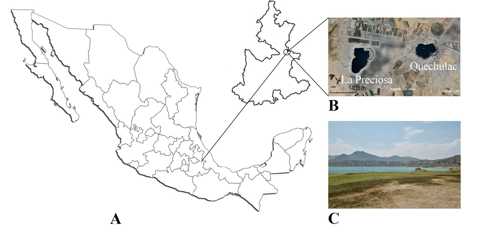
Located on the Central Plateau of Mexico, the Eastern Basin is one of the country’s largest endorheic basins, spanning 5,250 km2 and encompassing parts of Veracruz, Puebla, and Tlaxcala (Alcocer & Bernal-Brooks, 2019). Within this basin, to the east of Puebla State, lies the Llanos de San Juan Plain, where Lake La Preciosa is located at 19°22’17.91” N, 97°23’13.99” W, at an elevation of 2,330 m asl (Fig. 1). Lake La Preciosa is a warm, monomictic maar-type crater lake with a maximum depth of 46 m. It is characterized as subsaline according to Fritz (2013) with ~ 1.2 PSU and total dissolved solids ~ 1.7-1.8 g L-1 (athalassohaline type). The lake exhibits an alkaline pH (8.5-9.6) and is generally considered oligotrophic to low mesotrophic, with chlorophyll a concentration below 3 to 6 μg L-1. The eukaryotic plankton community responds dynamically to variations in dissolved oxygen levels in the water column (Havas et al., 2022, 2023; Iniesto et al., 2022; Sigala et al., 2017; Zeyen et al., 2021).
Sampling campaigns were conducted in September and October 2022 (during lake stratification) and January 2023 (during lake mixing). On September 22 and January 23, water samples were collected at 6 depths (2, 5, 10, 15, 20, and 30 m) using a 3-liter horizontal Van Dorn-La Motte bottle (Chestertown, USA). At each depth, environmental parameters including temperature, pH, dissolved oxygen, conductivity, underwater irradiance, and chlorophyll a concentration were measured with a multiparameter probe (Hydrolab, Kempten, Germany). We monitored underwater irradiance with a time-recording device (HOBO, Bourne, USA) attached to the probe. Surface irradiance values were estimated based on standard solar radiation conditions and converted to photon flux density (PPFD) using published lux-to-PPFD conversion factors (e.g., Thimijan & Heins, 1983). Calculations were performed using an online conversion tool (Waveform Lighting, https://www.waveformlighting.com/horticulture/convert-lux-to-ppfd-online-calculator), assuming full sunlight conditions. Due to the qualitative nature of the observations and the limited number of sampling points, statistical analyses were not applied.
Cell morphology and multiplication stages were examined on a sample of living material under a phase-contrast light microscope (Nikon Eclipse 80i, Melville, USA). Part of the sample was prepared for scanning electron microscopy (SEM) analyses by filtering 5 ml aliquots on 5 µm pore polycarbonate filters. The filters were fixed in 2% glutaraldehyde (GTA) and rinsed with phosphate-buffered saline (PBS); the cellular material was then dehydrated in an ascending series of ethanol (30, 50, 70, 90, 95, and 100%) for 15 min each. Subsequently, samples were transferred to a Petri dish with amyl acetate for 2-3 h and dried using CO2 critical-point drying (BAL-TEC CPD-030, New York, US). The dried material was sputter-coated with gold using a Denton Vacuum Desk II sputter coater (New Jersey, USA) and observed with a Jeol JSM-5310LV scanning electron microscope (SEM, MA, USA). The SEM was operated at 15 kV and secondary electrons as the detection mode.
Results
During the stratification period of the water column (October 2022), several stages of the reproductive process of Peridiniopsis borgei were observed, mainly in samples collected at depths of 5 and 10 m. In contrast, samples collected during the mixed water column period (January 2023) revealed a small population, consisting mainly of vegetative cells, with no reproductive stages observed.
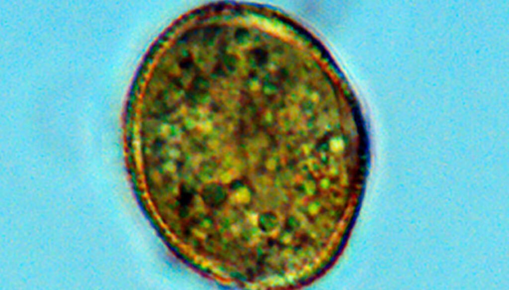
Peridiniopsis borgei cells display a gonyaulacoid/peridinioid morphology and thecal structure and are slightly flattened dorsoventrally and rounded toward the antapex. The apex displays a pronounced conical shape resulting from the union of the apical plates. The epitheca is triangular and symmetrical along the sagittal plane; the hypotheca is rounded. The dinokaryon-like nucleus is observed in the cell’s hypocone, and a red, vertically oriented stigma is visible on the ventral side (Fig. 2A, B). Numerous discoidal chloroplasts, a pusular sac, and a notably large extraplastidial pyrenoid are present in the cell’s epicone (Fig. 2C).
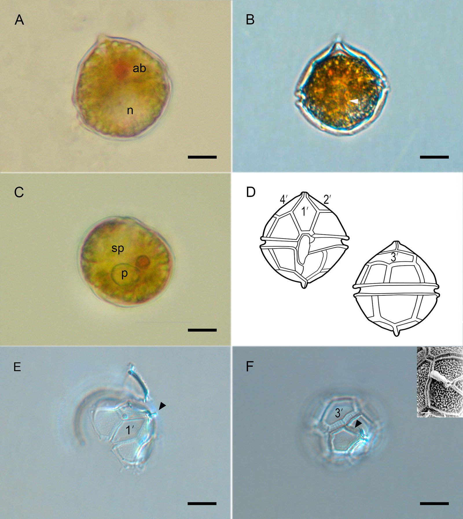
Cell dimensions from each observation (n = 20) range from 33.2-39.5 µm in length and 28.2-37.0 µm in transdiameter. The apex presents a complex apical pore consisting of an apical pore (Po) surrounded by a covering plate (cp) and a pore plate (pp). The thecal formula is Po (cp, pp), 4’, 6’, 5’’’, 6C, 5S, 2’’’’; the epitheca has 10 plates (Po, 4’, 6’’). (Fig. 2D-F). Plate 1’ is hexagonal and diamond-shaped, while plates 2’ and 4’ are pentagonal. We consider plate 3’ as the apical plate because it is in contact with the pore across the suture between 2’ and 4’ (Fig. 2F).
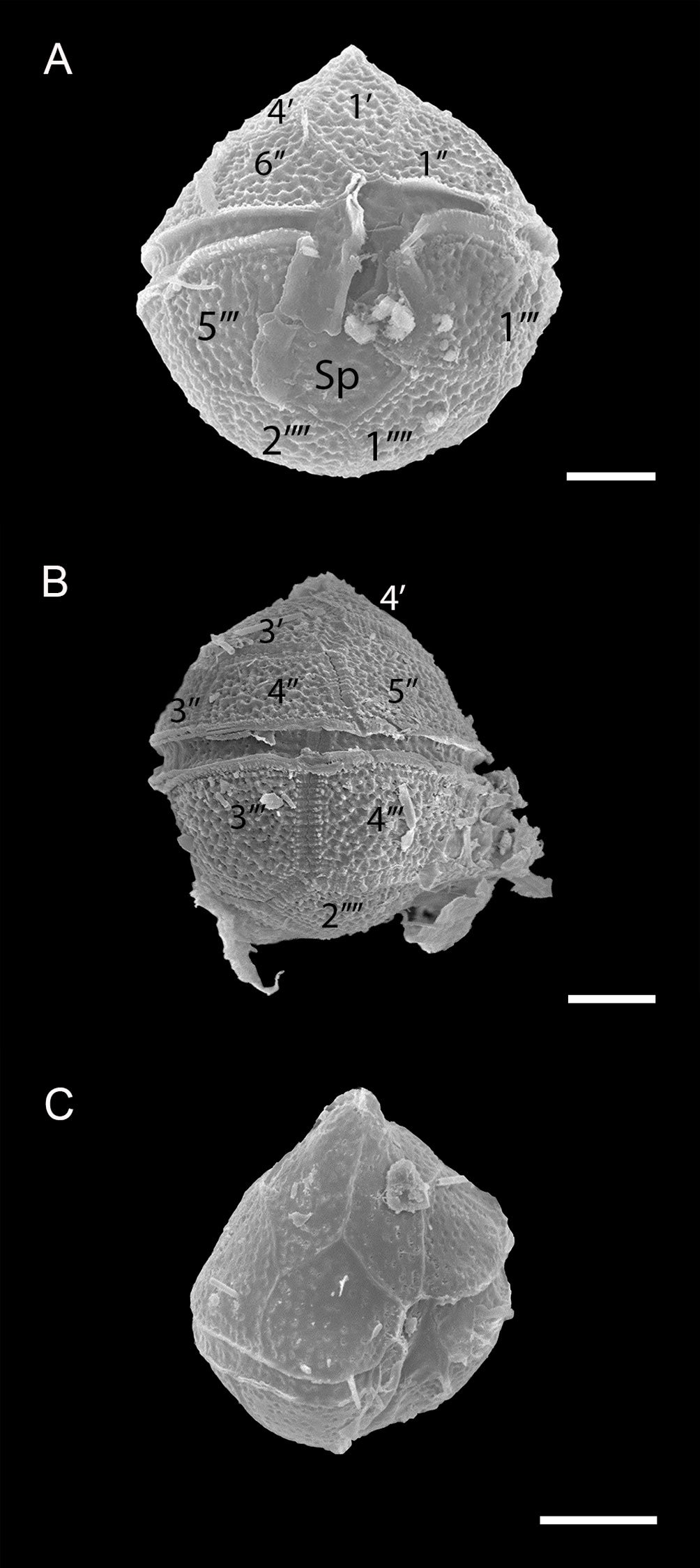
The cingulum is excavated and descends by approximately half its width; the sulcus appears deep and widens toward the bottom of the sulcal cleft; the left sulcal plate has a flap decorated with pores that cover part of the posterior sulcal plate. Another sulcal flap was observed in the posterior portion of the suture between plate 5’’’ and the posterior sulcal plate. The posterior sulcal plate appears wide and does not reach the antapex (Fig. 3A).
Only a few specimens exhibited a marginal ridge between the sutures of plates 2’ and 4’; conspicuous apical marginal ridges and intercalary bands with transverse striations between the sutures were observed. Coinciding with the presence or absence of intercalary bands, most specimens exhibited reticular ornamentation with a pore in each reticulum (Fig. 3B). Specimens lacking intercalary bands displayed a poorly developed reticulum and scattered pores over the theca (Fig. 3C).
Reproductive stages were recorded in samples collected during the stratified period (October 2022). The dominant stage was the motile vegetative cell. Before cell division, cells release from the theca (eleutheroschisis). Immediately, pyrenoid fission was observed (Fig. 4A). Nucleus division was also observed (Fig. 4B), followed by the formation of a cleavage furrow in the cell’s pellicular layer, which participated in cytokinesis (Fig. 4C). This division (desmoesquisis) resulted in 2 daughter cells that eventually separated (Fig. 4D).
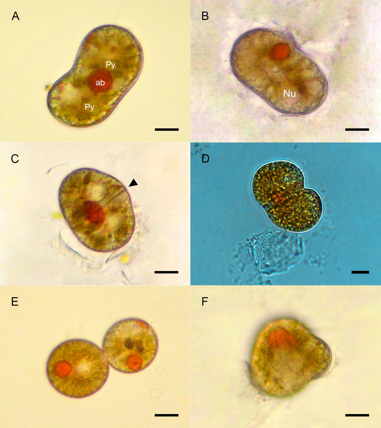
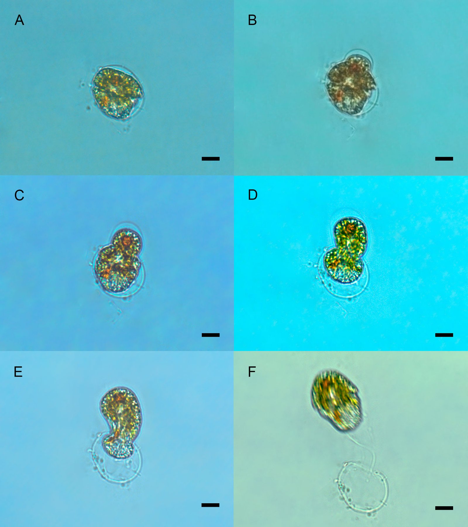
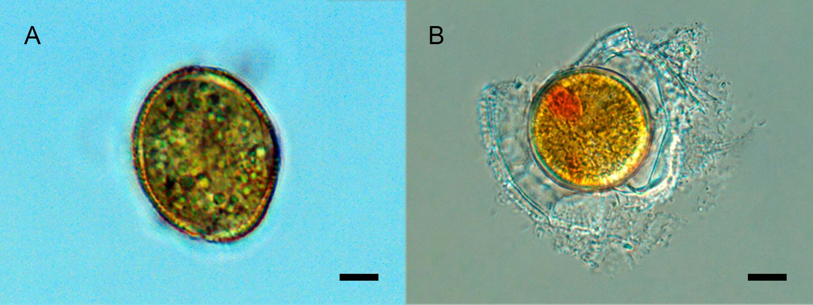
Small vegetative cells were also observed, lacking the theca, and surrounded by the pellicular layer (~ 35-50 µm long × 25-27 µm wide); the so-called peanut shape was recognized in these immobile cells (Fig. 4E). Other cells, also immobile, displayed an undefined morphology. They were rare and similarly lacked the theca and were also surrounded by the pellicular layer (Fig. 4F). There was no discernible sequence among these cellular shapes (Fig. 4E, F) and the stages observed during division (Fig. 4A-E). Moreover, a brief eleutheroschisis sequence was observed with cell ecdysis. Initially, the cell rearranged itself within the pellicular layer, demonstrating considerable flexibility and a partial loss of its typical shape. The cell, released through a pore in the pellicular layer, was immediately motile and of the size of the adult cell. Although theca was not visible, a shape close to that typical of a thecate cell was observed (Fig. 5A-F), indicating rapid synthesis of the cell’s new pellicular layer, which largely maintained its shape.
In the same sample, double-walled resistance cysts were observed (Fig. 6A). We also observed temporary cysts (Fig. 6B) approximately the same size as vegetative cells. Resistance cysts, however, were larger (40-60 µm long × 26-40 µm wide). No hatching events of any cystic stage were observed.
During the stratification of the water column in September 2022, temperature and dissolved oxygen profiles showed coinciding clines (Fig. 7A). In contrast, during the mixing period of January 2023, both parameters were homogeneous throughout the water column (Fig. 8A). Chlorophyll a concentration in the water column, measured in both periods (Table 1), had a median from 3.52 (September) to 3.54 (January) mg L-1. During water-column mixing in January 2023, chlorophyll a concentration exhibited a more homogeneous distribution across the depth profile and was consistent with irradiance values. However, during lake stratification in September 2022, a marked decrease in chlorophyll a concentration was observed over depth, consistent with the oxycline and thermocline, and especially also with irradiance/PAR values (Figs. 7B, 8B; Table 1).
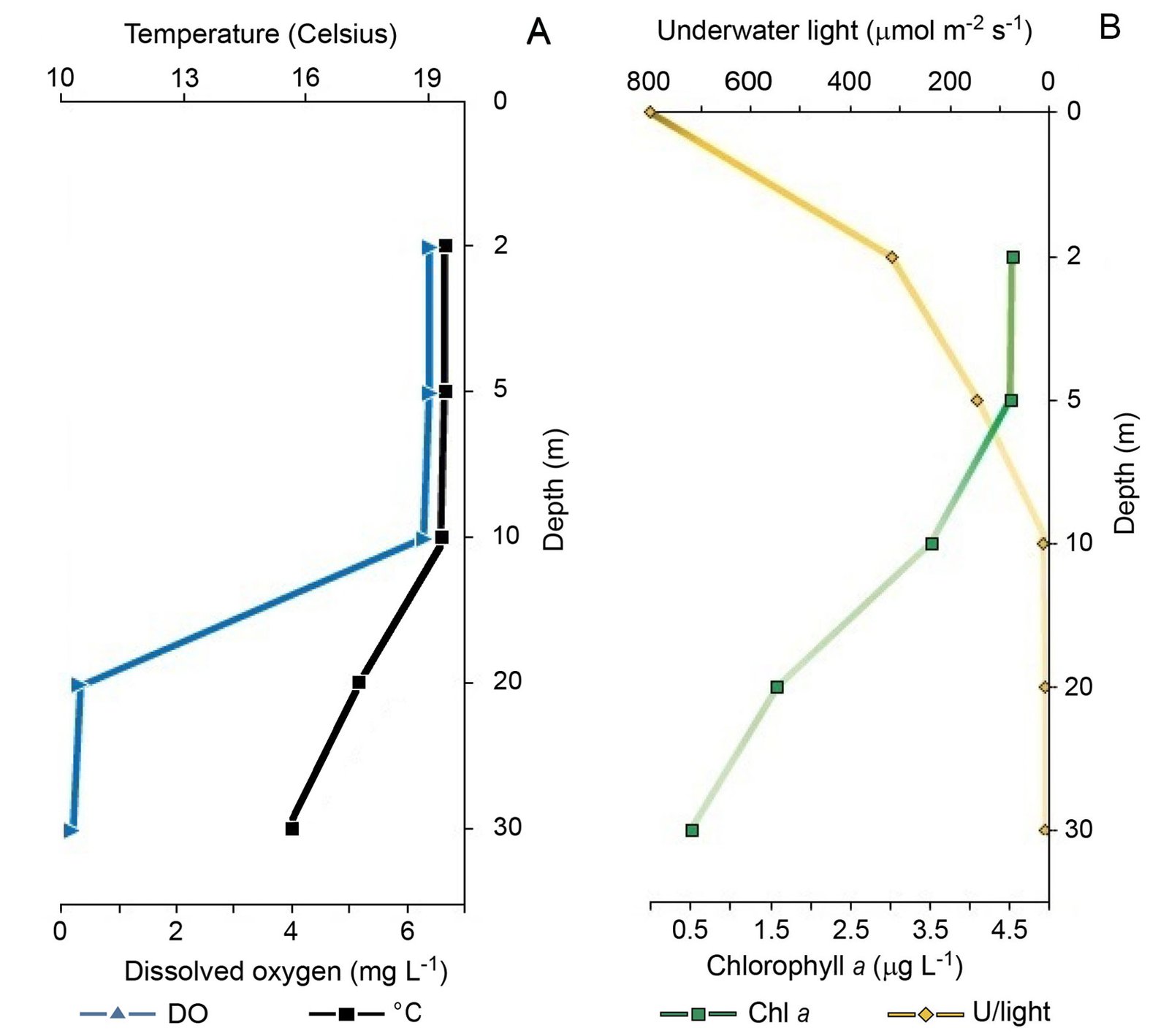
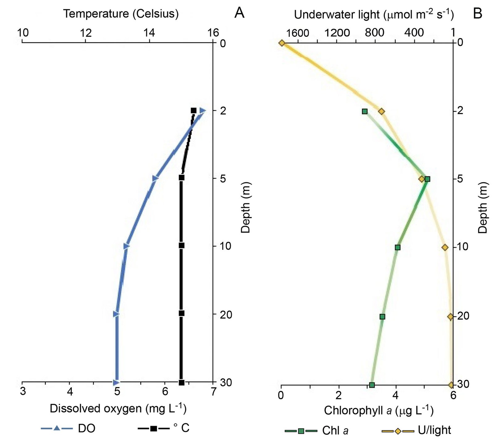
| Depth (m) | Temperature (°C) | pH | Conductivity (mS cm⁻¹) | Dissolved oxygen (mg L⁻¹) | Irradiance (µmol photons m⁻² s⁻¹) | Chlorophyll-a (µg L⁻¹) |
|---|---|---|---|---|---|---|
| September 2022 (stratified period) | ||||||
| 0* | — | — | — | — | 800 | — |
| 2 | 19.4 | 9.6 | 2.347 | 6.4 | 316.9 | 4.5 |
| 5 | 19.4 | 9.5 | 2.346 | 6.4 | 144 | 4.5 |
| 10 | 19.3 | 9.5 | 2.350 | 6.3 | 4.0 | 3.5 |
| 20 | 18.0 | 9.3 | 2.334 | 0.3 | 0 | 1.6 |
| 30 | 15.6 | 9.3 | 2.317 | 0.2 | 0 | 0.5 |
| January 2023 (mixing period) | ||||||
| 0* | — | — | — | — | 1762 | — |
| 2 | 15.4 | 8.8 | 2.315 | 6.8 | 738.6 | 3.0 |
| 5 | 15.0 | 8.8 | 2.313 | 6.0 | 320.9 | 5.1 |
| 10 | 14.9 | 8.8 | 2.313 | 5.2 | 31.77 | 4.0 |
| 20 | 14.8 | 8.8 | 2.312 | 5.0 | 8.17 | 3.5 |
| 30 | 14.8 | 8.8 | 2.310 | 5.2 | 1.06 | 3.2 |
Discussion
According to the literature, Peridiniopsis borgei is a common species in inland waters. However, the lack of information regarding its physiology and the environmental conditions under which it occurs prevents us from profiling its ecological responses. While P. borgei has occasionally been described as a bloom-forming species (e.g., Schagerl et al., 2010; Zohary et al., 1994), we have observed blooms of this species only in Lake La Preciosa, among the 10 lakes investigated by the TMVB. However, P. borgei has also occurred rarely, in small populations, in Lake Quechulac (located in the same eastern basin) during thermal stratification of the water column. The crater lakes of Puebla State share several hydrological characteristics and a specific geological origin, and they are geographically proximate. Despite these similarities, they also present marked differences, for example, in salinity and alkalinity. It has been established that the lakes of the TMVB, and in particular those in Puebla, exhibit a positive correlation between salinity and alkalinity (Zeyen et al., 2017, 2021). The values in La Preciosa, subsaline (PSU 1.17-1.41; TA 14.13-14.40 mM), are higher than in Quechulac, freshwater (PSU 0.45-0.41; TA 6.55-6.68 mM) and much lower than in Atexcac, hyposaline (PSU 2.4-8.73; TA 30.9-31.8 mM) and Alchichica, hyposaline (PSU 8.19-10.1; TA 42.81-43.94 mM) (Havas et al., 2023; Iniesto et al., 2022; Zeyen et al., 2019). Our observations suggest that although P. borgei occurs at low abundance in freshwater environments such as Lake Quechulac, it develops as blooms at salinity and alkalinity levels similar to those in Lake La Preciosa. These limnological differences are likely key factors determining the distribution and abundance of P. borgei in inland aquatic systems in Mexico. Furthermore, based on the existing literature on dinophytes and the conditions at La Preciosa, we propose that mainly during the stratified period, vertical migration occurs in response to gradients in light, nutrients, and iron availability, and constitutes a key ecological strategy for P. borgei (Raven & Lavoie, 2021; Stanković et al., 2024; Torres et al., 2023). Iron availability appears to be crucial for some microbial species at La Preciosa. For example, the cyanobacterium Cyanocatena sp. has been shown to localize iron as extracellular precipitates rich in Fe, Mn, and Si, contributing to iron cycling in the water column (Benzerara et al., 2023). Our observations suggest that P. borgei occurs under specific environmental conditions. In this context, previous metagenomic analyses from Lake La Preciosa have detected 18S rRNA gene sequences identical to those of Peridiniopsis borgei in earlier years (Supplementary material: Fig. S1), suggesting that this species may occur intermittently in the system, although a direct link to the present morphologically identified population remains to be confirmed.
The morphological diversity described in the literature for the apical region of P. borgei cells was also observed in our specimens. For example, according to Ascencio et al. (2020) and Boltovskoy (1999), we identified the 3’ plate as the apical plate. The extension of the suture crest that continues from the apex to the 3’ plate and which integrates it as part of the apical complex can be of variable length. In our specimens, it was relatively long. Accordingly, the studied population lacked intercalary plates. We also detected elongated cells with dividing nuclei and pyrenoids (Fig. 4A, B), as well as peanut-shaped cells (Fig. 4E). These forms resemble those described in species of the genus Parvodinium Carty, though their exact taxonomic significance remains uncertain. In both traditional and recent dinophyte literature, Parvodinium species with elongated and peanut-shaped cells are proposed to be yet unconfirmed reproductive stages within the sexual cycle, perhaps related to the germination of planozygotes or hypnozygotes, and which could represent morphological differences between meiosis 1 and meiosis 2 (Lehner et al., 2025; Pfiester et al., 1984; Sako et al., 1986).
Regarding the reproductive stages, we were unable to observe sexual processes such as syngamy or direct products of zygote germination. Thus, the different cell forms that we observed have been identified and described as part of vegetative multiplication (i.e., asexual reproduction). Classic works on the Dinophyta group have reported these forms, which appear to be common, although they have not yet been located in the life cycle (Entz, 1926). In P. borgei at La Preciosa, these might represent stages of the division process after the germination of hypnozygotes. Hence, only a few of these have been observed suspended in the water column (Fig. 6A, B). Therefore, it remains unclear whether these stages represent meiotic events. Consequently, the observed eleutheroschisis process will also have to wait until we obtain clonal cultures of Peridiniopsis borgei and determine its life cycle.
Overall, our observations suggest that P. borgei may have a short life cycle with overlapping generations under lake conditions associated with subsaline water (~ 1.2 PSU), alkaline water (~ 14 mM TA), and epilimnetic dissolved oxygen availability ≥ 6.0 mg L-1. The rarity of this species in Mexican limnological records could be attributed to its narrow ecological niche and the specific conditions required for bloom formation, if P. borgei in La Preciosa follows a vertical migration strategy driven by the availability of light, oxygen, nutrients, and iron. This lake satisfies the limiting factors that could restrict its growth. Therefore, its population dynamics are regulated by both microbial and geochemical processes.
Acknowledgments
We are grateful for A. Boltovskoy’s review and insightful comments, which substantially improved the article. To financial support from the MITI interdisciplinary programs of the CNRS and the French National Research Agency (ANR) under the Microbialite grant ANR-18-CE02-0013-01, and from the Postdoctoral Fellowship Program of the General Directorate of Academic Personnel Affairs of the UNAM, awarded to CBG. To Guadalupe Vidal’s (LACET) support with laboratory sample processing and Silvia Espinosa’s (Faculty of Sciences) assistance in obtaining electron micrographs.
References
Alcocer, J., & Bernal-Brooks, F. W. (2019). Physical and chemical characterization of inland waters. In A. L. Ibáñez (Ed.), Mexican aquatic environments (pp. 1–41). Ciudad de México: Springer. https://doi.org/10.1007/978-3-030-11126-7_1
Armienta, M. A., Vilaclara, G., De la Cruz-Reyna, S., Ramos, S., Ceniceros, N., Cruz, O. et al. (2008). Water chemistry of lakes related to active and inactive Mexican volcanoes. Journal of Volcanology and Geothermal Research, 178, 249–258. https://doi.org/10.1016/j.jvolgeores.2008.06.019
Ascencio, E., Mendoza-Carbajal, L., Baylón, M., Rivera, P., Krakhmalnyi, A. F., Cruces, F. et al. (2020). Reporte de Peridiniopsis borgei Lemmermann (Dinophyceae) en aguas continentales de Perú. Gayana Botánica, 77, 62–66. http://dx.doi.org/10.4067/S0717-66432020000100062
Ascencio, E., Rivera, P., & Cruces, F. (2015). Morfología de Peridiniopsis elpatiewskyi (Ostenfeld) Bourrelly (Dinophyceae) encontrada por primera vez en aguas continentales de Chile. Gayana Botánica, 72, 42–46. http://dx.doi.org/10.4067/S0717-66432015000100006
Ascencio, E., Rivera, P., Cruces, F., & Vila, I. (2018). Estudio morfológico de Naiadinium polonicum (Dinophyceae) reportado por primera vez en aguas continentales de Chile. Gayana Botánica, 75, 438–446. http://dx.doi.org/10.4067/S0717-66432018000100438
Benzerara, K., Elmaleh A., Ciobanu M., De Wever A., Bertolino P., Iniesto M. et al. (2023). Biomineralization of amorphous Fe-, Mn- and Si-rich mineral phases by cyanobacteria under oxic and alkaline conditions. Biogeosciences, 20, 4183– 4195. https://doi.org/10.5194/bg-20-4183-2023
Boltovskoy, A. (1973). Peridinium gatunense Nygaard. Estructura y estereoultraestructura tecal (Dinoflagellida). Physis B, 32, 331–344.
Boltovskoy, A. (1975). Estructura y estereoultraestructura tecal de dinoflagelados. II. Peridinium cinctum (Müller) Ehrenberg. Physis B, 34, 73–84.
Boltovskoy, A. (1999). Contribución al conocimiento de los dinoflagelados de la república Argentina (Tesis doctoral). Facultad de Ciencias Naturales y Museo, Universidad Nacional de La Plata. La Plata, Argentina.
Boltovskoy, A. (2003). Peridinium willei (Dinophyceae) y sus formas: ecología y distribución en la Argentina. Boletín de la Sociedad Argentina de Botánica, 38, 175–184.
Bourrelly, P. (1968) Notes sur les péridiniens d’eau douce. Protistologica, 4, 5–13.
Bustamante-Gil, C., Boltovskoy, A. Rengefors, K., Tavera, R., Amat, E., & Ramírez-Restrepo, J. J. (2021). Checklist, new records, and taxonomic annotations of freshwater thecate dinoflagellate (Dinophyceae) in Colombia. Phytotaxa, 509, 135–167. https://doi.org/10.11646/phytotaxa.509.2.1
Calado, A. J., & Moestrup, Ø. (2002). Ultrastructural study of the species of Peridiniopsis, Peridiniopsis borgei (Dinophyceae), with special reference to the peduncle and flagellar apparatus. Phycologia, 41, 567–584. https://doi.org/10.2216/i0031-8884-41-6-567.1
Carty, S. (2014). Freshwater dinoflagellates of North America. Ithaca: Cornell University Press.
Cavalcante, K. P., Craveiro, S. C., Calado, A. J., Ludwig, T. A. V., & Cardoso, L. S. (2017). Diversity of freshwater dinoflagellates in the State of Paraná, southern Brazil, with taxonomic and distributional notes. Fottea, 17, 240–263. https://doi.org/10.5507/fot.2016.026
Cruz-Aviña, J. R., Núñez-García, L. G., Cabrera, R., Díaz- Larrea, J., & Castañeda-Roldan, E. I. (2021). Microbiological and physicochemical quality of water in four crater lakes in the state of Puebla, Mexico. Nexo Revista Científica, 34, 1632–1648. https://doi.org/10.5377/nexo.v34i06.13124
Entz, G. (1926). Beiträge zur kenntnis der Peridineen. I. Zur morphologie und biologie von Peridinium borgei Lemmermann. Archiv für Protistenkunde, 56, 397–446.
Fritz, S. C. (2013). Salinity and climate reconstructions from continental lakes. In S. A. Elias, & C. J. Mock (Eds.), Encyclopedia of Quaternary Science, 2nd ed (pp. 507–515). Encyclopedia of Quaternary Science. Oxford: Elsevier.
Gätz, N., & Schagerl, M. (1990). Peridiniopsis borgei Lemm.- eine seltene Dinophyceae in ziegelteichen in Wien und Niederösterreich. Lauterbornia, 4, 24–29.
Gottschling, M., Čalasan, A. Ž., Kretschmann, J., & Gu, H. (2017). Two new generic names for dinophytes harbouring a diatom as an endosymbiont, Blixaea and Unruhdinium (Kryptoperidiniaceae, Peridiniales). Phytotaxa, 306, 296– 300. https://doi.org/10.11646/phytotaxa.306.4.6
Guiry, M. D., & Guiry, G. M. (2025). AlgaeBase. World-wide electronic publication, University of Galway. https://www.algaebase.org
Havas, R., Thomazo, C., Caumartin, J., Iniesto, M., Bert, H., Jézéquel, D. et al. (2025). Untangling the primary biotic and abiotic controls on oxygen, inorganic and organic carbon isotope signals in modern microbialites. Geobiology, 23, e70012. https://doi.org/10.1111/gbi.70012
Havas, R., Thomazo. C., Iniesto. M., Jézéquel. D., Moreira D., Tavera, R. et al. (2023). The hidden role of dissolved organic carbon in the biogeochemical cycle of carbon in modern redox-stratified lakes. Biogeosciences, 20, 2405–2424. https://doi.org/10.5194/bg-20-2405-2023
Iniesto, M., Moreira, D., Benzerara, K., Reboul, G., Bertolino, P., Tavera, R. et al. (2022). Planktonic microbial communities from microbialite-bearing lakes sampled along a salinity- alkalinity gradient. Limnology and Oceanography, 67, 2718–2733. https://doi.org/10.1002/lno.12233
Iniesto, M., Moreira, D., Reboum, G., Deschamps, P., Bertolino, P., Saghai, A. et al. (2020). Core microbial communities of lacustrine microbialites sampled along an alkalinity gradient. Environmental Microbiology, 23, 51–68. https://doi.org/10.1111/1462-2920.15252
Krakhmalny, A. F. (2014). New species of dinoflagellata (Dinoflagellate) for Ukraine. In Nature of Polessia: research and conservation (pp. 330–333). Ukraine: Rivnenska Daukarnia, Kiev.
Krakhmalny, A. F., Wasser, S. P., & Nevo, E. (2000). Dinophyta. In E. Nevo, & S. P. Wasser (Eds.), Biodiversity of Cyanoprocaryotes, algae and fungi of Israel: Cyanoprocaryotes and algae of continental Israel (pp. 177–183). Ruggell: A. R. A. Gantner Verlag K. G.
Lehner, P. M. E., Zhu, B., Stark, M., Knechtel, J., Nyoka, H., Owsianny, P. M. et al. (2025). Linking new DNA sequence data to old scientific names of Parvodinium (Peridiniopsidaceae, Dinophyceae). European Journal of Phycology, 60, 115–134. https://doi.org/10.1080/09670262.2025.2450678
Ling, H. U., Croome, R. L., & Tyler, P. A. (1989). Freshwater dinoflagellates of Tasmania, a survey of taxonomy and distribution. British Phycological Journal, 24, 111–129. https://doi.org/10.1080/00071618900650111
Ling, H. U., & Tyler, P. A. (2000). Australian freshwater algae (exclusive of diatoms). Berlin: J. Cramer in der Gebruder Borntraeger Verlagsbuchhandlung. Moestrup, Ø., & Calado, A. J. (2018). Dinophyceae. In B. Büdel, G. Gärtner, L. Krienitz, & M. Schagerl (Eds.), Süßwasserflora von Mitteleuropa, Bd. 6 – Freshwater Flora of Central Europe, Vol. 6 (pp. 1–561). Berlin: Springer Spektrum. https://doi.org/10.1007/978-3-662-56269-7
Novelo, E., & Tavera, R. (2019.2025). bdLACET Base de datos de algas continentales. Un buscador de algas pensado para ficólogos y no tan ficólogos. Facultad de Ciencias, UNAM. México. https://bdlacet.mx
Pfiester, L. A., Timpano, P., Skvarla, J. J., & Holt, J. R. (1984). Sexual reproduction and meiosis in Peridinium inconspicuum Lemmermann (Dinophyceae). American Journal of Botany, 71, 1121–1127. https://doi.org/10.2307/2443388
Pollingher, U., & Hickel, B. (1991). Dinoflagellate associations in a subtropical lake (Lake Kinneret, Israel). Archiv für Hydrobiologie, 120, 267–285. https://doi.org/10.1127/archiv-hydrobiol/120/1991/267
Raven, J. A., & Lavoie, M. (2021). Movement of aquatic oxygenic photosynthetic organisms. In U. Lüttge, F. M. Cánovas, M. C. Risueño, C. Leuschner, & H. Pretzsch (Eds.), Progress in Botany Vol. 83 (pp. 315–343). Cham: Springer International Publishing. https://doi.org/10.1007/124_2021_55
Rengenfors, K., & Kremp, A. (2018). The ecology of freshwater dinoflagellates. In Ø. Moestrup, & A. J. Calado (Eds.), Süßwasserflora von Mitteleuropa, Bd. 6 – Freshwater Flora of Central Europe, Vol. 6: Dinophyceae (pp. 27–36). Berlin: Springer Nature. https://doi.org/10.1007/978-3-662-56269-7_3
Rohrlack, T. (2020). The diel vertical migration of the nuisance alga Gonyostomum semen is controlled by temperature and by a circadian clock. Limnologica, 80, 125746. https://doi.org/10.1016/j.limno.2019.125746
Samanez, I. (2015). Primer registro de Lophodinium polylophum (Daday) Lemmermann 1910 (Dinophyceae: Lophodiniaceae) en el Perú. Revista Peruana de Biología, 22, 109–112. https://doi.org/10.15381/rpb.v22i1.11128
Sako, Y., Nakanishi, M., Konda, T., Ishida, Y., Kadota, H., Shrestha, K. et al. (1986). Life cycle of Peridinium sp. B3 (Dinophyceae) isolated from Lake Begnas, Nepal, Bulletin of Japanese Society of Microbial Ecology, 1, 19–27. https://doi.org/10.1264/microbes1986.1.19
Schagerl, M., Bloch, I., Angeler, D. G., & Fesl, C. (2010). The use of urban clay-pit ponds for human recreation: assessment of impacts on water quality and phytoplankton assemblages. Environmental Monitoring and Assessment, 165, 283–293. https://doi.org/10.1007/s10661-009-0945-2
Sigala, I., Caballero, M., Correa-Metrio, A., Lozano-García, S., Vázquez, G., Pérez, L. et al. (2017). Basic limnology of 30 continental waterbodies of the Transmexican Volcanic Belt across climatic and environmental gradients. Boletín de la Sociedad Geológica Mexicana, 69, 313–370. https://doi.org/10.18268/bsgm2017v69n2a3
Thimijan, R. W., & Heins, R. D. (1983). Photometric, radiometric, and quantum light units of measure: a review of procedures for interconversion. Hortscience, 18, 818–822. https://doi.org/10.21273/HORTSCI.18.6.818
Torres, R., Sherrell, R., Reid, B., Pizarro, G., Frangópulos, M., Alarcón, E. et al. (2023). Iron and silicic acid addition effects on early spring macronutrient drawdown and biogenic silica production of Patagonia estuarine waters. Progress in Oceanography, 214, 102982. https://doi.org/10.1016/j.pocean.2023.102982
Zhang, Q., Liu, G. X., & Hu, Z. Y. (2012). Study on freshwater genus Peridiniopsis (Dinophyta) from China. Acta Hydrobiologica Sinica, 36, 751–764.
Zeyen, N., Benzerara, K., Beyssac, O., Daval, D., Muller, E., Thomazo, C. et al. (2021). Integrative analysis of the mineralogical and chemical composition of modern microbialites from ten Mexican lakes: What do we learn about their formation? Geochimica et Cosmochimica Acta, 305, 148–184. https://doi.org/10.1016/j.gca.2021.04.030
Zeyen, N., Benzerara, K., Menguy, N., Brest, J., Templeton, A. S., Webb, S. M. et al. (2019). Fe-bearing phases in modern lacustrine microbialites from Mexico, Geochimica et Cosmochimica Acta, 253, 201–230. https://doi.org/10.1016/j.gca.2019.03.021
Zeyen, N., Daval, D., López-García, P., Moreira, D., Gaillardet, J., & Benzerara, K. (2017). Geochemical conditions allowing the formation of modern lacustrine microbialites, Procedia Earth and Planetary Science, 17, 380–383. https://doi.org/10.1016/j.proeps.2016.12.096
Zohary, T., Erez, J., Gophen, M., Berman-Frank, I., & Stiller, M. (1994). Seasonality of stable carbon isotopes within the pelagic food web of Lake Kinneret. Limnology and Oceanogra- phy, 39, 1030–1043. https://doi.org/10.4319/lo.1994.39.5.1030