a Bu-Ali Sina University, Department of Biology, 6517838695, Hamedan, Iran
b Kharazmi University, Faculty of Biological Sciences, Department of Plant Sciences, 4911-15719, Tehran, Iran
*Corresponding author: fgh@khu.ac.ir (F. Ghahremaninejad)
Received: 07 November 2024; accepted: 21 March 2025
Abstract
Leaf trichomes are vital for plant physiology, defense, and ecological interactions. This study utilized scanning electron microscopy to examine the leaf trichomes of 7 species in the family Cordiaceae, including 5 species from the genus Cordia and 2 from Varronia. Various types of trichomes, including glandular, non-glandular, and stellate, were observed. Our findings highlight differences in trichome types, density, and distribution both within and between genera. This research enhances the understanding of anatomical diversity and the significance of leaf trichomes in Cordiaceae, providing a foundation for future studies in plant biology. The high diversity of trichome structure in the studied species indicates their importance for their classification. Therefore, the results of this study provide a useful insight into the diversity of trichomes and their potential as a taxonomic tool.
Los tricomas de las hojas son vitales para la fisiología, la defensa y las interacciones ecológicas de las plantas. Este estudio utilizó microscopía electrónica de barrido para examinar los tricomas de las hojas de 7 especies de la familia Cordiaceae; se incluyeron 5 de los géneros Cordia y 2 de Varronia. Se observaron varios tipos de tricomas, incluidos glandulares, no glandulares y estrellados. Nuestros hallazgos resaltan las diferencias en los tipos de tricomas, la densidad y la distribución tanto dentro como entre géneros. Esta investigación mejora la comprensión de la diversidad anatómica, resalta la importancia de los tricomas de las hojas en Cordiaceae y proporciona una base para futuros estudios en biología vegetal. La gran diversidad de la estructura de los tricomas en las especies estudiadas indica su importancia para su clasificación. Por lo tanto, los resultados de este estudio proporcionan una buena visión de la diversidad de tricomas y su potencial como herramienta taxonómica.
Palabras clave: Indumento; Glandular; No glandular; Tricomas estrellados; Taxonomía
Contribución al valor sistemático de los tricomas foliares en las Cordiaceae (Boraginales)
Introduction
The family Cordiaceae in Boraginales includes 2 large genera, Cordia L. and Varronia P. Browne with a total of 368 accepted species (POWO, 2024) or 350 (APG 4, 2016). Its members are usually shrubs or trees that are distributed in tropical and subtropical regions (Luebert et al., 2016). The classification of this family and its subfamilies has been debated by botanists for years. In most references Cordiaceae has been recognized as a family or as part of Heliotropiaceae and Boraginaceae. Finally, based on the results of Luebert et al. (2016), this family was accepted as a separate family in Boraginales. Cordiaceae is the only family in Boraginales with plicate cotyledons and generally a twice-dichotomous style with 4 stigmatic branches (Luebert et al., 2016). Johnston (1951) introduced the genus Cordia with 5-7 sections for Cordiaceae and molecular work placed Varronia as the sister genus of Cordia (Miller & Gottschling, 2007). Species of Cordia are found principally in tropical and subtropical regions of the American, Asian, and African continents. In this genus, there are many species cultivated as ornamentals, and for wood and medicinal applications, with extensive use by traditional communities (Matias et al., 2015). Varronia is a Neotropical genus with most of its species naturally distributed in Brazil (Silva & Melo, 2019).
Leaf trichomes are specialized structures that grow on the epidermis of leaves. They play an important role in how plants adapt to their environment and interact with it (Wagner, 1991); also, they are a powerful tool in solving taxonomic problems (e.g., Ghahremaninejad et al., 2012). Trichomes play a significant role in the leaf texture of Cordiaceae species, contributing to their diagnostic value within the family. Studies have shown that the family possesses both glandular and non-glandular leaf trichomes, which influence their rough texture (Amer et al., 2016). Notably, in some regions, the abrasive surface of these leaves (including those of Cordia sebestena L.) has led to their traditional use as natural sandpaper (Flowers of India, 2025). Cordiaceae exhibits a wide range of leaf trichome morphologies, yet comprehensive studies elucidating their diversity and functional significance remain limited. Trichome morphology, density, and distribution are known to vary extensively even within closely related taxa, reflecting adaptation to diverse ecological niches and selective pressures (Rodríguez & Healey, 2000). In addition to their functional roles, the structural and functional diversity of leaf indument in Cordiaceae are indispensable taxonomic tools. Trichome characteristics often serve as apomorphic characters that assist botanists in resolving complex taxonomic issues (e.g., Beilstein et al., 2006; Ghahremaninejad, 2004; Kong & Hong, 2019; Steyn & Van Wyk, 2021). However, many studies within this family have paid insufficient attention to trichome features (e.g., Amer et al., 2016; Taroda, 1984).
Trichome morphology exhibits remarkable diversity across the plant kingdom, playing a significant role in the taxonomy and ecology of various plant families, including the Boraginaceae and Cordiaceae (Selvi & Bigazzi, 2001). Limited studies on Cordiaceae trichomes reveal a high diversity of trichomes both among closely related species (Tölke et al., 2013) and at the genus level (Silva et al., 2023). This diversity in trichome morphology suggests intricate evolutionary adaptations that may serve various ecological functions.
Given the existing literature on Cordia and Varronia, trichomes have critical taxonomic features in Cordiaceae, particularly for differentiating closely related genera. This study aims to examine the trichome morphology of both genera using SEM to clarify their taxonomic relationships. Scanning electron microscopy enhances trichome trait comparison by providing greater detail (Gonçales et al., 2023). We anticipated identifying distinct trichome characteristics to resolve existing taxonomic ambiguities. By focusing on these morphological characters, we seek to enhance the understanding of Cordiaceae taxonomy.
Material and methods
Samples were obtained from Herbarium specimens of the Naturhistorisches Museum Wien (W) and Botanical Museum Berlin-Dahlem (B), acronyms according to Thiers (2025). Trichome morphology was studied using SEM at the Natural History Museum (NHM), Vienna. Leaves were soaked in 40-degree water for 10 minutes to restore trichomes and then air-dried for 1 hour. Leaf samples were attached to SEM stubs with conductive adhesive tabs and coated with a thin layer of gold for conductivity, applied with a sputter coater. Imaging was performed with a JEOL JXA 6610LV SEM at the Vienna Museum. Digital images from various regions were captured, documenting trichome characteristics such as type, shape, size, density, and distribution for each species. Additionally, a voucher table listing the information and codes of the specimens used was prepared (Table 1).
Table 1
The voucher table of studied specimens.
No.
Species
Locality
Collector and number
QR code
Herbarium acronym
Type of trichome
1
Cordia africana Lam.
Ethiopia
U. i. 285
W0254121
W
Non-glandular
2
Cordia alliodora (Ruiz & Pav.) Oken
Brazil
s.c. 893
W0058028
W
Non-glandular
3
Cordia bicolor A.DC.
Brazil
Schomburgk 678
W0010617
W
Stellate
4
Cordia decandra Hook. & Arn.
Chile
K.H. & Rechinger 63378
W0254122
W
Non-glandular
5
Cordia myxa L.
Greece
Rainer KARL
W0254123
W
Non-glandular
6
Varronia bullata L.
–
–
W0254157
W
Glandular, non-glandular
7
Varronia guanacastensis (Standl.) J.S.Mill.
Costa Rica
Zamora et al. 6340
B101137807
B
Glandular, non-glandular (short pyramidal shape)
Results
Electron microscopy studies of 5 species of Cordia and 2 species of Varronia illustrate the diversity in the size, shape, and distribution of trichomes in Cordiaceae. Variation was observed both within and between genera. Two main types of trichomes were identified among the studied species, glandular and non-glandular. Glandular trichomes were observed only in Varronia species, while non-glandular were observed in species of both genera. In addition, a special type of stellate trichome, which mostly has more than 10 arms, as a first-time report, was observed in Cordia bicolor A. DC.
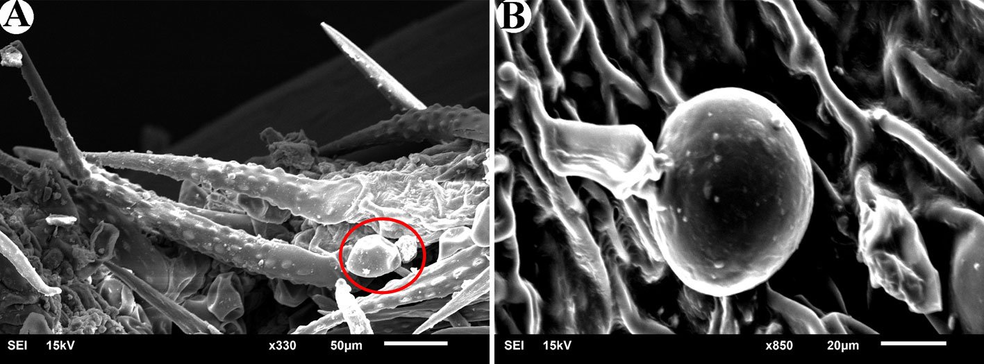
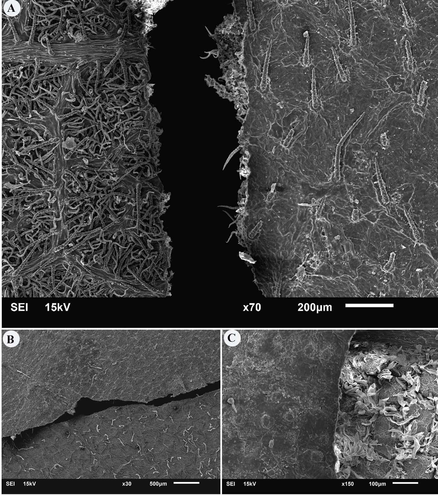
Among the investigated species, glandular trichomes with a size of 100 to 120 μm were observed only in Varronia bullata L. and V. guanacastensis (Standl.) J.J. Mill. (Fig. 1). A type of trichome was observed in Cordia myxa L., which is bag-like and different from non-glandular trichomes. The samples used in this research were herbarium specimens, but for accurate diagnosis of this trichome, fresh material is needed.
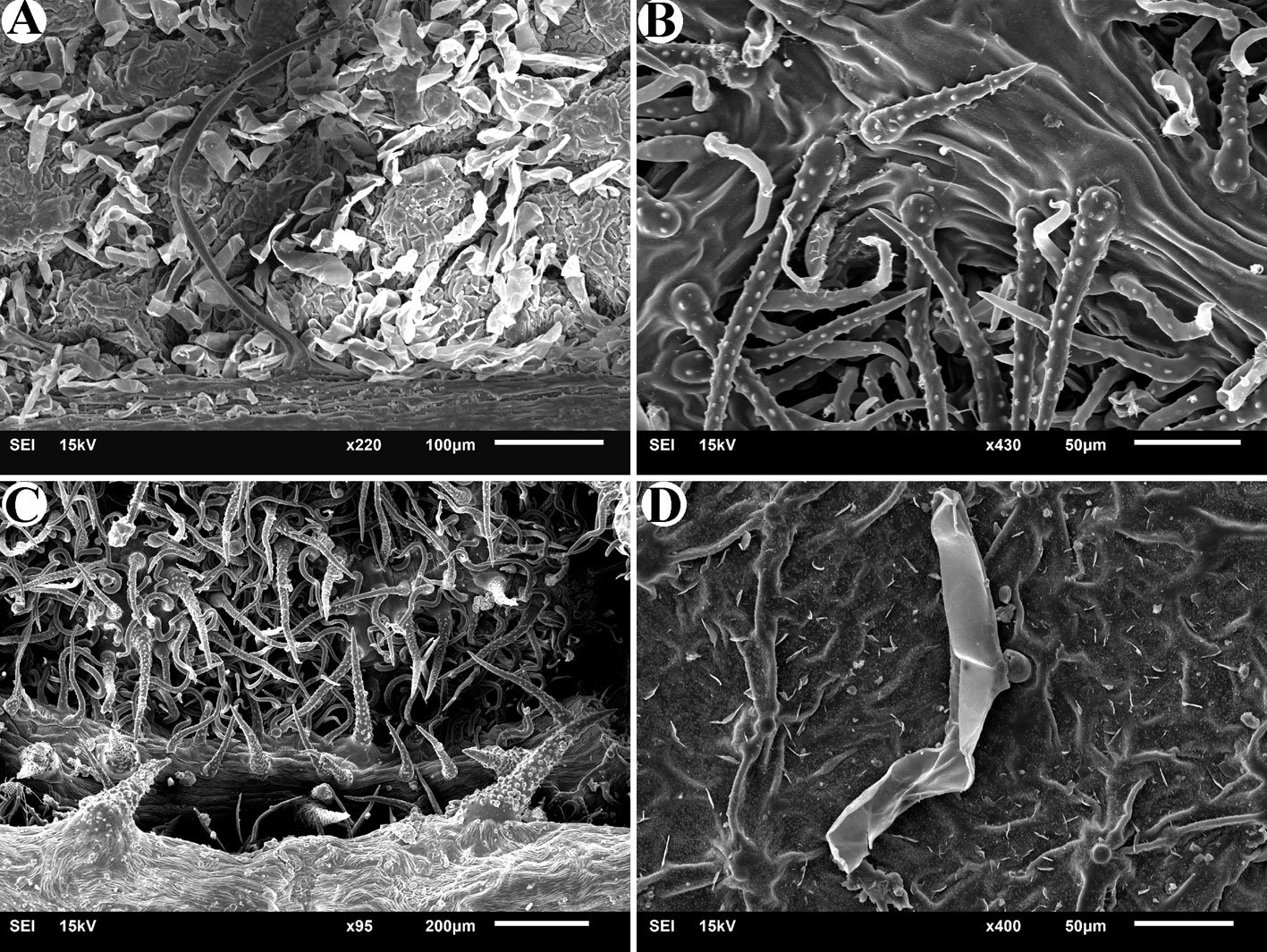
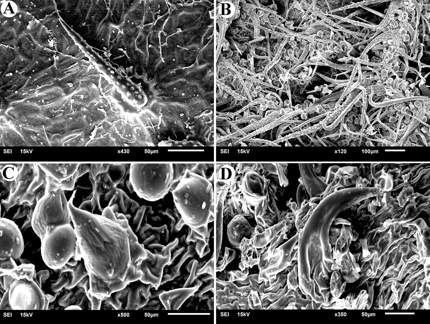
In all 7 studied species, non-glandular trichomes were observed on both adaxial and abaxial leaf surfaces. In Cordia africana Lam., the upper surface of the leaf has scattered short trichomes that exist in the form of small protrusions distant from one another, while the back surface of the leaf in this species (Fig. 2A) has depressions where different trichomes are densely present in these depressions. In C. alliodora (Ruiz & Pav.) Oken., simple trichomes are observed on both surfaces. On the abaxial surface of the leaf, in addition to this type of trichome, there are smaller and curved trichomes that do not have small projections on their surface (Fig. 2B). On the abaxial surface of C. decandra Hook. & Arn. leaves, in addition to short and long simple trichomes, which are smooth and sometimes curved, horn-shaped hard trichomes are also observed (Fig. 2C). In C. myxa, in addition to the specific type of trichomes mentioned (referring to glandular trichomes), very small trichomes that include a widened base and a protrusion (Fig. 2D) are observed in nearly regular rows on the adaxial surface of the leaf. On the abaxial surface, long trichomes with small projections are observed. Varronia bullata also has simple trichomes, which are similar to the trichomes of the mentioned species, except that on the adaxial surface, trichomes are completely dormant (Fig. 3A), but on the abaxial surface, in addition to greater diversity and density, some standing or twisted trichomes are also seen (Fig. 3B). In Varronia guanacastensis, a special type of trichome was observed, which covered the adaxial surface of the leaf in addition to many glandular trichomes. These trichomes are short and have a conical end (Fig. 3C). On the abaxial surface, a type of horn-like non-glandular trichome is also seen (Fig. 3D).
Figure 1. Glandular trichomes. A, Glandular trichomes (Icircled) next to non-glandular trichomes in Varronia bullata; B, glandular trichomes in Varronia guanacastensis.
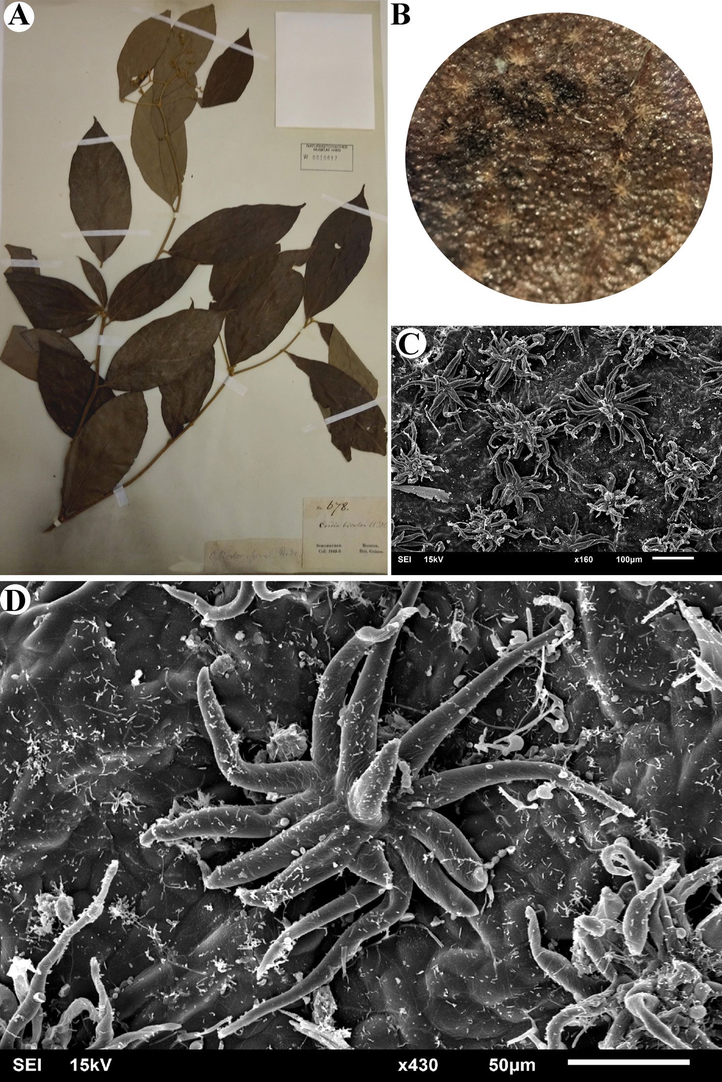
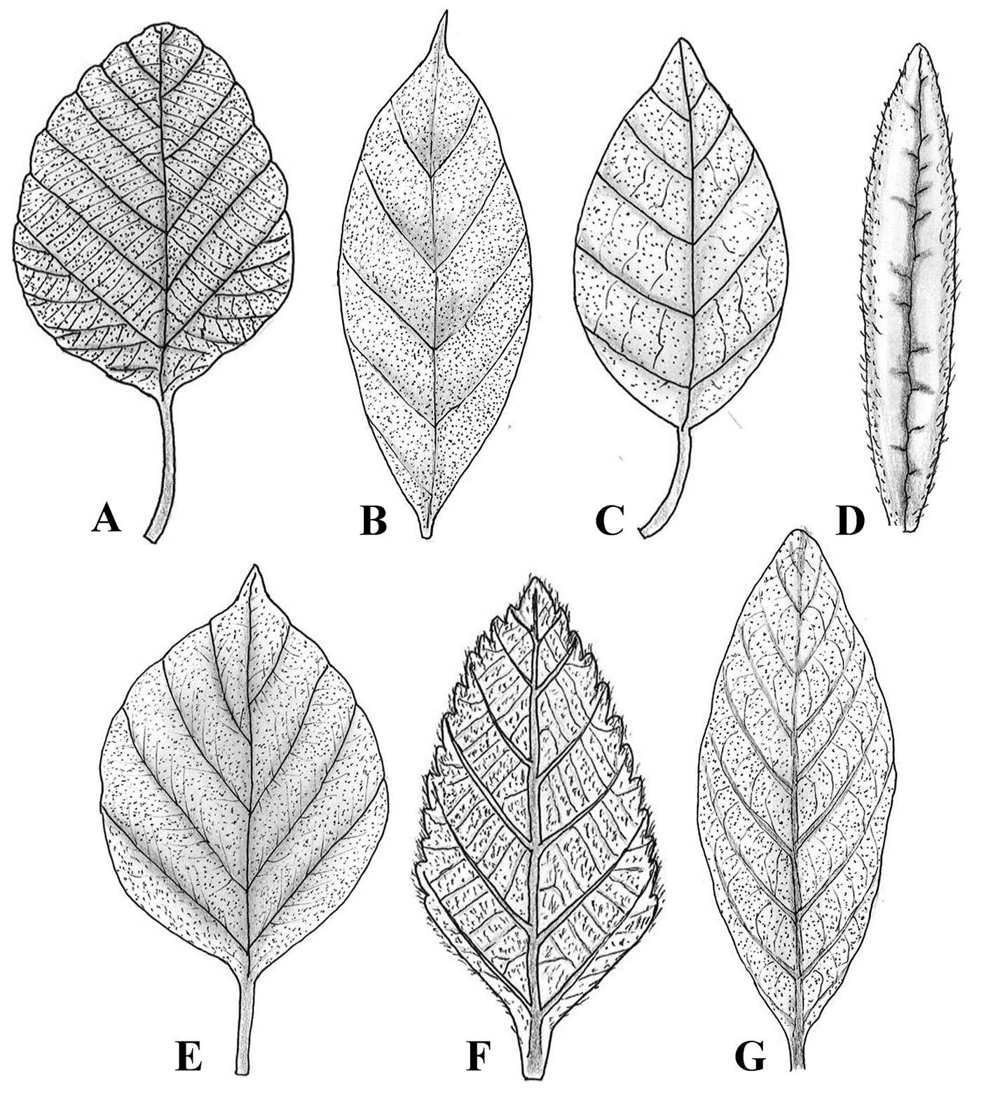
Stellate trichomes are a type of branched trichome that have several arms arising from a common base (either stalked or sessile). This type of trichome was observed only in Cordia bicolor. The number of arms of these trichomes is between 10 and 17, and the length of each arm is between 50 and 100 μm. Figure 4 shows the studied specimen and the observed stellate trichome. The distribution and density of trichomes on the surface of the leaf can be seen with the naked eye most of the time. Figure 5 shows the leaves of the studied species where trichome density can be seen on their surface.
Microscopic examination of these 7 species revealed that the adaxial leaf surface generally has a lower density of trichomes compared to the abaxial surface, which has a higher density. On the adaxial surface of the leaf of Cordia africana, there are trichomes in the form of small protrusions that are sparse, while on the abaxial surface, the trichomes have a different shape and are densely placed inside the depressions, while long trichomes are rarely seen. In Cordia alliodora, C. bicolor, C. decandra, and C. myxa the type of trichome is similar on both surfaces, with the difference that they are scattered on the adaxial side and dense on the abaxial side. In the case of the 2 Varronia species, the trichomes are scattered on the adaxial surface of V. bullata and dense on the abaxial surface, but in V. guanacastensis, the trichomes are dense on both leaf surfaces, with the difference being that the type of trichome is different between the surfaces. Figure 6 shows the difference between trichome distribution on adaxial and abaxial leaf surfaces in all studied species.
Figure 2. Non-glandular trichomes in studied species of Cordia. A, Abaxial surface in Cordia africana; B, abaxial surface in Cordia alliodora; C, abaxial surface in Cordia decandra; D, abaxial surface in Cordia myxa.
Figure 3. Non-glandular trichomes in studied species of Varronia. A, Adaxial surface in Varronia bullata; B, abaxial surface in Varronia bullata; C, adaxial surface in Varronia guanacastensis; D, abaxial surface in Varronia guanacastensis.
Discussion
Our study on leaf trichomes within Cordiaceae highlights significant morphological diversity, underscoring the diagnostic value of trichomes in taxonomy. Trichomes were categorized as either glandular (exclusively in Varronia species) or non-glandular (present in both genera), establishing clear taxonomic distinctions between the 2 genera. The restriction of glandular trichomes to Varronia supports the value of trichomes as reliable taxonomic markers, reinforcing molecular evidence that separates Varronia from Cordia. Despite the limited number of species examined, these findings offer a foundation for further studies across Cordiaceae. A notable finding was the discovery of stellate trichomes in Cordia bicolor, a first report for this species and probably rare within Boraginales. Stellate trichomes, characterized by multiple arms emerging from a common base, were observed solely in Cordia bicolor after an examination of over 100 Boraginales species (details in preparation). Figure 5 illustrates this unique stellate trichome, underscoring its taxonomic significance and its potential adaptive function.
Previous studies align with our findings on trichome diversity. Silva et al. (2023) examined leaf anatomy in 10 Varronia species from Brazil, identifying secretory and non-secretory trichomes as key taxonomic indicators. Similarly, Tölke et al. (2013) demonstrated variation in glandular and non-glandular trichomes among V. globosa Jacq. and V. leucocephala (Moric.) J.S. Mill., with unique glandular types and micropapillae-covered trichomes distinguishing each species. Demétrio et al. (2020) reported distinctive non-glandular trichomes and a single glandular type in V. polycephala Lam., a medicinal species, which also contained cystoliths and crystal sand idioblasts. These variations highlight trichomes as valuable taxonomic features and adaptive characters. While this study suggests a difference between Varronia and Cordia, the limited scope relative to the genera’s size precludes definitive conclusions. Further sampling of more species is needed to confirm this distinction in leaf trichomes.
Figure 4. Stellate trichome in Cordia bicolor. A, studied specimen (W0010617); B, leaf surface visible with a normal magnification (X4); C, stellate trichomes are densely distributed on the leaf surface; D, stellate trichome with more than 10 arms. Figure 5. The distribution of trichomes on the adaxial leaf surface of the studied species of Cordiaceae. A, Cordia africana; B, Cordia alliodora; C, Cordia bicolor; D, Cordia decandra; E, Cordia myxa;F, Varronia bullata; G, Varronia guanacastensis.
Moreover, the distribution of trichomes, denser on the abaxial leaf surfaces, suggests adaptations to environmental pressures and provides additional taxonomic insight. Consistent trichome types on particular leaf surfaces across species indicate conserved characters, useful for classification within the family. This diversity, particularly in non-glandular trichomes that vary in shape, size, and density, emphasizes the evolutionary lability of trichomes and their dual roles in adaptation and taxonomy.
Our use of SEM enabled detailed trichome analysis, offering precise structural insights and enhancing taxonomic resolution. Nazari and Ghahremaninejad (2024, 2025) also found that trichome examination can verify generic separations within Boraginales, as demonstrated in their study on Heliotropiaceae. Collectively, these findings underscore the morphological and taxonomic significance of trichomes, establishing them as key indicators in botanical classification.
Figure 6. Differences in trichome distribution on adaxial and abaxial surfaces of some Cordia species. A, Cordiaalliodora; B, Cordia myxa; C, Cordia africana.
Acknowledgments
We sincerely thank all the invaluable contributors to this work. Special thanks to Heimo Rainer, head of the Botany Department at the Natural History Museum of Vienna, for his extensive support. Additionally, we appreciate Astrid Hille, Andreas Berger, Tanja Schuster, and Johannes Walter from the same department for their assistance. We are also grateful to Wenke Wegner, a SEM specialist, for supervising the microscopic imaging of the fragments. Our thanks extend to W herbarium and B herbarium for sharing their extensive botanical resources.
References
Amer, W. M., Hamdy, R. S., & Mahdy, R. A. (2016). Taxonomic revision of genus Cordia L. (Boraginaceae) in Egypt. Egypt Journal of Botany, 6th International Conference, Menoufia University, 375–408.
APG IV. (2016). An update of the Angiosperm Phylogeny Group classification for the orders and families of flowering plants: APG III. Botanical Journal of the Linnean Society, 181, 1–20. https://doi.org/10.1111/boj.12385
Beilstein, M. A., Al-Shehbaz, I. A., & Kellogg, E. A. (2006). Brassicaceae phylogeny and trichome evolution. American Journal of Botany, 93, 607–619. https://doi.org/10.3732/ajb.93.4.607
Demétrio, A. M., Lusa, M. G., Lima, D. F., & Rodrigues, C. (2020). Leaf anatomy of Varronia polycephala Lam. (Cordiaceae). Flora, 271, 151677. https://doi.org/10.1016/j.flora.2020.151677
Flowers of India. (2025). Scarlet Cordia (Cordia sebestena). Retrieved March 7, 2025, from https://www.flowersofindia.net/catalog/slides/Scarlet%20Cordia.html
Ghahremaninejad, F. (2004). Value of trichome characteristics for separating of bifurcating Astragalus (Fabaceae) on sectional level. Turkish Journal of Botany, 28, 241–245.
Ghahremaninejad, F., Khalili, Z., Maassoum, A. A., Mirzaie-Nodoushan, H., & Riahi, M. (2012). Leaf epidermal features of Salix species (Salicaceae) and their significance. American Journal of Botany, 99, 769–777. https://doi.org/10.3732/ajb.1100019
Gonçales, R., Do Carmo, A. A. O., dos Santos, A. F., & de Souza, L. A. (2023). Floral indumentum of Miconia: In search of diagnostic characters for the tribe Miconieae. Revista Mexicana de Biodiversidad, 94, e945272. https://doi.org/10.22201/ib.20078706e.2023.94.5272
Johnston, I. M. (1951). Studies in the Boraginaceae XX. Representatives of three subfamilies in eastern Asia. Journal of the Arnold Arboretum, 32, 99–122. https://doi.org/10.5962/bhl.part.9730
Kong, M. J., & Hong, S. P. (2019). Leaf micromorphology of the Persicaria sect. Cephalophilon (Polygonaceae) and its systematic re-evaluation. Phytotaxa, 391, 167–184. https://doi.org/10.11646/phytotaxa.391.3.1
Luebert, F., Cecchi, L., Frohlich, M. W., Gottschling, M., Guilliams, C. M., Hasenstab-Lehman, K. E. et al. (2016). Familial classification of the Boraginales. Taxon, 65, 502–522. https://doi.org/10.12705/653.5
Matias, E. F. F., Alves, E. F., Silva, M. K. N., Carvalho, V. R. A., Coutinho, H. D. M., & da Costa, J. G. M. (2015). The genus Cordia: Botanists, ethno, chemical and pharmacological aspects. Revista Brasileira de Farmacognosia, 25, 542–552. https://doi.org/10.1016/j.bjp.2015.05.012
Miller, J. S., & Gottschling, M. (2007). Generic classification in the Cordiaceae (Boraginales): Resurrection of the genus Varronia R.Br. Taxon, 56, 163–169. https://doi.org/10.2307/25065747
Nazari, H., & Ghahremaninejad, F. (2024). A SEM study of leaf trichomes in Heliotropiaceae. Iranian Journal of Botany, 30, 173–183. https://doi.org/10.22092/ijb.2024.366196.1475
POWO. (2024). Plants of the World. Online. https://powo.science.kew.org/
Rodríguez, A., & Healey, P. L. (2000). Morphological diversity of trichomes in Loasaceae and its systematic significance. Annals of the Missouri Botanical Garden, 87, 71–86. https://doi.org/10.1093/aobpla/plab071
Selvi, F., & Bigazzi, M. (2001). Leaf surface and anatomy in Boraginaceae tribe Boragineae with respect to ecology and taxonomy. Flora, 196, 269–285. https://doi.org/10.1016/S0367-2530(17)30056-7
Silva, T. S., & de Melo, J. I. M. (2019). A new species and a new record of Varronia (Cordiaceae) from Brazil. Systematic Botany, 44, 692–696. https://doi.org/10.1600/036364419×15620113920752
Silva, T. S., Tölke, E. E. D., & de Melo, J. I. M. (2023). A contribution of leaf anatomy to the taxonomy of Varronia P. Browne (Cordiaceae, Boraginales). Flora, 304, 152299. https://doi.org/10.1016/j.flora.2023.152299
Steyn, H. M., & Van Wyk, A. E. (2021). Taxonomic significance of trichomes in the genus Acanthopsis Harv. (Acanthaceae, tribe Acantheae). Adansonia, 43, 163–176. https://doi.org/10.5252/adansonia2021v43a14
Taroda, N. (1984). Taxonomic studies on Brazilian species of Cordia L. (Boraginaceae)(Ph.D. Thesis). University of St. Andrews. St. Andrews, UK.
Thiers, B. M. (2025). Index Herbariorum: a global directory of public herbaria and associated staff (Continuously updated). New York Botanical Garden’s Virtual Herbarium. http://sweetgum.nybg.org/ih/
Tölke, E. E. D., de Melo, J. I. M., Carmello-Guerreiro, S. M., & Lacchia, A. P. S. (2013). Leaf anatomy with emphasis on separation of two species of Varronia P.Br. (Cordiaceae) of the Brazilian semi-arid region. Brazilian Journal of Botany, 36, 189–201. https://doi.org/10.1007/s40415-013-0022-5
Wagner, G. J. (1991). Secreting glandular trichomes: more than just hairs. Plant Physiology, 96, 675–679. https://doi.org/10.1104/pp.96.3.675
Eduardo Reyes-Grajales a, b, *, Christian Rico c, John Iverson d, Luis Díaz-Gamboa e, Marco A. López-Luna f, Wilfredo A. Matamoros g
a El Colegio de la Frontera Sur, Departamento Conservación de la Biodiversidad, Doctorado en Ciencias en Ecología y Desarrollo Sustentable, Carretera Panamericana y Periférico Sur s/n, Barrio María Auxiliadora, 29290 San Cristóbal de Las Casas, Chiapas, Mexico
b Turtle Survival Alliance, 1030 Jenkins Road, Charleston, 29407 South Carolina, USA
c Universidad de Ciencias y Artes de Chiapas, Instituto de Ciencias Biológicas, Maestría en Ciencias en Biodiversidad y Conservación de Ecosistemas Tropicales, Libramiento Norte Poniente, 1150, Lajas Maciel, 29039 Tuxtla Gutiérrez, Chiapas, Mexico
d Earlham College, Department of Biology, 801 National Road West, 47374 Richmond, Indiana, USA
e Red para la Conservación de los Anfibios y Reptiles de Yucatán, Km. 5.5 Carr. Sierra Papacal-Chuburná Pto. Tablaje, 31257 Sierra Papacal, Yucatán, Mexico
f Universidad Juárez Autónoma de Tabasco, División Académica de Ciencias Biológicas, Carretera Villahermosa-Cárdenas Km 0.5, 86039 Villahermosa, Tabasco, Mexico
g Universidad de Ciencias y Artes de Chiapas, Instituto de Ciencias Biológicas, Libramiento Norte Poniente, 1150, Lajas Maciel, 29039 Tuxtla Gutiérrez, Chiapas, Mexico
*Corresponding author: eduardo.reyes.grajales@gmail.com (E. Reyes-Grajales)
Abstract
Mexico has the second highest species richness of turtles in the world. However, there are several research gaps within this group compared to other reptiles, especially in the southeastern region, which has been described as the area with the highest herpetofaunal diversity. These data deficiencies have led to much uncertainty regarding the identification, occurrence, and status of the area’s turtles. Given this context, we provide an update for all the native turtle taxa present in each state in southeastern Mexico. We include maps with historical and unpublished distribution records for each taxon, along with a dichotomous key based on external morphology. We discuss the native turtle taxa documented in southeastern Mexico, the possible presence of some species in certain states, the identification of the turtles in this region, and their conservation status according to national, historical and current contexts.
Tortugas continentales del sureste de México: una actualización sobre la identificación, composición, distribución y conservación
Resumen
México tiene la segunda mayor riqueza de especies de tortugas en el mundo. Sin embargo, en este país existen varios vacíos de investigación dentro de este grupo en comparación con otros reptiles, especialmente en la región sureste, que ha sido descrita como el área con mayor diversidad herpetofaunística. Esta deficiencia de datos ha generado mucha incertidumbre en cuanto a la identificación, distribución y estado de las tortugas en la región. Dado este contexto, proporcionamos una actualización de todos los taxones nativos de tortugas presentes en cada estado del sureste de México. Incluimos mapas con registros históricos y no publicados de distribución para cada taxón, junto con una clave dicotómica basada en la morfología externa. Discutimos los taxones de tortugas nativas documentados en el sureste de México, la posible presencia de algunos taxones en ciertos estados, la identificación de las tortugas en esta región y su estado de conservación según los contextos nacional, histórico y actual.
Palabras clave: Prioridades de conservación; Tortugas dulceacuícolas; Región neotropical; Reptiles; Testudines
Introduction
The order Testudines includes 2 extant suborders, comprising 14 families, 97 genera, 7 marine turtle species, and 346 continental land and freshwater turtles (TTWG, 2021). Mexico supports the second richest turtle fauna per country in the world (after the USA) with 7 families, 13 genera, and 49 species of continental terrestrial and freshwater turtles (Hurtado-Gómez et al., 2024; Loc-Barragán et al., 2020; López-Luna et al., 2018; TTWG, 2021). When subspecies are included, a total of 63 distinct taxa are recognized in Mexico, with nearly half (49%, 31 taxa) being endemic (Hurtado-Gómez et al., 2024; Loc-Barragán et al., 2020; López-Luna et al., 2018; TTWG, 2021). Of those, 5 taxa have been described since 2010: Gopherus evgoodei, G. morafkai, Kinosternon cora, K. vogti, and Trachemys venusta iversoni (Edwards et al., 2016; Loc-Barragán et al., 2020; López-Luna et al., 2018; McCord et al., 2010; Murphy et al., 2011), and 5 taxa were taxonomically rearranged: K. cruentatum, K. mexicanum, K. stejnegeri, Terrapene mexicana, and T. yucatana (Hurtado-Gómez et al., 2024; Iverson & Berry, 2024; Martin et al., 2013; McCord, 2016;). It is noteworthy to mention that ~ 50% of Mexican continental turtles lack basic natural history data, which makes it cumbersome to comprehensively assess their actual conservation status (Flores-Villela & García-Vázquez, 2014; Macip-Ríos et al., 2015). Legler and Vogt (2013) stated that to solve “Mexican turtle conservation issues”, an integrative conservation research approach is necessary, to build knowledge about life history and ecology of these organisms.
Within Mexico, the southeastern region (Campeche, Chiapas, Oaxaca, Quintana Roo, Tabasco, Veracruz, and Yucatán) is characterized by high turtle endemism, with high presence of human induced alterations at different ecosystem levels (Ennen et al., 2020). Historically, many indigenous cultures in this region incorporated turtles (e.g., Dermatemys mawii, and those in the genera Trachemys and Kinosternon) into their diets as ceremonial or complementary foods (Beauregard et al., 2010; González-Porter et al., 2011; Legler & Vogt, 2013; Velázquez-Nucamendi et al., 2021). More recently, southeastern Mexico has become one of the hotspots where turtles are extracted for illegal trade worldwide, especially from Chiapas, Tabasco, and Veracruz (Legler & Vogt, 2013; Macip-Ríos et al., 2015; TTWG, 2021). In response to these actions, standards have been established at both the national level (e.g., the list of Mexican federal government protected species of flora and fauna) and international levels (e.g., the CITES appendices) to regulate the use of turtles. However, the application of protocols for the study or protection of this group of vertebrates in southeastern Mexico has shown inconsistencies in the processes of identification and/or systematization of information on their distribution (Flores-Villela & García-Vázquez, 2014; Legler & Vogt, 2013; Macip-Ríos et al., 2015). Therefore, there is a recognized need to design tools and update information on these aspects to safeguard these turtles.
To contribute to the resolution of this situation, in this study we set 2 main goals: 1) to provide an up-to-date checklist, with general comments on the distribution of the continental turtles occurring in southeastern Mexico, and 2) to present a practical dichotomous key for their identification that encompasses the current turtle taxonomy in southeastern Mexico. Our findings can be implemented immediately in activities related to the study and protection of continental turtles in this region of Mexico.
Materials and methods
We define southeastern Mexico as the states of Campeche, Chiapas, Oaxaca, Quintana Roo, Tabasco, Veracruz, and Yucatán (Fig. 1). The region is delimited by the Isthmus of Tehuantepec as the initial dividing line, given its significance as a major biogeographic barrier (Huidobro et al., 2006; Quiroz-Martínez et al., 2014). However, considering that several turtle taxa extend west and north just beyond this barrier, we chose to include Mexican states that comprise part of the isthmus region. This region comprises ~ 415,000 km2 originally vegetated with tropical evergreen forest and tropical deciduous forest with warm humid and warm subhumid climates (Leopold, 1950). In this region, turtles typically occur from sea level to approximately 1,200 m (Legler & Vogt, 2013). Major geographic features of the region are: the Gulf Coastal Plain, Sierra Madre de Oaxaca, Sierra Madre del Sur, Pacific Coastal Plain, Isthmus of Tehuantepec, Chiapas Highlands, Sierra Madre de Chiapas, Chiapas Coastal Plain, Central Depression of Chiapas, and the Yucatán Peninsula (Contreras-Balderas et al., 2008; Legler & Vogt, 2013). The main rivers are: Candelaria, Usumacinta, Grijalva, Tonalá, Coatzacoalcos, Papaloapan, Jamapa, Tehuantepec, and Ometepec (INEGI, 2021) (Fig. 1).
Georeferenced point locations were obtained for native continental turtles of southeastern Mexico from a combination of our own fieldwork records, the Turtle Taxonomy Working Group (TTWG, 2021, TTWG, 2025), and data repositories, including the Global Biodiversity Information Facility (GBIF, 2024), Sistema Nacional de Información sobre la Biodiversidad (SNIB-Conabio, 2024), Áreas Naturales y Vida Silvestre of the Secretaría del Medio Ambiente e Historia Natural (Semahn), iNaturalist (2024), and published scientific literature (Supplementary material: Table S1).
We used a systematic approach, establishing the TTWG shapefile (2021, 2025) as our base dataset. All newly identified occurrence points from additional sources were stored in a georeferenced database for the subsequent creation of maps using QGIS 3.34.2 (QGIS, 2024). Distribution polygons were based on TTWG (2025) and were expanded to include extralimital records verified through our compilation.
To ensure spatial accuracy, only points that provided open, non-obscured coordinates with reported location uncertainties of ≤ 10 km were incorporated. In the case of iNaturalist data, we exclusively used “research-grade” observations and excluded any records with obscured or low-precision coordinates (e.g., obscured within ~ 400 square miles). Records without supporting metadata or located significantly outside of known ranges were critically assessed, and when appropriate, excluded from analyses.
Special attention was given to isolated or potentially non-native records. For instance, we included K. flavescens from Veracruz based on the report of Auth et al. (2000) and the corroboration of voucher specimens (BCB 7489) by JBI at the Strecker Museum. We also carefully evaluated records of other species commonly subject to anthropogenic translocation, considering only those that plausibly represent native occurrences based on geographic and ecological context (see Discussion section). This rigorous validation process aimed to minimize the inclusion of records originating from village collections, pet trade, or other human-mediated movements, unless justified by the locality’s proximity to known native ranges or supporting documentation.
Our checklist includes valid names, authorities, and year of publication as in TTWG (2025). The dichotomous key we created based initially on the taxon descriptions provided in Legler and Vogt (2013). We then updated the key with the taxa recognized subsequent to Legler and Vogt (2013), and verified and added pertinent meristic and morphometric characters, and pigmentation patterns from specimens deposited in the Herpetology Section at the Zoológico Regional Miguel Álvarez del Toro (ZooMAT, Tuxtla Gutiérrez, Chiapas), the Colección herpetológica of El Colegio de la Frontera Sur (ECOSUR, San Cristóbal, Chiapas), and our own field data.
We refer to the plastral formula as the relative lengths of the midline sutures of each plastral scute (Supplementary material: Fig. S1). Due to inconsistencies (conventional vs. new) in the nomenclature of plastral scutes (Hutchison & Bramble, 1981; Legler & Vogt, 2013), we followed the recommendation of Legler and Vogt (2013) to use the numerical nomenclature for the plastral formula, where the counts start anteriorly (Supplementary material: Fig. S1). For the nomenclature of the head stripes, we followed Ernst (1978) for Rhinoclemmys spp., and Legler (1990) for Trachemys spp. We used the term “tomiodonts” according to Moldowan et al. (2015), and “tubercles” according to Winokur (1982).
For conservation criteria at the national level, we consulted the Mexican Government list in the NOM-059 (Semarnat, 2010, 2025), and for international criteria, we referred to the IUCN Red List of threatened species (IUCN, 2024) and the CITES appendices (CITES, 2024). Additionally, we used the EDGE metrics, as it prioritizes species based on their evolutionary distinctiveness (ED), which measures the relative contribution of a species to the total evolutionary history of their taxonomic group, and global endangerment (GE), or extinction risk (Gumbs et al., 2018). This metric has been recognized as useful to meaningful priorities for conservation.
Results
Native continental turtles in southeastern Mexico are represented by 5 families, 8 genera and 22 species with 9 recognized subspecies, for a total of 25 extant continental taxa (Table 1). The most speciose families in southeastern Mexico are the Kinosternidae, with a total of 13 taxa and Emydidae with 6 taxa (Table 1). The states with the greatest species richness are Oaxaca (16 taxa), followed by Chiapas (15), and Veracruz (13), and those with the least species richness were Quintana Roo (11), Tabasco (9) and Yucatán (7) (Table 2). Ten continental turtles in 4 genera are endemic to the region, as follows: Kinosternon (4 taxa), Terrapene (2 taxa), Trachemys (2 taxa) and Rhinoclemmys (2 taxa) (Table 3). The states with the highest number of Mexican endemic taxa are Oaxaca (2 species and 2 subspecies), Quintana Roo (2 species and 1 subspecies), Veracruz (2 species and 1 subspecies), and Yucatán (2 species and 1 subspecies) (Tables 1, 3).
Table 1
Diversity of continental turtles in southeastern Mexico. Ca = Campeche, Ch = Chiapas, Oa = Oaxaca, QR = Quintana Roo, Ta = Tabasco, Ve = Veracruz, Yu = Yucatán; CSP = count of states present; % S = percentage considering all the taxa distributed in southeastern Mexico; X = presence; – = undocumented.
Family
Genus
Species
Subspecies
Ca
Ch
Oa
QR
Ta
Ve
Yu
CSP
% S
Chelydridae
Chelydra
C. rossignonii
X
X
X
–
–
X
–
5
71.4
Dermatemydidae
Dermatemys
D. mawii
X
X
–
X
X
X
–
5
71.4
Emydidae
Terrapene
T. mexicana
–
–
–
–
–
X
–
1
14.3
T. yucatana
X
–
–
X
–
–
X
3
42.9
Trachemys
T. grayi
T. g. grayi
–
X
X
–
–
–
–
2
28.6
T. venusta
T. v. cataspila
–
–
–
–
–
X
–
1
14.3
T. v. iversoni
–
–
–
X
–
–
X
2
28.6
T. v. venusta
X
X
X
X
X
X
–
6
85.7
Geoemydidae
Rhinoclemmys
R. areolata
R. areolata
X
X
X
X
X
X
X
7
100
R. pulcherrima
R. p. incisa
–
X
X
–
–
–
–
2
28.6
R. p. pulcherrima
–
–
X
–
–
–
–
1
14.3
R. rubida
R. r. rubida
–
X
X
–
–
–
–
2
28.6
Kinosternidae
Claudius
C. angustatus
X
X
X
X
X
X
X
7
100
Kinosternon
K. abaxillare
–
X
–
–
–
–
–
1
14.3
K. acutum
X
X
X
X
X
X
–
6
85.7
K. creaseri
X
–
–
X
–
–
X
3
42.9
K. cruentatum
X
X
X
X
X
X
X
7
100
K. flavescens
–
–
–
–
–
X
–
1
14.3
K. herrerai
–
–
–
–
–
X
–
1
14.3
K. integrum
–
–
X
–
–
–
–
1
14.3
K. leucostomum
X
X
X
X
X
X
X
7
100
K. mexicanum
–
X
X
–
–
–
–
2
28.6
K. oaxacae
–
–
X
–
–
–
–
1
14.3
Staurotypus
S. salvinii
–
X
X
–
–
–
–
2
28.6
S. triporcatus
X
X
X
X
X
X
–
6
85.7
Totals by state
11
15
16
11
9
13
7
Table 2
Summary of taxonomic diversity of continental turtles by state in southeastern Mexico. The number of continental turtles found in Mexico was established according to TTWG (2021, 2025). Hurtado-Gómez et al. (2024), and Iverson and Berry (2024). The percentage with respect to diversity at the national level (left) and in southeastern Mexico (right) is in parentheses.
Place
Families
Genera
Species
Taxa
Mexico
7
13
50
65
Southeastern Mexico
5 (71.4)
8 (61.5)
22 (45)
25 (39.7)
Campeche
5 (71.4 / 100)
8 (61.5 / 100)
11 (22.4 / 50)
11 (17.5 / 44)
Chiapas
5 (71.4 / 100)
7 (53.8 / 87.5)
15 (30.6 / 68.2)
15 (23.8 / 60)
Oaxaca
4 (57.1 / 80)
6 (46.2 / 75)
15 (30 / 68.2)
16 (25.4 / 64)
Quintana Roo
4 (57.1 / 80)
6 (46.2 / 75)
10 (20 / 45.5)
11 (17.5 / 44)
Tabasco
5 (71.4 / 100)
7 (53.8 / 87.5)
9 (18.4 / 40.9)
9 (14.3 / 36)
Veracruz
5 (71.4 / 100)
8 (61.5 / 100)
13 (26.5 / 59.1)
14 (22.2 / 56)
Yucatán
3 (42.9 / 60)
4 (30.8 / 50)
5 (10.2 / 22.7)
5 (7.9 / 20)
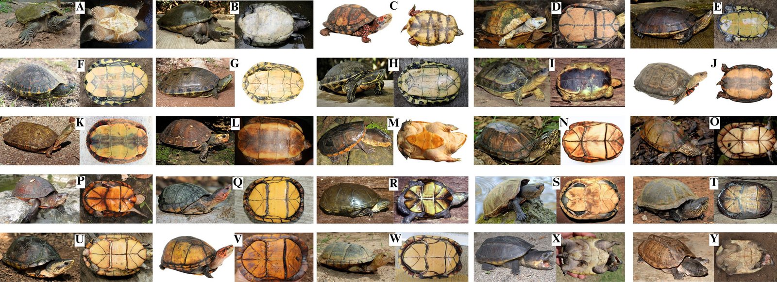
Dichotomous key to the general and plastron view of each turtle (Fig. 2), the plastral nomenclature and the names of the head stripes (Supplementary material: Figs. 1S, S2). The Spanish translation is presented in Supplementary material.
1A. Posterior plastral lobe ends in a point …………………………………… 2 (Fig. 2A)
1B. Posterior plastral lobe does not end in a point …………………………………… 5 (Fig. 2B)
2A. Reduced bridge almost the same length size as plastral scute 2. Kinosternidae: Staurotypus, …………………………………… 3
2B. Extremely narrow, reduced bridge shorter than the length of plastral scute 2 …………………………………… 4
3A. Top and sides of head boldly marked with irregular dark and pale reticulations; 3 distinctive carapace keels, well developed posteriorly, especially the middle keel ……………………………………S. triporcatus (Fig. 2Y)
3B. Top of head dark, usually unicolored, lacking bold pattern; 3 distinctive carapace keels of similar size, anterior to posterior ……………………………………S. salvinii (Fig. 2X)
4A. Three pointed tomiodonts on the upper tomium; neck not ornamented with cutaneous tubercles; tail not as long as the plastron …………………………………… Kinosternidae: Claudius angustatus (Fig. 2M)
4B. One medial pointed extension on the upper tomium; neck ornamented with long, flat, pointed cutaneous tubercles; tail about as long as the plastron …………………………………… Chelydridae: Chelydra rossignonii (Fig. 2A)
5A. Interdigital membranes not present on forefeet or hind feet …………………………………… 6
5B. Interdigital membranes present on fore and hind feet ……………………………………11
6A. One plastral hinge, allowing movement of anterior and posterior plastral lobes; the posterior end of the plastron rounded ……………………………………Emydidae: Terrapene, 7
6B. A transverse plastral hinge is absent; the posterior end of the plastron is notched …………………………………… Geoemydidae: Rhinoclemmys, 8
7A. Plastral scute 5 averaging 15% the straight-midline length of the posterior plastral lobe; plastral scute 3 averaging 23% the straight-midline length of the anterior plastral lobe ……………………………………T. mexicana (Fig. 2C)
7B. Plastral scute 5 averaging 21% the straight-midline length of the posterior plastral lobe; plastral scute 3 averaging 33% the straight-midline length of the anterior plastral lobe …………………………………… T. yucatana (Fig. 2D)
8A. Yellow markings predominate on the forefeet, hind feet, neck and/or head ……………………………………9
8B. Red markings predominate on the forefeet, hind feet, neck and/or head …………………………………… 10
9A. Red/orange dorsolateral head stripes continuous or discontinuous; each costal scute uniformly colored ……………………………………R. areolata (Fig. 2I)
9B. Red/orange dorsolateral head stripes are absent; each lateral scute with 1 pale areolar spot with a dark outline, possibly with obvious light-colored concentric rings around it ……………………………………R. rubida rubida (Fig. 2L)
10A. Carapace is elevated; inferior surface of each marginal scute with 1 transverse pale mark ……………………………………R. pulcherrima incisa (Fig. 2J)
10B. Carapace is depressed; inferior surface of each marginal scute with 2 transverse, dark-bordered pale marks ……………………………………R. p. pulcherrima (Fig. 2K)
Table 3
Conservation status of the continental turtles of southeastern Mexico. NOM-059 = List of Mexican federal government protected species of flora and fauna; P = endangered (in danger of extinction), Pr = under special protection, A = threatened; Red List = the IUCN Red list of threatened species; CITES = Convention on International Trade in Endangered Species of Wild Fauna and Flora; En = national endemism; Y = endemic to Mexico; N = not endemic to Mexico; EDGE = EDGE score (Gumbs et al., 2018; higher threat = higher score).
Taxa
NOM-059
Red List
CITES
EDGE
En
Chelydra rossignonii
Pr
Vulnerable A2d
–
5.22
N
Dermatemys mawii
P
Critically endangered A2abd+4d
II
7.15
N
Terrapene mexicana
Pr
Vulnerable A2bcde+4bcde
II
4.19
Y
Terrapene yucatana
Pr
Vulnerable A2bcde+4bcde
II
4.19
Y
Trachemys grayi grayi
–
–
–
–
N
Trachemys venusta cataspila
–
–
–
–
Y
Trachemys venusta iversoni
–
–
–
–
Y
Trachemys venusta venusta
–
–
–
–
N
Rhinoclemmys areolata
A
Near threatened
II
3.77
N
Rhinoclemmys pulcherrima incisa
A
–
II
–
N
Rhinoclemmys pulcherrima pulcherrima
A
–
II
–
Y
Rhinoclemmys rubida rubida
Pr
Near threatened
II
3.87
Y
Claudius angustatus
P
Near threatened
II
4.36
N
Kinosternon abaxillare
–
Vulnerable A2cd+4cd
II
–
N
Kinosternon acutum
Pr
Near threatened
II
4.36
N
Kinosternon creaseri
–
Least concern
II
3.67
Y
Kinosternon cruentatum
–
–
II
–
N
Kinosternon flavescens
–
Least concern
II
3.67
N
Kinosternon herrerai
Pr
Near threatened
II
4.36
Y
Kinosternon integrum
Pr
Least concern
II
3.67
Y
Kinosternon leucostomum
Pr
–
II
–
N
Kinosternon mexicanum
–
–
II
–
N
Kinosternon oaxacae
Pr
–
II
4.36
Y
Staurotypus salvinii
Pr
Near threatened
II
4.69
N
Staurotypus triporcatus
A
Near threatened
II
4.69
N
Total of taxa in the categories considered
15
15
20
15
11A. There are no transversely oriented plastral hinges allowing plastral movement ……………………………………12
11B. There are 1 or 2 transversely oriented plastral hinges, allowing 1 or both plastral lobes to move around the scute 4…………………………………… Kinosternidae: Kinosternon, 16
12A. The head typically has a pale-colored patch that covers part or all of the dorsal region of the head but is never striped ……………………………………Dermatemydidae: Dermatemys mawii (Fig. 2B)
12B. The head is colored with obvious yellow stripes…………………………………… Emydidae: Trachemys, 13
13A. Carapace ocelli centered on each costal scute and occupying most of scute…………………………………… 14
13B. Carapace ocelli not centered on costal scutes ……………………………………15
14A. Carapace ocelli have an obvious large dark center to each ocellus ……………………………………T. venusta iversoni (Fig. 2G)
14B. Carapace ocelli lack an obvious dark center to each ocellus ……………………………………T. v. venusta (Fig. 2H)
15A. No light-colored ocelli on carapace; most of the central secondary orbitocervical stripes are narrow, of roughly equal width, and lacking black borders…………………………………… T. grayi grayi (Fig. 2E)
15B. Ocelli not centered in lateral scutes or broken; The postorbital stripe is thin where it contacts the orbit, then widens to about the diameter of the orbit; the central secondary orbitocervical stripes vary in width ……………………………………T. v. cataspila (Fig. 2F)
16A. Only the anterior plastral lobe is movable ……………………………………K. herrerai (Fig.2S)
16B. Both plastral lobes are movable relative to plastral scute 4 ……………………………………17
19B. Formula plastral is 4 > 6 > 1 > 5 > 2 > 3 ……………………………………K. oaxacae (Fig. 2W)
20A. Eyes with red pigment ……………………………………K. acutum (Fig. 2O)
20B. Eyes do not have red pigment…………………………………… 21
21A. Tricarinate carapace (less obvious in older individuals) ……………………………………22
21B. Unicarinate carapace ……………………………………23
22A. Plastral formula is 4 > 6 ……………………………………K. integrum (Fig. 2T)
22B. Plastral formula is 6 > 4 ……………………………………24
23A. Plastral formula is 1 > 2 ……………………………………K. creaseri (Fig. 2P)
23B. Plastral formula is 2 > 1 ……………………………………K. leucostomum (Fig. 2U)
24A. Maximum carapace width averages 77.1% (standard deviation [S.D.] 2.8%) of maximum plastron length in females and 74.6% (S.D. 2.8%) in males; the length of the plastral suture 4 averages 55.3% (S.D. 3.9%) of maximum shell height in females and 61.7% (S.D. 4.9%) in males ……………………………………K. mexicanum (Fig. 2V)
24B. Maximum carapace width averages 69.8% (S.D. 3.5%) of maximum plastron length in females and 68.9% (S.D. 2.8%) in males; the length of the plastral suture 4 averages 61.7% (S.D. 4.9%) of maximum shell height in females and 64.4% (S.D. 4.5%) in males ……………………………………K. cruentatum (Fig. 2Q)
Figure 2. Photos of the continental turtles of southeastern Mexico. All photos (except when indicated, see Acknowledgments) by Eduardo Reyes-Grajales or John B. Iverson. A. Chelydra rossignonii, B. Dermatemys mawii, C. Terrapene mexicana, D. T. yucatana, E. Trachemys grayi grayi, F. T. venusta cataspila, G. T. v. iversoni, H. T. v. venusta, I. Rhinoclemmys areolata, J. R. pulcherrima incisa, K. R. p. pulcherrima, L. R. rubida rubida, M. Claudius angustatus, N. Kinosternon abaxillare, O. K. acutum, P. K. creaseri, Q. K. cruentatum, R. K. flavescens, S. K. herrerai, T. K. integrum, U. K. leucostomum, V. K. mexicanum, W. K. oaxacae, X. Staurotypus salvinii, Y. S. triporcatus.
Distributions of continental turtles of southeastern Mexico
In this section the abbreviations are: Ca = Campeche, Ch = Chiapas, Oa = Oaxaca, QR = Quintana Roo, Ta = Tabasco, Ve = Veracruz, Yu = Yucatán.
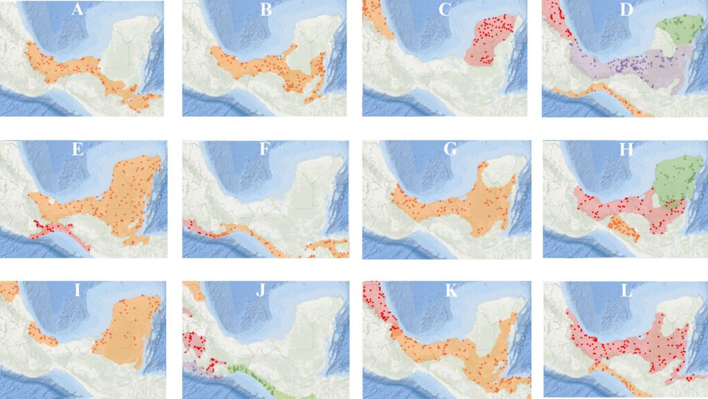
Chelydra rossignonii (Bocourt, 1868; Figs. 2A, 3A): along the Gulf of Mexico versant from the Jamapa River (Ve) to the Usumacinta River (Ta and Ch), including Laguna de Terminos (Ca). Other rivers include the Papaloapan, Coatzacoalcos (both in Oa and Ve), and lower Grijalva (Ch).
Dermatemys mawii Gray, 1847 (Figs. 2B, 3B): from the Jamapa River (Ve) to Champoton River (Ca) on the Gulf of Mexico versant, including the Usumacinta (Ca, Ch and Ta) and Lacantun Rivers (Ch). In Chetumal Bay (QR) the distribution extends to and beyond the Belize border.
Terrapene mexicana (Gray, 1849; Figs. 2C, 3C): from the Tamesi River to the Jamapa River, including the Tamiahua Lake area; on the Gulf of Mexico versant in Veracruz and northward into Tamaulipas.
Terrapene yucatana (Boulenger, 1895; Figs. 2D, 3C): most of the western Yucatán Peninsula (primarily Yucatán and Campeche), with rare and localized occurrences in the northern region of Quintana Roo.
Trachemys grayi grayi (Bocourt, 1868; Figs. 2E, 3D): from La Arena River (Oa) to the Suchiate River on the Pacific versant (Ch), and southward to El Salvador.
Trachemys venusta cataspila (Günther, 1885; Figs. 2F, 3D): from the Jamapa River to the Panuco River (both in Ve) on the Gulf of Mexico versant, and northward in Tamaulipas.
Trachemys venusta iversoni McCord, Joseph-Ouni, Hagen, and Blanck, 2010 (Figs. 2G, 3D): most of the northern portions of Yucatán and Quintana Roo.
Trachemys venusta venusta (Gray, 1856; Figs. 2H, 3D): from the Jamapa River (Ve) to the Champoton River (Ca) on the Gulf of Mexico versant, including the Usumacinta (Ca, Ch and Ta) and Lacantun Rivers (Ch). In Chetumal Bay (QR) the distribution continues across the border into Belize and Guatemala.
Rhinoclemmys areolata (Duméril and Bibron in Duméril and Duméril, 1851; Figs. 2I, 3E): from the Jamapa River eastward (Ve) on the Gulf of Mexico versant throughout the north portions of Oaxaca and Chiapas and extended across Tabasco and the Yucatán peninsula (Ca, Yu and QR), and into Guatemala and Belize.
Rhinoclemmys pulcherrima incisa (Bocourt, 1868; Figs. 2J, 3F): from the west portion of the Copalita River (Oa) to the Suchiate River (Ch) on the Pacific versant, and southeast to Nicaragua.
Rhinoclemmys pulcherrima pulcherrima (Gray, 1856; Figs. 2K, 3F): from the Tehuantepec River (Oa) westward on the Pacific versant to the Balsas River, Guerrero.
Rhinoclemmys rubida rubida (Cope, 1870; Figs. 2L, 3E): from the Ometepec River (Oa) to the Suchiate River (Ch) on the Pacific versant, including the Atoyac and Tehuantepec basins (both in Oa).
Claudius angustatus Cope, 1865 (Figs. 2M, 3G): from the Jamapa River (Ve) to the western and southern Yucatán peninsula on the Gulf of Mexico versant, including the Usumacinta (Ca, Ch and Ta) and Lacantun Rivers (Ch), and southward into Guatemala and Belize.
Kinosternon abaxillare Baur in Stejneger, 1925 (Figs. 2N, 3H): restricted to the Grijalva River basin in the Central Depression and Plateau of Chiapas.
Kinosternon acutum Gray, 1831 (Figs. 2O, 3H): from the Jamapa River (Ve) to the Champoton River (Ca) on the Gulf of Mexico versant, including the Papaloapan, Coatzacoalcos (both in Oa and Ve) and lower Grijalva Rivers (Ch), and Laguna de Terminos (Ca). Also found on the southern Yucatán peninsula (Ca and QR), northern Guatemala and Belize.
Kinosternon creaseri Hartweg, 1934 (Figs. 2P, 3H): from the Champoton River (Ca) across the Yucatán peninsula (QR and Yu).
Kinosternon cruentatum Duméril and Bibron in Duméril and Duméril, 1851 (Figs. 2Q, 3I): from the Pánuco River (Ve) across the Tabasco lowlands to the entire Yucatán Peninsula on the Gulf of Mexico versant and into Guatemala and Belize.
Kinosternon flavescens Agassiz, 1857 (Figs. 2R, 3J): in southeastern Mexico it is restricted to the Panuco River basin (Ve) but ranges north across the Great Plains of the USA.
Kinosternon herrerai Stejneger, 1925 (Figs. 2S, 3K): from the Jamapa to the Panuco River (both in Ve) and northward on the Gulf of Mexico versant.
Kinosternon integrum Le Conte, 1854 (Figs. 2T, 3J): in southern Mexico from the Atoyac (Oa and Ve) and Tlapaneco River basins westward (Oa).
Kinosternon leucostomum Duméril and Bibron in Duméril and Duméril, 1851 (Figs. 2U, 3K): from the Jamapa River (Ve) to the Champoton River (Ca) on the Gulf of Mexico versant, including the Usumacinta (Ca, Ch and Ta) and Lacantun rivers (Ch). Also found from Chetumal Bay through eastern Quintana Roo, and southward through Central America.
Kinosternon mexicanum Le Conte, 1854 (Figs. 2V, 3J): restricted to the Pacific versant from the Tehuantepec River (Oa) to the Suchiate River (Ch), and southeast to El Salvador.
Kinosternon oaxacae Berry and Iverson, 1980 (Figs. 2W, 3J): restricted to the Ometepec, La Arena, Colotepec and Copalita River basins on the Pacific versant in Oaxaca; also, westward into Guerrero.
Figure 3. Distribution maps of the continental turtles of southern Mexico. The shaded color represents the distribution of each taxon (taken from TTWG, 2025). Maps with 2 or more taxa present dots and shade ranges with similar colors, and maps with a single taxa dots and shade ranges are orange. A. Chelydra rossignonii, B. Dermatemys mawii, C. Terrapene mexicana (orange), T. yucatana (red), D. Trachemys grayi grayi (orange), T. venusta cataspila (red), T. v. iversoni (green), T. v. venusta (purple), E. Rhinoclemmys areolata (orange), R. rubida rubida (red), F. R. pulcherrima incisa (orange), R. p. pulcherrima (red), G. Claudius angustatus, H. Kinosternon abaxillare (orange), K. acutum (red), K. creaseri (green), I. K. cruentatum (orange), J. K. flavescens (orange), K. integrum (red), K. mexicanum (green), K. oaxacae (purple) K. K. herrerai (red), K. leucostomum (orange), L. Staurotypus salvinii (orange), S. triporcatus (red). Maps by Eduardo Reyes-Grajales.
Staurotypus salvinii Gray, 1864 (Figs. 2X, 3L): from the Copalita River (Oa) to the Suchiate River (Ch) on the Pacific versant, and southeast to El Salvador.
Staurotypus triporcatus (Wiegmann, 1828; Figs. 2Y, 3L): from the Jamapa River (Ve) to Campeche state on the Gulf of Mexico versant, including the Usumacinta (Ca, Ch and Ta) and Lacantun River (Ch). Also, in southern Quintana Roo to Chetumal Bay and into Guatemala, Belize and northwestern Honduras. Insertar fig 3:12cm
There was a wide range of conservation status for most of the native turtle taxa under national and international criteria. For example, the Mexican Government list (NOM-059 list; Semarnat, 2010) includes 16 species (64% of all turtle taxa found in southeastern Mexico), the IUCN Red List includes 18 (72%), and 20 are CITES listed (80%; Table 3). The turtles listed by NOM-059 and IUCN Red List as most threatened are D. mawii, C. angustatus, and those within the genus Terrapene (Table 3). However, the taxa whose conservation status is totally unknown includes all species in the genus Trachemys (Table 3). The average EDGE score for southeastern Mexican turtle taxa was 4.41 with a minimum value of 3.67 (K. creaseri, K. flavescens, and K. integrum), and a maximum of 7.15 (D. mawii) (Table 3).
Discussion
This is the first dichotomous key based on external characteristics to consider all native continental turtles that are distributed in southeastern Mexico, in accordance with advances in turtle taxonomy. Other dichotomous keys, although effective, are based on the review of internal characteristics that require the sacrifice of the organisms —e.g., for cranial measurements (Legler & Vogt, 2013). These criteria may not be useful when carrying out population ecology studies, or when required in forensic procedures, as for the identification of confiscated turtles. The inclusion of a Spanish translation may facilitate the work of early-career professionals and local authorities in the preparation of expert reports on seized specimens, contributing to a more accurate assessment of the extent of illegal trade affecting certain taxa. Generally, when there are national reports of seizures involving mud turtles, this is limited to filing a report at the genus level (Profepa, 2020).
According to our results, southeastern Mexico supports 3/4 of the families —and more than half of the genera— found nationwide in Mexico (Table 2). These findings highlight southeastern Mexico as a region of high importance for the composition of land and freshwater turtles at the national level. This fact is consistent with studies focused on analyzing the composition of the Mexican herpetofauna (Flores-Villela & García-Vazquez, 2014; Johnson et al., 2017; Ramírez-Bautista et al., 2023). In addition to this, Oaxaca, Chiapas, and Veracruz stand out as the states with the highest species richness and/or endemism, respectively. These 3 states are located within the region considered the richest in total herpetofauna, a result of complex orography, diverse habitats and environments, and the biogeographic history of Mexico (Ramírez-Bautista et al., 2023).
Previously published checklists of reptile diversity in Mexico often included taxonomic and distributional inconsistencies for turtles, possibly due to the disposition of the taxonomic status at that time (e.g., Flores-Villela & García-Vazquez, 2014; Hernández-Ordoñez et al., 2015; Johnson et al., 2015; Wilson et al., 2013). For example, Johnson et al. (2015) reported the presence of T. ornata in Chiapas, but according to the TTWG (2021, 2025) this species occurs only in western Mexico, from Culiacan, Sinaloa to Puerto Vallarta, Jalisco. Likewise, R. areolata and C. serpentina have been reported from the southeastern part of the Lacandona rainforest, Chiapas (Hernández-Ordoñez et al., 2015). However, experts agree that there is no evidence for native populations of R. areolata in that region (TTWG, 2021, 2025). Velázquez-Nucamendi et al. (2021) suggested that the records might involve animals transported from Tabasco to this region of Chiapas. Furthermore, the closely related C. serpentina and C. rossignonii are commonly confused in southeastern Mexico, despite the former species being found only in the United States and Canada (TTWG, 2021, 2025).
It is important to note that the Berlandier’s tortoise (Testudinidae: Gopherus berlandieri) has been reported from the northern portion of Veracruz in recent checklists (Torres-Hernández et al., 2021; Vásquez-Cruz et al., 2021) and in virtual repositories (GBIF, 2024; iNaturalist, 2024; SNIB-Conabio, 2024). However, the inclusion of this turtle in these reports is not confirmed by collection data, and in the case of virtual repositories, it is based on individuals in captivity near the state border with Tamaulipas, where G. berlandieri is known to occur naturally. As no wild populations have been reported, and no wild individuals have been formally collected in the northern portion of Veracruz, we do not consider this species native to Veracruz. However, this issue needs to be addressed in the future to clarify the total richness of continental turtles in southeastern Mexico.
Southeastern Mexico has been categorized globally as an area with high to moderate endemism (Ennen et al., 2020). Our data indicates that this region contains 33% of the endemic continental turtle taxa at the national level within the genera Terrapene, Trachemys, Rhinoclemmys, and Kinosternon (Table 3). However, there are suggestions that in this region the probability remains high for potential new taxonomic discoveries or upgrades from subspecies to species, especially within Trachemys and Kinosternon, 2 genera that have been poorly studied in Mexico (Flores-Villela & García-Vázquez, 2014; Legler & Vogt, 2013). The T. venusta complex has historically presented many inconsistencies in its taxonomic relationships. Despite prominent color pattern differences among species, genetic divergences within this complex are shallow, and the taxonomic diversity of each species with several currently recognized subspecies could be overestimated (Fritz et al., 2011, 2023; Parham et al., 2013; Seidel & Ernst, 2017). Another scientific priority is to clarify the genetically distinct group of D. mawii located in the Papaloapan River in Veracruz (González-Porter et al., 2011), and into the Lacandona rainforest (Martínez Gómez et al., 2017).
Historically, turtles in southeastern Mexico have been widely exploited at unsustainable rates, resulting in negative repercussions on their populations and pushing some species to the brink of near local extinctions (Legler & Vogt, 2013). Examples include C. angustatus and D. mawii, both recognized as most threatened by national criteria (Table 3). For the first species, in a study conducted in Lerdo de Tejada, Veracruz (Mexico), Espejel-González (2004) reported a capture of 254 individuals. However, due to the high rates of extraction per year (Espejel-González, 2004), Reynoso et al. (2016) found no individuals in the same locality. For D. mawii in the Lacandona rainforest (Chiapas), a systematic evaluation that started in the 1980s and concluded 20 years later inferred a reduction of ~ 80% of the native population in the area (Guichard-Romero, 2006; Legler & Vogt, 2013). Other current events that provide a glimpse of the crisis facing Mexican turtles include 2 trade confiscations in 2020. In just 2 seizures, approximately 30,000 Mexican turtles were confiscated, mainly Trachemys, Kinosternon, Claudius, and Staurotypus (Profepa, 2020; Excelsior, 2020).
We found that D. mawii, T. mexicana, and T. yucatana were the only species in which their risk categories were consistent under national and international criteria. For the remainder of the turtles distributed in southeastern Mexico, these categorizations were inconsistent or nonexistent (Table 3). The genus Trachemys is the one that is not under any national or international protection criteria (Table 3). The absence of protection criteria does not indicate a lesser concern for the conservation of these turtles, but rather a potential neglect of an approach to diagnose the conservation status through their distribution, even though these turtles are highly demanded for local consumption and the illegal pet trade (Beauregard-Solís et al., 2010; Profepa, 2020; Velázquez-Nucamendi et al., 2021). One main problem for the conservation of turtles in Mexico is the lack of concordance between the lists issued by the national Government and those by international organizations (Macip-Ríos et al., 2015). This leads to confusion by the Mexican public administration regarding the data available for decisions related to management and permitting, especially for species poor studied and highly demanded as Trachemys and Kinosternon (Macip-Ríos et al., 2015; Profepa, 2020).
Mexico is an active country in the trade of wild species. Just in the period 2007-2011, Mexico was the second-largest exporter worldwide, with approximately 750,000 specimens of live reptiles removed, of which continental turtles stood out (Semarnat, 2012). Therefore, agreement between national and international standards is a high priority to conserve Mexican turtle species. More research and data are needed on least Rhinoclemmys, K. cruentatum, K. herrerai, K. leucostomum, K. oaxacae, K. mexicanum, and S. salvinii. The need for updates in national and international legal regulations is recognized as essential to the protection of the different species of turtles that are distributed in southeastern Mexico. We encourage the assessment of population trends and potential threats to provide a more comprehensive conservation status for these turtles under national and international criteria.
The information provided in this work should help to improve and focus future research and conservation in southeastern Mexico. However, we recognize that more information is required on the population status of the turtles in each state to face the new challenges that we need to consider for their conservation. Indeed, addressing the remaining gaps in the distribution and systematics of certain turtle species would be a valuable avenue for future research. Continued efforts to enhance our understanding of these aspects will contribute to more accurate assessments of conservation status. Finally, we encourage the resolution of the discrepancies between international and Mexican conservation priorities to ensure the protection of the turtles that occur in this region. Aligning priorities will help create more unified and impactful strategies, ensuring better protection for these turtles and their habitats (Macip-Ríos et al., 2015). This underscores the importance of international collaboration and coordination to address conservation challenges comprehensively.
Acknowledgments
Fieldwork was conducted under the Mexican Federal Government permit numbers: SGPA/DGVS/01156/19 and SGPA/DGVS/06570/21 issued by Semarnat. We thank the authorities of the Áreas Naturales y Vida Silvestre from SEMAHN for providing essential information on turtle locations; Carlos A. Guichard Romero and Antonio Ramirez for providing access to the ZooMAT facilities; Antonio Muñoz Alonso for providing access to the reptile collection from ECOSUR, and Anders G. J. Rhodin for providing all Mexican turtle localities from the Turtle Taxonomy Working Group’s distributional database (TTWG 2021, 2025). For photographs in Fig. 2, we thank Ernesto Eduardo Perera Trejo (A, general view and plastron), Julio Gonzalez (C, plastron), Mike Jones (D and P, general view and plastron), Taggert Butterfield (L plastron), Alejandra Monsivais (K general view and plastron), Antonio Ramírez (M, general view), and Erasmo Cázares (S, general view and plastron). Anders G. J. Rhodin, Peter Paul van Dijk, Oscar Flores-Villela, Taggert Butterfield, Jacobo Reyes-Velasco and Donald McKnight made important comments to improve this manuscript. We thank the Turtle Survival Alliance, Mohamed bin Zayed Species Conservation Fund (242536595), Turtle Conservation Fund, Chelonian Research Foundation (TCF-0790), and Turtle Taxonomy Fund for their support during field work and laboratory analysis. Finally, ERG is grateful to the scholarship “Becas de Preparación de Posgrado” from El Colegio de la Frontera Sur for support during the writing of this work.
References
Auth, D. L., Smith, H. M., Brown, B. C., & Lintz, D. (2000). A description of the Mexican amphibian and reptile collection of the Strecker Museum. Bulletin of the Chicago Herpetological Society, 35, 65–85.
Barão-Nóbrega, J. A. L., Nahuat-Cervera, P. E., Avella, I., Capehart, G., Garcia, B., Oakley, J. et al. (2022). Herpetological diversity in Calakmul, Campeche, Mexico: species list with new distribution notes. Revista Mexicana de Biodiversidad, 93, e933927. https://doi.org/10.22201/ib.20078706e.2022.93.3927
Barragán-Vázquez, M. D. R., Ríos-Rodas, L., Fucsko, L. A., Mata-Silva, V., Porras, L. W., Rocha, A. et al. (2022). The herpetofauna of Tabasco, Mexico: composition, distribution, and conservation status. Amphibian and Reptile Conservation, 16, 1–61.
Beauregard-Solís, G., Zenteno-Ruiz, C., Armijo-Torres, R., & Guzmán-Juárez, E. (2010). Las tortugas de agua dulce: patrimonio zoológico y cultural de Tabasco. Kuxulkab´, 17, 5–19.
Calderón, R., Cedeño-Vázquez, J. R., & Pozo, C. (2003). New distributional records for amphibians and reptiles from Campeche, Mexico. Herpetological Review, 34, 269–272.
Casas-Andreu, G., Méndez-De la Cruz, F. R., & Camarillo, J. L. (1996). Anfibios y reptiles de Oaxaca. Lista, distribución y conservación. Acta Zoológica Mexicana, 69, 1–35. https://doi.org/10.21829/azm.1996.69691928
Cázares-Hernández, E. (2015). Guía de las tortugas dulceacuícolas de Veracruz. Zongolica, Veracruz: Instituto Tecnológico Superior de Zongolica.
Charruau, P., Morales-Garduza, M. A., López-Luna, M. A., Reyes- Trinidad, J. G., Ramírez-Pérez, M. A., López-Hernández, J. A. et al. (2023). Herpetofauna de los humedales de la laguna de Chaschoc, Tabasco, México. Revista Latinoamericana de Herpetología, 6, 75–92. https://doi.org/10.22201/fc.25942158e.2023.2.616
Chrapliwy, P. S. (1956). Extensions of known range of certain amphibians and reptiles of Mexico. Herpetologica, 12, 121–124.
CITES (Convention on International Trade in Endangered Species of Wild Fauna and Flora). (2024). The CITES Appendices, 2024. Retrieved April 7, 2023, from: https://cites.org/eng
Conabio (Comisión Nacional para el Conocimiento y Uso de la Biodiversidad). (2024). Comisión Nacional para el Conocimiento y Uso de la Biodiversidad. Retrieved April 7, 2024, from: https://www.gob.mx/conabio
Contreras-Balderas, S., Ruiz-Campos, G., Schmitter-Soto, J. J., Díaz-Pardo, E., Contreras-Macbeath, T., Medina-Soto, M. et al. (2008). Freshwater fishes and water status in México: a country-wide appraisal. Aquatic Ecosystem Health and Management, 11, 246–256.
Díaz-Gamboa, L., May-Herrera, D., Gallardo-Torres, A., Cedeño-Vázquez, R., González-Sánchez, V., Chiappa-Carrara, X. et al. (2020). Catálogo de reptiles de la península de Yucatán. Mérida, Yucatán: Escuela Nacional de Estudios Superiores Unidad Mérida, UNAM.
Edwards, T., Karl, A. E., Vaughn, M., Rosen, P. C., Torres, C. M., & Murphy, R.W. (2016). The Desert Tortoise trichotomy: Mexico hosts a third, new sister-species of tortoise in the Gopherus morafkai–G. agassizii group. Zookeys, 562, 131–158. https://doi.org/10.3897/zookeys.562.6124
Ennen, J. R., Agha, M., Sweat, S. C., Matamoros, W. A., Lovich, J. E., Rhodin, A. G. J. et al. (2020). Turtle biogeography: global regionalization and conservation priorities. BiologicalConservation, 241, 108323. https://doi.org/10.1016/j.biocon.2019.108323
Ernst, C. H. (1978). A revision of the Neotropical turtle genus Callopsis (Testudines: Emydidae: Batagurinae). Herpetologica, 34, 113–134.
Espejel-González, V. E. (2004). Aspectos biológicos del manejo del chopontil, Claudius angustatus (Testudines: Staurotypinae) (M. Sc. Thesis). Universidad Nacional Autónoma de México, Xalapa, Veracruz.
EXCELSIOR. (2020). Estos son los animales rescatados tras decomiso histórico en Iztapalapa. Retrieved April 7, 2023, from: https://www.excelsior.com.mx/comunidad/estos-son-los-animales-rescatados-tras-decomiso-historico-en-iztapa lapa/1419311
Ferreira-García, M. E., & Canseco-Márquez, L. (2006). Estudio de la herpetofauna del Monumento Natural Yaxchilán, Chiapas, México. In A. Ramírez-Bautista, L. Canseco-Máquez, & F. Mendoza-Quijano (Eds.), Inventarios herpetofaunísticos de México: avances en el conocimiento de su biodiversidad (pp. 293–310). México D.F.: Sociedad Herpetológica Mexicana, A.C.
Flores-Villela, O., & García-Vázquez, U. O. (2014). Biodiversidad de reptiles en México. Revista Mexicana de Biodiversidad, 85 (Suplem.), S467–S475. https://doi.org/10.7550/rmb.43236
Fritz, U., Kehlmaier, C., Scott, R. J., Fournier, R., McCranie, J. R., & Gallego-García, N. (2023). Central American Trachemys revisited: new sampling questions current understanding of taxonomy and distribution (Testudines: Emydidae). Vertebrate Zoology, 73, 513–523. https://doi.org/10.3897/vz.73.e104438
Fritz, U., Stuckas, H., Vargas-Ramírez, M., Hundsdöfer, A. K., Maran, J., & Päckert, M. (2011). Molecular phylogeny of Central and South American slider turtles: implications for biogeography and systematics (Testudines: Emydidae: Trachemys). Journal of Zoological Systematics and Evolutionary Research, 50, 125–136. https://doi.org/10.1111/j.1439-0469.2011.00647.x
GBIF (Global Biodiversity Information Facility). (2024). Retrieved April 7, 2023, from: https://www.gbif.org/
González-Porter, G. P., Hailer, F., Flores-Villela, O., García-Anleu, R., & Maldonado, J. E. (2011). Patterns of genetic diversity in the critically endangered Central American river turtle: human influence since the Mayan age? Conservation Genetics, 12, 1229–1242. https://doi.org/10.1007/s10592-011-0225-x
González-Sánchez, V. H., Johnson, J. D., García-Padilla, E., Mata-Silva, V., DeSantis, D. L., & Wilson. L.D. (2017). The herpetofauna of the Mexican Yucatán Peninsula: composition, distribution, and conservation. Mesoamerican Herpetology, 4, 264–380.
Guichard-Romero, C. A. (2006). Situación actual de las poblaciones de tortuga blanca (Dermatemys mawii) en el sureste de México (Informe final del Proyecto AS003-Conabio). Mexico City: Comisión Nacional para el Conocimiento y Uso de la Biodiversidad.
Gumbs, R., Gray, C. L., Wearn, O. R., & Owen, N. R. (2018). Tetrapods on the EDGE: Overcoming data limitations to identify phylogenetic conservation priorities. Plos One, 13, e0194680. https://doi.org/10.1371/journal.pone.0194680
Hernández-Ordoñez, O., Arroyo-Rodríguez, V., González-Hernández, A., Russildi, G., Luna-Reyes, R., Martínez-Ramos, M. et al. (2015). Extensión del área de distribución de anfibios y reptiles en la parte sureste de la selva Lacandona, México. Revista Mexicana de Biodiversidad, 86, 457–468. https://doi.org/10.1016/j.rmb.2015.04.005
Hernández-Ordóñez, O., Martínez-Ramos, M., Arroyo-Rodríguez, V., González-Hernández, A., González-Zamora, A., Zárate, D. A. et al. (2014). Distribution and conservation status of amphibian and reptile species in the Lacandona rainforest, Mexico: an update after 20 years of research. Tropical Conservation Science, 7, 1–25. https://doi.org/10.1177%2F194008291400700101
Huidobro, L., Morrone, J. J., Villalobos, J. L., & Álvarez, F. (2006). Distributional patterns of freshwater taxa (fishes, crustaceans, and plants) from the Mexican transition zone. Journal of Biogeography, 33, 731–741. https://doi.org/10.1111/j.1365-2699.2005.01400.x
Hurtado-Gómez, J. P., Vargas-Ramírez, M., Iverson, J. B., Joyce, W., Mccranie, J. R., & Fritz, U. 2024. Diversity and biogeography of South American Mud Turtles elucidated by multilocus DNA sequencing (Testudines: Kinosternidae). Molecular Phylogenetics and Evolution, 197, 108083. https://doi.org/10.1016/j.ympev.2024.108083
Hutchison, J. H., & Bramble, D. M. (1981). Homology of the plastral scales of the Kinosternidae and related turtles. Herpetologica, 37, 73–85.
iNaturalist. (2024). iNaturalist. Retrieved June 6, 2024, from: https://www.inaturalist.org/
IUCN (International Union for the Conservation of Nature). 2024. IUCN Red List of Threatened Species. Retrieved June 6, 2024, from: http://www.iucnredlist.org
INEGI (Instituto Nacional de Estadística y Geografía). (2021). Instituto Nacional de Estadística y Geografía. Retrieved March 3, 2021: https://www.inegi.org.mx/
Iverson, J. B., & Berry, J. F. (2024). Morphometric variation in the Red-cheeked Mud Turtle (Kinosternon cruentatum) and its taxonomic significance. Chelonian Conservation and Biology, 23, 13–22. https://doi.org/10.2744/CCB-1589.1
Iverson, J. B., Le, M., & Ingram, C. (2013). Molecular phylogenetics of the mud and musk turtle family Kinos- ternidae. Mollecular Phylogenetics and Evolution, 69, 929–939. https://doi.org/10.1016/j.ympev.2013.06.011
Johnson, J. D., Mata-Silva, V., García-Padilla, E., & Wilson, L. D. (2015). The herpetofauna of Chiapas, Mexico: compo- sition, distribution, and conservation. Mesoamerican Herpetology, 2, 272–329.
Johnson, J. D., Wilson, L. D., Mata-Silva, V., García-Padilla, E., & DeSantis, D. L. (2017). The endemic herpetofauna of Mexico: organisms of global significance in severe peril. Mesoamerican Herpetology, 4, 544–620.
Lee, J. C. (1996). The amphibians and reptiles of the Yucatán Peninsula. Ithaca: Cornell University Press.
Legler, J. M. (1990). The genus Pseudemys in Mesoamerica: taxonomy, distribution, and origins. In J. W. Gibbons (Ed.), Life history and ecology of the slider turtle (pp. 82–105). Washington D.C: Smithsonian Institution Press.
Legler, J. M., & Vogt, R. C. (2013). The turtles of Mexico: land and freshwater forms. Berkeley, California: University of California Press.
Leopold, A. S. (1950). Vegetation zones of Mexico. Ecology, 31, 507–518.
Loc-Barragán, J. A., Reyes-Velasco, J., Woolrich-Piña, G. A., Grünwald, C. T., Venegas-de Anaya, M., Rangel-Mendoza, J. et al. (2020). A new species of mud turtle of genus Kinosternon (Testudines: Kinosternidae) from the Pacific coastal plain of northwestern Mexico. Zootaxa, 4885, 509–529. https://doi.org/10.11646/zootaxa.4885.4.3
López-Luna, M. A., Cupul-Magaña, F. G., Escobedo-Galván, A. H., Gonzáles-Hernández, A. J., Centenero-Alcalá, E., Rangel-Mendoza, J. A. et al. (2018). A distinctive new species of mud turtle from western Mexico. Chelonian Conservation and Biology, 17, 2–13. https://doi.org/10.2744/CCB-1292.1
Luna-Reyes, R., Canseco-Márquez, L., & Hernández-García, E. (2013). Los reptiles. In A. Cruz-Angón, E. D. Melgarejo, F. Camacho-Rico y K. C. Nájera-Cordero (Ed.), La biodiversidad en Chiapas, Estudio de Estado (pp.319–328). México D.F.: Comisión Nacional para el Conocimiento y Uso de la Biodiversidad/ Gobierno del Estado de Chiapas.
Macip-Ríos, R., Ontiveros, R., López-Alcaide, S., & Casas-Andreu, G. (2015). The conservation status of the freshwater and terrestrial turtles of Mexico: A critical review of biodiversity conservation strategies. Revista Mexicana de Biodiversidad, 86, 1048–1057. https://doi.org/10.1016/j.rmb.2015.09.013
Martin, B. T., Bernstein, N. P., Birkhead, R. D., Koukl, J. F., Mussmann, S. M., & Placyk, J. S. (2013). Sequence-based molecular phylogenetics and phylogeography of the American box turtles (Terrapene spp.) with support from DNA barcoding. Molecular Phylogenetics and Evolution, 68, 119–134. https://doi.org/10.1016/j.ympev.2013.03.006
Martínez-Gómez, J. (2017). Sistemática molecular e historia evolutiva de la Familia Dermatemydidae en Mesoamérica (Bachelor Thesis). Universidad de Ciencias y Artes de Chiapas. Chiapas, México.
Mata-Silva, V., García-Padilla, E. L., Rocha, A., Desantis, D. L., Johnson, J. D., Ramírez-Bautista, A. et al. (2021). A reexamination of the herpetofauna of Oaxaca, Mexico: composition update, physiographic distribution, and conservation commentary. Zootaxa, 4996, 201–252. https://doi.org/10.11646/zootaxa.4996.2.1
Mata-Silva, V., Johnson, J. D., Wilson, L. D., & García-Padilla, E. (2015). The herpetofauna of Oaxaca, Mexico: composition, physiographic distribution, and conservation status. Mesoamerican Herpetology, 2, 6–62.
McCord, W. P., Joseph-Ouni, M., Hagen, C., & Blanck, T. (2010). Three new subspecies of Trachemys venusta (Testudines: Emydidae) from Honduras, northern Yucatán (Mexico), and Pacific coastal Panama. Reptilia, 71, 39–49.
McCord, R. D. (2016). What is Kinosternon arizonense? Historical Biology, 28, 310–315. https://doi.org/10.1080/08912963.2015.1053879
Moldowan, P. D., Brooks, R. J., & Litzgus, J. D. (2015). Turtles with “teeth”: beak morphology of Testudines with a focus on the tomiodonts of Painted Turtles (Chrysemys spp.). Zoomorphology, 35, 121–135. https://doi.org/10.1007/s00435-015-0288-1
Moreno-Avendaño, V., & Reyes-Grajales, E. (2022). Distribution. Central Chiapas Mud Turtle. Kinosternon abaxillare. Herpetological Review, 53, 261.
Murphy, R. W., Berry, K. H., Edwards, T., Leviton, A. E., Lathrop, A., & Riedle, J. D. (2011). The dazed and confused identity of Agassiz’s land tortoise, Gopherus agassizii (Testudines, Testudinidae) with the description of a new species, and its consequences for conservation. Zookeys, 113, 39–71. https://doi.org/10.3897/zookeys.113.1353
Muñoz-Alonso, L. A., Rodiles-Hernández, R., López-León, N. P., González-Navarro, A., Chau-Cortés, A. M., & Nieblas-Camacho, J. A. (2018). Diversidad de la herpetofauna en la cuenca del Usumacinta, México. Revista Mexicana de Biodiversidad, 89 (Suplem.), 79–99. https://doi.org/10.22201/ib.20078706e.2018.0.2447
Ortiz-Medina, J. A., Cabrera-Cen, D. I., Nahuat-Cervera, P. E., & Chablé-Santos, J. B. (2020). New distributional records for the herpetofauna of Campeche and Yucatán, Mexico. Herpetological Review, 51, 83–87.
Parham, J. F., Papenfuss, T. J., van Dijk, P. P., Wilson, B. S., Marte, C., Rodríguez-Schettino, L. et al. (2013). Genetic introgression and hybridization in Antillean freshwater turtles (Trachemys) revealed by coalescent analyses of mitochondrial and cloned nuclear markers. Molecular Phylogenetics and Evolution, 67, 176–187. https://doi.org/10.1016/j.ympev.2013.01.004
Profepa (Procuraduría Federal de Protección al Ambiente). 2020. Profepa asegura precautoriamente más de 15 mil tortugas que pretendían exportarse de manera ilegal a China. Retrieved March 3, 2021: https://www.gob.mx/profepa/prensa/profepa-asegura-precautoriamente-mas-de-15-mil-tortugas-que-pretendian-exportarse-de-manera-ilegal-a-china?idiom=es
Quiroz-Martínez, B., Álvarez, F., Espinosa, H., & Salgado-Maldonado, G. (2014). Concordant biogeographic patterns among multiple taxonomic groups in the Mexican freshwater biota. Plos one, 9, e105510. https://doi.org/10.1371/journal.pone.0105510
Ramírez-Bautista, A., Torres-Hernández, L. A., Cruz-Elizalde, R., Berriozábal-Islas, C., Hernández-Salinas, U., Wilson, L. D. et al. (2023). An updated list of the Mexican herpetofauna: with a summary of historical and contemporary studies. Zookeys, 1166, 287–306. https://doi.org/10.3897/zookeys.1166.86986
Ramírez-González, C. G., & Canseco Márquez, L. (2015). Chelydra rossignonii, confirmación de su presencia en el estado de Oaxaca. Revista Mexicana de Biodiversidad, 86, 832–834. https://doi.org/10.1016/j.rmb.2015.05.007
Reynoso-Rosales, V. H., Vázquez-Cruz, M. L., & Rivera-Arroyo, R. C. (2016). Estado de conservación, uso, gestión, comercio y cumplimiento de los criterios de inclusión a los Apéndices de la CITES para las especies Claudius angustatus y Staurotypus triporcatus. Mexico City: Conabio-CITES.
Ríos-Solís, J. A., Lavariega, M. C., García-Padilla, E., & Mata-Silva, V. (2021). Noteworthy records of freshwater turtles in Oaxaca, Mexico. Revista Latinoamericana de Herpetología, 4, 184–191.
Semarnat (Secretaría de Medio Ambiente y Recursos Naturales). (2010). Norma Oficial Mexicana NOM-059-SEMARNAT-2010, Protección ambiental – Especies nativas de México de flora y fauna silvestres – Categorías de riesgo y especificaciones para su inclusión, exclusión o cambio – Lista de especies en riesgo. Diario Oficial de la Federación. 30 de diciembre de 2010, Segunda Sección, México.
Semarnat (Secretaría de Medio Ambiente y Recursos Naturales). (2012). Informe anual PROFEPA 2012. México D.F.: Semarnat.
Semarnat (Secretaría de Medio Ambiente y Recursos Naturales). (2025). Proyecto de Norma Oficial Mexicana PROY-NOM-059-SEMARNAT-2025, Protección ambiental – Especies nativas de México de flora y fauna silvestres – Categorías de riesgo y especificaciones para su inclusión, exclusión o cambio – Lista de especies en riesgo. Diario Oficial de la Federación. 14 de abril de 2025, Segunda Sección, México.
Seidel, M. E., & Ernst, C. H. (2017). A systematic review of the turtle family Emydidae. Vertebrate Zoology, 67, 1–122. https://doi.org/10.3897/vz.67.e31535
Spencer, R. J., Van Dyke, J. U., & Thompson, M. B. (2017). Critically evaluating best management practices for preventing freshwater turtle extinctions. Conservation Biology, 6, 1340–1349. https://doi.org/10.1111/cobi.12930
Spinks, P. Q., Thomson, R. C., Gidiş, M., & Shaffer, H. B. (2014). Multilocus phylogeny of the New-World mud turtles (Kinosternidae) supports the traditional classification of the group. Molecular Phylogenetics and Evolution, 76, 254–260. https://doi.org/10.1016/j.ympev.2014.03.025
Torres-Hernández, L. A., Ramírez-Bautista, A., Cruz-Elizalde, R., Hernández-Salinas, U., Berriozábal-Islas, C., DeSantis, D. L. et al. (2021). The herpetofauna of Veracruz, Mexico: composition, distribution, and conservation status. Amphibian and Reptile Conservation, 15, 72–155.
Turtle Conservation Coalition (TCC: Stanford, C. B., Rhodin, A. G. J., van Dijk, P. P., Horne, B. D., Blanck, T., Goode, E. V., Hudson, R., Mittermeier, R. A., Currylow, A., Eisemberg C. et al.). (2018). Turtles in Trouble: The World’s 25+ Most Endangered Tortoises and Freshwater Turtles—2018. Ojai, California: IUCN SSC Tortoise and Freshwater Turtle Specialist Group, Turtle Conservancy, Turtle Survival Alliance, Turtle Conservation Fund, Chelonian Research Foundation, Conservation International, Wildlife Conservation Society, and Global Wildlife Conservation.
Turtle Taxonomy Working Group (TTWG: Rhodin, A. G. J., Iverson, J. B., Bour, R., Fritz, U., Georges, A., Shaffer, H. B., & van Dijk, P. P.). (2021). Turtles of the World: annotated checklist and atlas of taxonomy, synonymy, distribution, and conservation status. 9th Edition. Chelonian Research Monographs 8. Rochester, NY.
Turtle Taxonomy Working Group (TTWG: Rhodin, A. G. J., Iverson, J. B., Fritz, U., Gallego-García, N., Georges, A., Ihlow, F., Shaffer, H. B., & van Dijk, P. P.). (2025). Turtles of the World: Annotated Checklist and Atlas of Taxonomy, Synonymy, Distribution, and Conservation Status (10th Ed.). Chelonian Research Monographs 9.
Vásquez-Cruz, V., Cazares-Hernández, E., Reynoso-Martínez, A., Kelly-Hernández, A., Fuentes-Moreno A., & Lara-Hernández, F. A. (2021). New distributional records of freshwater turtles from west-central Veracruz, Mexico. Reptiles and Amphibians, 28, 146–151. https://doi.org/10.17161/randa.v28i1.15373
Velázquez-Nucamendi, I. A., García-del Valle, Y., Reyes-Grajales, E., Sánchez-Cortés, M. S., & Ruan-Soto, F. (2021). Usos, prácticas y conocimiento local sobre las tortugas continentales (Testudines: Cryptodira) de la comunidad de Playón de la Gloria, Chiapas, México. Revista de Etnobiología, 19, 46–61.
Wilson, L. D., Mata-Silva, V., & Johnson, J. D. (2013). A conservation reassessment of the reptiles of Mexico based on the EVS measure. Amphibian and Reptile Conservation, 7, 1–47.
Winokur, R. M. (1982). Integumentary appendages of chelonians. Journal of Morphology, 172, 59–74. https://doi.org/10.1002/jmor.1051720106
Ingrid Yutzil Ruiz-Nuñez a, Cristian M. Galván-Villa a, *, Omar Domínguez-Domínguez b, Rebeca Granja-Fernández a, c, Miriam Hueytletl-Pérez d y Francisco Alonso Solís-Marín e
a Universidad de Guadalajara, Centro Universitario de Ciencias Biológicas y Agropecuarias, Departamento de Ecología Aplicada, Laboratorio de Ecología, Conservación y Taxonomía, Camino Ramón Padilla Sánchez Núm. 2100, 45200 Zapopan, Jalisco, México
b Universidad Michoacana de San Nicolás de Hidalgo, Facultad de Biología, Laboratorio de Biología Acuática, Av. Francisco J. Múgica s/n, Ciudad Universitaria, 58030 Morelia, Michoacán, México
c Universidad de Guadalajara, Centro Universitario de Ciencias Biológicas y Agropecuarias, Investigación posdoctoral (SECIHTI)-Programa de Maestría en Biosistemática y Manejo de Recursos Naturales y Agrícolas, Camino Ramón Padilla Sánchez Núm. 2100, 45200 Zapopan, Jalisco, México
d Instituto Politécnico Nacional, Centro Interdisciplinario de Ciencias Marinas, Programa de Doctorado en Ciencias Marinas, Av. Instituto Politécnico Nacional s/n, Playa Palo de Santa Rita, 23096 La Paz, Baja California Sur, México
e Universidad Nacional Autónoma de México, Instituto de Ciencias del Mar y Limnología, Laboratorio de Sistemática y Ecología de Equinodermos, Circuito Exterior s/n, Ciudad Universitaria, Coyoacán, 04510 Ciudad de México, México
*Autor para correspondencia: cristian.galvan@academicos.udg.mx (C.M. Galván-Villa)
Resumen
Compuesto por 4 islas volcánicas tropicales, el archipiélago de Revillagigedo cuenta con ecosistemas únicos y bien preservados que lo han incluido en la lista de patrimonio mundial de la UNESCO. El objetivo de este trabajo fue dar a conocer un inventario actualizado de equinodermos del Parque Nacional Revillagigedo, incluidos registros nuevos de especies de las clases Asteroidea y Ophiuroidea, y un análisis de su composición. La diversidad de las islas se evaluó mediante análisis ecológicos (riqueza de especies, similitud y distintividad taxonómica). En total, se obtuvieron 94 especies de equinodermos de las cuales 5 son nuevos registros, 2 estrellas de mar (Heliaster kubiniji y Astrometis sertulifera) y 3 ofiuros (Ophiolepis variegata, Ophiopsila californica y Ophiothela mirabilis). La clase con mayor número de especies en todo el parque resultó ser Ophiuroidea con 31, seguida de Asteroidea, Echinoidea y Holothuroidea con 21 cada una. Las islas Clarión (60 spp.) y Socorro (58 spp.) presentaron la mayor riqueza de especies y diversidad taxonómica. Sin embargo, la riqueza de especies en San Benedicto y Roca Partida puede estar subestimada debido a un menor esfuerzo de muestreo histórico realizado en estas islas.
Diversity analysis and new records of echinoderms (Echinodermata) for Revillagigedo National Park, Mexico
Abstract
Consisting of 4 tropical volcanic islands, the Revillagigedo Archipelago has unique and well-preserved ecosystems that have placed it on the UNESCO World Heritage List. The objective of this work was to present the updated inventory of echinoderms of Revillagigedo National Park, including new records of species of the classes Asteroidea and Ophiuroidea, and to analyze their composition. The diversity of the islands was evaluated by ecological analysis (species richness, similarity, and taxonomic distinctiveness). A total of 94 records of echinoderm species were obtained, of which 5 are new records, 2 sea stars (Heliasterkubiniji and Astrometissertulifera) and 3 brittle stars (Ophiolepisvariegata, Ophiopsilacalifornica, and Ophiothelamirabilis). The class with the highest number of species in the park was Ophiuroidea with 31, followed by Asteroidea, Echinoidea, and Holothuroidea with 21 each. Clarion (60 spp.) and Socorro (58 spp.) islands had the highest species richness and taxonomic diversity. However, species richness in San Benedicto and Roca Partida may be underestimated due to less historical sampling effort on these islands.
El Parque Nacional Revillagigedo (PNR) es un archipiélago de 4 islas volcánicas tropicales (Clarión, San Benedicto, Socorro y Roca Partida) en el Pacífico mexicano, que están geográficamente aisladas del continente. En 1994, el archipiélago ingresó a la lista de las Áreas Naturales Protegidas de México bajo la categoría de Reserva de la Biosfera. En 2016, sus islas fueron incluidas en la lista del patrimonio mundial de la UNESCO debido a sus ecosistemas únicos y bien preservados. Para asegurar la conservación del área, el 24 de noviembre de 2017 se decretó la creación del PNR, otorgándole la mayor protección posible, y convirtiéndose en el Parque Nacional más grande de América del Norte (DOF, 2017). El aislamiento geográfico y la protección que se le ha dado a la zona la han convertido en un refugio para las distintas especies que la habitan (Becerril-García et al., 2020; Ruiz-Sakamoto et al., 2018). Asimismo, en el Parque se han desarrollado procesos evolutivos que han dado como resultado un alto grado de endemismos, tanto en la parte terrestre como en la marina, reflejando así su relevancia biológica, geológica y ecológica, así como la necesidad de su adecuada protección y manejo (Conanp-Semarnat, 2015; Semarnat-Conanp, 2019).
Los equinodermos son uno de los muchos grupos de fauna marina presentes en el PNR. El primer estudio realizado sobre los equinodermos del PNR data de 1907, cuando H. L. Clark describió al equinoideo Hesperocidaris perplexa, con material recolectado por la expedición Albatrossen isla Clarión (Clark, 1907). Resultado de la misma expedición y en la misma isla, Fisher (1911) reportó el primer registro de un asteroideo, Henricia clarki. Posteriormente, como producto de las recolectas de la expedición Zaca en isla Clarión, se generaron los primeros registros de ofiuroideos (Ophiacantha pyriformis, Ophiactis savignyi, Ophioderma variegatum, Ophiocoma aethiops, Ophionereis annulata y Ophiothrix galapagensis) (Ziesenhenne, 1937) y holoturoideos (Holothuria (Cystipus) inhabilis y Holothuria (Platyperona) difficilis) (Deichmann, 1937).
Algunos trabajos han reportado, de manera particular para cada isla, escasos equinodermos (e.g., Bautista-Romero et al., 1994; Reyes-Bonilla, 1995), y otros se han dado a la tarea de compilar listas más completas para la región del Pacífico mexicano, incluyendo en estas listas el PNR (Granja-Fernández et al., 2015, 2021; Honey-Escandón et al., 2008; Solís-Marín et al., 2013). El inventario más reciente para el PNR es el presentado por Granja-Fernández et al. (2021), quienes incluyeron un total de 85 especies presentes en el archipiélago, de las cuales 19 pertenecen a la clase Asteroidea, 25 a Ophiuroidea, 21 a Echinoidea y 20 a Holothuroidea. Sin embargo, en ninguno de estos trabajos se analiza la diversidad de especies para cada una de las islas que conforman el archipiélago. Además, recientemente y producto de la revisión de ejemplares depositados en colecciones científicas biológicas, se han encontrado nuevos registros para el área. Lo anterior hace necesario una actualización de información, por lo que este trabajo tuvo como objetivo sumar al conocimiento taxonómico de las islas Revillagigedo la incorporación de nuevos registros de especies de las clases Asteroidea y Ophiuroidea, así como realizar un análisis de la diversidad mediante índices cualitativos de similitud y de diversidad taxonómica para cada una de las islas.
Materiales y métodos
El PNR se localiza en el Pacífico mexicano, a unos 400 km de Cabo San Lucas, Baja California Sur y a 540 km del puerto de Manzanillo, Colima (fig. 1). La superficie total del archipiélago es de 14,808,780 ha, de las cuales 14,793,261 ha corresponden a la parte marina y 15,518 ha corresponden a la parte insular. El archipiélago está compuesto por 4 islas: Clarión (20 km2), San Benedicto (6 km2), Socorro (132 km2) y Roca Partida (0.014 km2) (Semarnat-Conanp, 2019). Las islas están compuestas por un conjunto de acantilados, playas rocosas y arenosas, bahías y manantiales (Conabio-Conanp-TNC-Pronatura, 2007). Su origen se asocia con la actividad de 3 placas tectónicas (Pacífico, Rivera y Cocos) y fenómenos volcánicos (Pardo y Suárez, 1995). Las islas tienen un origen volcánico común, pero, cada una de ellas presenta una morfología distinta que a su vez impacta, en mayor o menor grado, a la biodiversidad presente en ellas (Conanp-Semarnat, 2017).
El PNR converge entre 2 extensas regiones biogeográficas: la del Pacífico nororiental templado y la del Pacífico oriental tropical (Spalding et al., 2007). Algunas de las características oceanográficas que se presentan en las aguas cercanas al archipiélago son la influencia de las aguas templadas y ricas en nutrientes de la corriente de California y de las aguas cálidas de la corriente Norecuatorial y corriente costera de Costa Rica; asimismo, se tiene una marea mixta predominantemente semidiurna, un oleaje alto, un intervalo de temperatura de 20-28 °C, la presencia de surgencias estacionales, erupciones volcánicas ocasionales, eventos ENSO, tormentas tropicales y huracanes. En la zona del archipiélago se ha registrado una profundidad máxima de 4,856 m (Conanp-Semarnat, 2017).
Con la finalidad de conocer la riqueza específica de equinodermos para cada una de las islas del PNR, se llevó a cabo la revisión de literatura histórica, abarcando registros desde 1907 hasta 2024. Posteriormente, se revisó el material depositado en la Colección Nacional de Equinodermos “Dra. Ma. Elena Caso Muñoz” (ICML-UNAM), Ciudad de México, México y Colección Biológica del Laboratorio de Ecología Molecular y Taxonomía (LEMITAX) del Departamento de Ecología Aplicada del Centro Universitario de Ciencias Biológicas y Agropecuarias (CUCBA) de la Universidad de Guadalajara (UdeG), Jalisco, México. La determinación taxonómica de los ejemplares se llevó a cabo con las descripciones originales tomando en cuenta las características morfológicas externas diagnósticas para cada especie (Clark, 1921; Lütken, 1856; Verrill, 1867; Xantus, 1860). El material se examinó utilizando un microscopio estereoscópico Olympus ® SZX7 y revisando, tanto ejemplares preservados en seco, como en alcohol etílico al 70%.
Con los registros obtenidos para cada una de las islas, se construyó una matriz de incidencia (binaria), ya que los datos provenían de distintas fuentes y métodos de muestreo. Se revisaron y actualizaron los nombres válidos de las especies en WoRMS (2024). La similitud de especies se estimó con el índice de Jaccard, con base en la matriz de incidencia. Se realizó un análisis de escalonamiento multidimensional no métrico (nMDS) y un análisis de clasificación (dendrograma) con la finalidad de identificar agrupaciones entre las islas con base en su riqueza. El dendrograma se construyó con el método de agrupamiento de pares con la media aritmética no ponderada (UPGMA) y la identificación de grupos se hizo con la prueba de perfiles de similitud (Simprof) basada en promedio en 10,000 permutaciones y 9,999 simulaciones con un nivel de significancia de 0.05 (Clarke et al., 2008).
Figura 1. Localización del Parque Nacional Revillagigedo. El límite del área natural protegida está indicado con el recuadro punteado. Mapa por Miriam Hueytletl-Pérez.
Para medir el grado en el cual las especies están relacionadas taxonómicamente unas con otras y el grado por el cual los taxones están alta o pobremente representados entre las islas, se estimó la distinción taxonómica promedio (Δ+) y su variación (Λ+) (Clarke y Warwick, 1999). Se usaron 5 categorías taxonómicas jerárquicas: especie, género, familia, orden y clase, las cuales se tomaron con base en la clasificación para equinodermos propuesta en WoRMS (2024). Los niveles taxonómicos fueron ponderados de la siguiente manera: w1, especies dentro del mismo género; w2, especies dentro de la misma familia, pero en diferente género; w3, especies dentro del mismo orden, pero en diferente familia, y así sucesivamente (Warwick y Clarke, 1995). Los embudos de la Δ+ y Λ+ se crearon con un intervalo de confianza de 95%. Todos los análisis estadísticos se realizaron con el programa PRIMER v6 (Clarke y Gorley, 2006).
Las abreviaturas utilizadas fueron, para asteroideos, R: radio mayor (medida del disco al brazo), r: radio menor (medida del disco al interadio), R/r: radio mayor entre el radio menor, AD: alto del disco. Para ofiuroideos, DD: diámetro del disco, LB: largo del brazo, AB: ancho del brazo (tomando la medida siempre en la vértebra 15).
Descripciones
Nuevos registros
Phylum Echinodermata Klein, 1778
Subphylum Asterozoa Zittel, 1895
Clase Asteroidea de Blainville, 1830
Orden Forcipulatida Perrier, 1884
Familia Asteriidae Gray, 1840
Género Astrometis Fisher, 1923
Astrometis sertulifera (Xantus, 1860)
Fig. 2
Material examinado: 1 individuo. Bahía Eclipse, isla Roca Partida, islas Revillagigedo, México, 19°00’32” N, 112°04’55.9” O: 1 ind. (ICML-UNAM 2.125.5), preservado en seco.
Descripción: R = 20.11 mm; r = 15.4 mm; R/r = 1.30 mm; AD = 16.98 mm. Disco pequeño, bien definido, sobresaliente, delimitado por placas abactinales que soportan cada espina medianamente larga, cónica y lisa. Cuerpo de aspecto espinoso (fig. 2A). El surco ambulacral es amplio, los pies ambulacrales presentan ventosa terminal (fig. 2B). Por cada mandíbula se encuentran 4 espinas orales y 2 suborales (fig. 2C). Del disco salen 5 radios estrechos en su base, angulares, moderadamente cónicos y con punta roma, ornamentados con espinas (fig. 2D). Madreporita redonda, con estrías irregulares. Las espinas abactinales son grandes, con la base más ancha que la punta, de aspecto cónico con punta roma. Las espinas marginales son de menor longitud que todas las demás de la superficie abactinal. Entre las placas abactinales se encuentran las áreas papulares en forma grupal e individualmente entre las espinas ambulacrales y adambulacrales de la superficie actinal. Las espinas adambulacrales se distinguen claramente, son largas, lisas, planas, presentes en 1 sola fila y con punta roma. Las espinas ambulacrales son similares, pero de menor tamaño. Cada espina abactinal y superomarginal se encuentra rodeada en su base o en algún punto longitudinal por un collar de pedicelarios no pedunculados cruzados, de tamaño mediano, bivalbados, dentados en la punta con la base empalmada (fig. 2E, F). Los pedicelarios se encuentran también dispersos en la superficie abactinal, diferenciados por los de alrededor de las espinas principalmente por un mayor tamaño.
Familia Heliasteridae Viguier, 1879
Género Heliaster Gray, 1840
Heliaster kubiniji Xantus, 1860
Fig. 3
Material examinado: 11 individuos. Bahía Eclipse, isla Roca Partida, islas Revillagigedo, México, 19°00’32” N, 112°04’55.9” O: 7 ind. (ICML-UNAM 2.62.2); 4 ind. (ICML-UNAM 2.62.3), preservado en seco.
Descripción: R = 22.24-68.37 mm; r = 12.33-37.43 mm; R/r (x̄) = 1.83 mm; AD = 4.69-25.98 mm. Disco grande con relación a los brazos, no elevado pero abultado en el centro, ornamentado con espinas abactinales cilíndricas robustas y de punta roma, unido a varios radios (de 21 a 24) deprimidos actinalmente, con espinas gruesas, cortas y con espineletas en el extremo distal, dispuestas en 4-6 hileras (fig. 3A, B). Una madreporita pequeña y estriada irregular y onduladamente (fig. 3C). Espinas que ornamentan la placa carinal son las más cilíndricas y gruesas de todas las espinas actinales. Placas marginales soportan espinas de aspecto cilíndrico con la punta aplanada. Superficie actinal plana, con espinas aplanadas en el extremo distal. Espinas ambulacrales en 1 hilera, surco ambulacral más amplio en la base del brazo que en la punta, podios con ventosa terminal (fig. 3D). Cuatro espinas orales por mandíbula, centrales aplanadas, largas, cónicas, con punta aguda, laterales del mismo aspecto, pero de menor longitud (fig. 3E). Pedicelarios en la superficie abactinal; cruzados y rectos, bivalbados, dispersos en todo el disco y más concentrados conforme se acerca la parte distal del radio, algunos cuantos dispersos también entre las espinas orales (fig. 3F).
Figura 2. Astrometis sertulifera (ICML-UNAM 2.125.5). A, Ejemplar completo; vista abactinal; B. ejemplar completo; vista actinal; C, detalle de la mandíbula; espinas orales; D, detalle del disco; espinas que rodean el ano; E, detalle del brazo; espinas con montículos de pedicelarios en la base; F, valvas de pedicelarios rectos.
Clase Ophiuroidea Gray, 1840
Orden Amphilepidida O´Hara, Hugall, Thuy, Stöhr et Martynov, 2017
Familia Ophiolepididae Ljungman, 1867
Género Ophiolepis Müller et Troschel, 1840
Ophiolepis variegata Lütken, 1856
Fig. 4
Material examinado: 2 individuos. Bahía Eclipse, isla Roca Partida, islas Revillagigedo, México, 19°00’32” N, 112°04’559” O: 2 ind. (ICML-UNAM 3.26.3), preservado en seco.
Descripción: DD = 5.36-6.94 mm; LB = 12.00-20.00 mm; AB = 1.21-1.48 mm. Disco pentagonal. Disco dorsal (fig. 4A) cubierto por placas grandes rodeadas por placas más pequeñas, todas con arreglo muy definido; placa central del disco casi redonda, rodeada por 5 placas pentagonales más o menos regulares que forman una roseta; de este sistema de placas centrales irradian 5 hileras que abarcan hasta los márgenes interradiales. Gran placa cuadrangular entre la base de los radios y la parte distal de los escudos radiales. Escudos radiales grandes, en forma de gota. Interradio cubierto por placas grandes de forma y tamaño irregular (fig. 4B). Escudos orales pentagonales con el borde distal alargado, más largos que anchos, angostos en su parte media (fig. 4C). Escudos adorales pequeños, en contacto entre sí (fig. 4C). Cuatro papilas orales triangulares a cada lado de la mandíbula (fig. 4C). Cinco brazos robustos. Placas dorsales trapezoidales con bordes rectos, más anchas que largas (fig. 4D). Placas accesorias pequeñas, segmentadas en 2 piezas. Placas ventrales heptagonales, más anchas que largas (fig. 4E). Dos escamas tentaculares grandes, predominando en tamaño la adradial. De 3 a 4 espinas de los brazos, cortas y puntiagudas. Coloración del disco beige con algunas manchas gris oscuro dispersas; brazos en vista dorsal con bandas beige y gris oscuro, abarcando 1-3 segmentos (fig. 4A).
Famlia Ophiopsilidae Matsumoto, 1915
Género Ophiopsila Forbes, 1843
Ophiopsila californica A. H. Clark, 1921
Fig. 5
Material examinado: 1 individuo. Isla Roca Partida, islas Revillagigedo, México, 19°00’32” N, 112°04’55” O: 1 ind. (ICML-UNAM 3.102.2), preservado en alcohol al 70%.
Descripción: DD = 6.08 mm; LB = 48.48 mm; AB = 1.48 mm. Disco cubierto dorsal y ventralmente por numerosas escamas pequeñas, redondeadas e imbricadas, con apariencia de piel (fig. 5A, B). Escudos radiales largos y delgados, con aspecto triangular. Escudos orales casi tan largos como anchos, triangulares, ángulos laterales redondeados, ligeramente cóncavos. Escudos adorales pequeños, difíciles de distinguir (fig. 5C). Dos papilas orales puntiagudas a cada lado de la mandíbula, la más distal de mayor tamaño (fig. 5C). Cinco brazos delgados. Placas dorsales de los brazos casi tan largas como anchas, ovaladas, en contacto unas con otras (fig. 5D). Placas ventrales de los brazos más largas que anchas, cuadrangulares (fig. 5E). Dos escamas tentaculares, la adradial con forma de hoja, muy larga, llegándose a cruzar distalmente con la adradial contigua, la abradial de menor tamaño. Cinco espinas de los brazos no tan largas, con punta roma, la de en medio la más larga y robusta. Coloración del disco amarillenta con puntos negros tanto dorsal como ventralmente (fig. 5B). Brazos dorsalmente amarillos con bandas transversales marrón (usualmente compuesto por 3 placas), placas claras; brazo con una línea clara que lo recorre longitudinalmente desde la base hasta la punta (fig. 5B). Algunas placas dorsales y espinas de los brazos presentan puntos.
Figura 3. Heliaster kubiniji (ICML-UNAM 2.62.2). A, Ejemplar completo; vista abactinal; B, ejemplar completo; vista actinal; C, detalle del disco; espinas que rodean el ano y madreporita; D, detalle del brazo; canal ambulacral, espinas ambulacrales y adambulacrales; espinas superomarginales e infoeromarginales; E, detalle de la mandíbula; espinas orales; F, valva aislada de un pedicelario recto y un pedicelario recto ensamblado.
Familia Ophiotrichidae Ljungman, 1867
Género Ophiothela Verrill, 1867
Ophiothela mirabilis (Verrill, 1867)
Fig. 6
Material examinado: 4 individuos. Caleta Norte, isla Clarión, islas Revillagigedo, México, 19°22’14.3” N, 114°41’40.7” O: 1 ind. (LEMA-EQ 829), 23/abril/2023, 20 m, preservado en alcohol al 96%; 3 ind. (LEMA-EQ 831), Punta Tosca, isla Socorro, islas Revillagigedo, México, 18°46’47.8” N, 111°02’49.7” O, 25/abril/2023, 20 m, preservado en alcohol al 96%.
Descripción: DD = 1.45 mm; LB = 4.54-6.04 mm; AB = 0.20-0.24 mm. Disco rosetado, dorsalmente cubierto con piel y gránulos (fig. 6A). Escudos radiales en contacto, cubriendo casi todo el disco, parcialmente cubiertos por gránulos. Interradio cubierto con piel (fig. 6B). Escudos orales y adorales unidos formando un anillo continuo alrededor de la boca, cubiertos totalmente por piel (fig. 6C). Sin papilas orales (fig. 6C). Cerca de 10 papilas dentales. Seis brazos prensiles. Placas dorsales de los brazos cubiertas por pocos gránulos de tamaño irregular, espacio de piel entre las placas (fig. 6D). Placas ventrales de los brazos cubiertas por piel (fig. 6E). De 5 a 6 espinas de los brazos, aserradas en la punta y en forma de gancho, la más dorsal es la de menor tamaño. Sin escamas tentaculares. Coloración dorsal del disco y brazos morado-rosáceo (fig. 6A).
Figura 4. Ophiolepis variegata (ICML-UNAM 3.26.3). A, Ejemplar completo, vista dorsal; B, ejemplar completo, vista ventral; C, detalle de la mandíbula; D, detalle del brazo, vista dorsal; E, detalle del brazo, vista ventral.
Riqueza de especies
Como resultado de la revisión histórica y los nuevos registros previamente mencionados, en total se registraron 94 especies de equinodermos (tabla 1), de las cuales 21 corresponden a la clase Asteroidea, 31 a Ophiuroidea, 21 a Echinoidea y 21 a Holothuroidea (tabla 2). La mayor riqueza de equinodermos se encontró en las islas Clarión (60 spp.) y Socorro (58 spp.), mientras que la menor en Roca Partida (20 spp.) y San Benedicto (10 spp.). El mayor número de especies de crinoideos, asteroideos, ofiuroideos y equinoideos se tuvo en la isla Clarión, y para holoturoideos se presentó en la isla Socorro (tabla 2).
El 4% de las especies (Asteroidea: Acanthaster planci, Mithrodia bradleyi; Echinoidea: Echinometra vanbrunti y Eucidaris thouarsii) han sido registradas en todas las islas. En contraste, el 51% (49 spp.) habitan en solo 1 de las 4 islas. El 7% (7 spp.) (Asteroidea: Mediaster transfuga;Ophiuroidea: Ophiacantha moniliformis, Ophiopholis bakeri, Ophiothrix (Ophiothrix) rudis y Ophiothrix (Ophiothrix) spiculata;Holothuroidea: Euthyonidiella zacae y Holothuria (Mertensiothuria) hilla)han sido reportadas para el archipiélago, pero no se especifica en qué isla fueron encontradas (Granja-Fernández et al., 2015, 2021; Honey-Escandón et al., 2008; Solís-Marín et al., 2013).
Tabla 1
Lista de especies de equinodermos del Parque Nacional Revillagigedo. IS = Isla Socorro, IC = isla Clarión, IB = isla San Benedicto, IR = isla Roca Partida, NE = isla no especificada. *Nuevos registros para cada isla. Especies en negritas corresponden a registros nuevos para el Parque Nacional Revillagigedo.
Clase
Especie
Asteroidea
Acanthaster planci (Linnaeus, 1758) IS, IC, IB, IR*
Asteropsis carinifera (Lamarck, 1816) IS, IC
Astrometis sertulifera(Xantus, 1860) IR*
Astropecten armatus Gray, 1840 IC
Heliaster kubinijiXantus, 1860 IR*
Henricia clarki Fisher, 1910 IC
Henricia seminudus (A.H. Clark, 1916) IC
Linckia columbiae Gray, 1840 IS, IC
Luidia bellonae Lütken, 1864 IC
Luidia columbia (Gray, 1840) IC
Mediaster transfuga Ludwig, 1905 NE
Meridiastra modesta (Verrill, 1867) IS
Mithrodia bradleyi Verrill, 1867 IS, IC, IB, IR*
Nearchaster (Nearchaster) aciculosus (Fisher, 1910) IC
Nidorellia armata (Gray, 1840) IB
Patiria miniata (Brandt, 1835) IS
Pauliastra aenigma (Ludwig, 1905) IC
Pentaceraster cumingi (Gray, 1840) IS, IC, IR*
Tabla 1. Continúa
Clase
Especie
Pharia pyramidata (Gray, 1840) IS
Phataria unifascialis (Gray, 1840) IS
Sclerasterias heteropaes Fisher, 1924 IC
Ophiuroidea
Amphipholis pugetana (Lyman, 1860) IC
Amphiura seminuda Lütken et Mortensen, 1899 IC
Astrodictyum panamense (Verrill, 1867) IR
Ophiacantha diplasia H.L. Clark, 1911 IC
Ophiacantha moniliformis Lütken et Mortensen, 1899 NE
Ophiacantha pyriformis Ziesenhenne, 1937 IC
Ophiactis savignyi (Müller et Troschel, 1842) IS*, IC, IR*
Ophiactis simplex (Le Conte, 1851) IS*
Ophiocoma aethiops Lütken, 1859 IS, IC
Ophiocomella alexandri (Lyman, 1860) IS, IC, IR*
Ophiocomella schmitti A.H. Clark, 1939 IS, IC
Ophiocomella sexradia (Duncan, 1887) IC
Ophioderma aija Humara-Gil, Granja-Fernández, Bautista-Guerrero, Solís-Marín et Rodríguez-Troncoso, 2024 IS Ophioderma bichi Humara-Gil, Granja-Fernández, Bautista-Guerrero, Solís-Marín et Rodríguez-Troncoso, 2024 IR Ophioderma hendleri Granja-Fernández, Pineda-Enríquez, Solís-Marín et Laguarda-Figueras, 2020 IS
Ophioderma occultum Humara-Gil, Granja-Fernández, Bautista-Guerrero et Rodríguez-Troncoso, 2022 IS*, IR
Ophioderma panamense Lütken, 1859 IS, IC
Ophioderma variegatum Lütken, 1856 IS, IC
Ophiolepis pacifica Lütken, 1856 IS
Ophiolepis variegataLütken, 1856 IR*
Ophiomyxa panamensis Lütken et Mortensen, 1899 IS
Ophionereis annulata (Le Conte, 1851) IS, IC, IR*
Ophiopholis bakeri McClendon, 1909 NE
Ophiophragmus papillatus Ziesenhenne, 1940 IS
Ophiopsila californicaA.H. Clark, 1921 IR*
Ophiosphalma variabile (Lütken et Mortensen, 1899) IC
Ophiothela mirabilis(Verrill, 1867) IS*, IC*
Ophiothrix galapagensis Lütken et Mortensen, 1899 IC
Ophiothrix (Ophiothrix) rudis Lyman, 1874 NE
Ophiothrix (Ophiothrix) spiculata Le Conte, 1851 NE
Ophiuroconis bispinosa Ziesenhenne, 1937 IS
Echinoidea
Astropyga pulvinata (Lamarck, 1816) IS
Brissopsis pacifica (A. Agassiz, 1898) IS, IC
Clypeaster europacificus H.L. Clark, 1914 IC
Tabla 1. Continúa
Clase
Especie
Clypeaster ochrus H.L. Clark, 1914 IC
Clypeaster rotundus (A. Agassiz, 1863) IC
Clypeaster speciosus Verrill, 1870 IS, IC
Diadema mexicanum A. Agassiz, 1863 IS, IC, IB
Echinometra insularis H. L. Clark, 1912 IS
Echinometra oblonga (Blainville, 1825) IS, IC
Echinometra vanbrunti A. Agassiz, 1863 IS, IC, IB, IR*
Encope micropora insularis H.L. Clark, 1948 IS, IC
Eucidaris thouarsii (L. Agassiz et Desor, 1846) IS, IC, IB, IR*
Heterocentrotus mamillatus (Linnaeus, 1758) IS, IC, IB
Hesperocidaris asteriscus H.L. Clark, 1948 IS
Hesperocidaris perplexa (H.L. Clark, 1907) IC
Lovenia cordiformis A. Agassiz, 1872 IS, IC
Meoma ventricosa grandis Gray, 1851 IS, IC
Rhyncholampas pacificus (A. Agassiz, 1863) IS, IC
Toxopneustes roseus (A. Agassiz, 1863) IS, IC
Tripneustes depressus A. Agassiz, 1863 IS, IC, IB
Tripneustes gratilla (Linnaeus, 1758) IC
Holothuroidea
Euapta godeffroyi (Semper, 1868) IS, IR*
Euthyonidiella zacae (Deichmann, 1938) NE
Holothuria (Cystipus) inhabilis Selenka, 1867 IC
Holothuria (Halodeima) inornata Semper, 1868 IS, IC
Holothuria (Halodeima) kefersteinii (Selenka, 1867) IS, IC
Holothuria (Lessenothuria) coronata Yánez Villanueva, Solís-Marín et Laguarda-Figueras, 2022 IS
Holothuria (Mertensiothuria) hilla Lesson, 1830 NE
Holothuria (Mertensiothuria) leucospilota (Brandt, 1835) IS, IC
Holothuria (Platyperona) difficilis Semper, 1868 IS, IC
Holothuria (Selenkothuria) lubrica Selenka, 1867 IS, IC, IR*
Holothuria (Selenkothuria) portovallartensis Caso, 1954 IS, IC
Holothuria (Semperothuria) imitans Ludwig, 1875 IS, IC, IR*
Holothuria (Theelothuria) paraprinceps Deichmann, 1937 IC
Holothuria (Thymiosycia) arenicola Semper, 1868 IS, IC
Holothuria (Thymiosycia) impatiens (Forsskål, 1775) IS, IR*
Isostichopus fuscus (Ludwig, 1875) IS, IB*, IR*
Labidodemas americanum Deichmann, 1938 IS*
Leptosynapta albicans (Selenka, 1867) IS
Lisacucumis gibber (Selenka, 1867) IS
Pentamera chierchiae (Ludwig, 1886) IS
Tabla 2
Riqueza de especies para cada clase de equinodermos del Parque Nacional Revillagigedo y para cada una de sus islas. * Registros que no tienen una isla especificada.
Clase
Total
Isla Socorro
Isla Clarión
Isla San Benedicto
Isla Roca Partida
*No especificado
Asteroidea
21
9
13
3
5
1
Ophiuroidea
31
16
18
0
8
4
Echinoidea
21
16
18
5
2
0
Holothuroidea
21
17
11
2
5
3
Total
94
58
60
10
20
8
Similitud entre islas
La comparación de riqueza con los análisis de clasificación y nMDS entre las islas del PNR mostró 2 grupos bien definidos, un primer grupo formado por las islas Socorro y Clarión con 40% de similitud (Simprof, π = 0, p > 0.05) y el segundo formado por San Benedicto y Roca Partida con 20% de similitud (Simprof, π = 0, p > 0.05) (fig. 7A).
Figura 5. Ophiopsilacalifornica (ICML-UNAM 3.102.2). A, Ejemplar completo, vista dorsal; B, ejemplar completo, vista ventral; C, detalle de la mandíbula; D, detalle del brazo, vista dorsal; E, detalle del brazo, vista ventral.
Diversidad taxonómica de las islas
El modelo global de Δ+ y Λ+ entre las islas del archipiélago mostró que todas las islas poseen una diversidad taxonómica y variación dentro del intervalo de confianza del 95% esperado (p > 0.05) (fig. 7B). Roca Partida tuvo la mayor Δ+, a pesar de que Socorro y Clarión son las islas con una mayor riqueza de especies. San Benedicto presentó la menor Δ+. Las estimaciones de Λ+ para cada una de las islas estuvieron dentro de las estimaciones de probabilidad (fig. 7C).
Discusión
La revisión taxonómica de los especímenes depositados en colecciones científicas del presente trabajo permitió encontrar 2 nuevos registros de asteroideos (Astrometis sertulifera y Heliaster kubiniji) y 3 de ofiuroideos (Ophiolepis variegata, Ophiopsila californica y Ophiothela mirabilis) para el PNR. Con lo anterior se actualiza el inventario a 94 especies y con los nuevos registros se incrementa la riqueza de especies a 21 para la clase Asteroidea y 31 para Ophiuroidea. Anteriormente, el ofiuroideo Ophioderma teres se reportó para el PNR (Granja-Fernández et al., 2021); sin embargo, la reciente revisión taxonómica de la especie permitió determinar que su identidad corresponde a Ophioderma aija (Humara-Gil et al., 2024), por lo tanto, el registro de O. teres para el PNR se invalida. Al igual que lo reportado por Granja-Fernández et al. (2021), no se encontraron registros verificados de ejemplares de la clase Crinoidea para Revillagigedo. El único registro que se tenía de un crinoideo es el descrito por Roux y Pawson (1999), que corresponde a la especie Hyocrinusfoelli, una especie encontrada entre Clarión y la zona de fractura de Clipperton a una profundidad de entre 4,300 y 4,700 m; sin embargo, este no se considera válido, ya que al verificar las coordenadas geográficas del ejemplar recolectado, se encontró distante del límite del parque (Granja-Fernández et al., 2021). La diferencia en riqueza de especies entre trabajos se debe a la actualización del inventario mediante la consulta de literatura, nuevas descripciones y la revisión taxonómica de material depositado en colecciones científicas.
Figura 6. Ophiothela mirabilis (LEMA-EQ 831). A, Ejemplar completo, vista dorsal; B, ejemplar completo, vista ventral; C, detalle de la mandíbula; D, detalle del brazo, vista dorsal; E, detalle del brazo, vista ventral.
El registro del asteroideo Astrometis sertulifera para Roca Partida corresponde a un nuevo registro para el PNR, así como para el Pacífico central mexicano, ya que la especie solo había sido reportada en México para la costa oeste de Baja California y el golfo de California (Honey-Escandón et al., 2008; Solís-Marín et al., 2005). Por otro lado, Heliaster kubiniji fue reportada por Bautista-Romero et al. (1994) para isla Clarión, sin embargo, al no poder ser confirmado por la falta de un ejemplar que permitiera la validación, no fue incluida en el recuento de Granja-Fernández et al. (2021). La revisión de material depositado en la colección del ICML-UNAM permitió corroborar su presencia en el PNR, específicamente en isla Roca Partida. La distribución de esta estrella es muy amplia, abarcando México, las islas Galápagos y Perú (Solís-Marín et al., 2013). La especie es más común en el golfo de California en México, sin embargo, se ha reportado que se presentan registros ocasionales dispersos hacia el noroeste de Baja California y el sur de California (EUA), los cuales probablemente están asociados a eventos de El Niño (Kerstitch y Bertsch, 2007).
Tres nuevos registros de ofiuros se lograron identificar con base en ejemplares de las colecciones ICML-UNAM y LEMITAX. Ophiolepis variegata ha sido previamente registrada a lo largo de la costa del Pacífico mexicano (Granja-Fernández et al., 2015) y específicamente en el Pacífico central mexicano, en Nayarit, Jalisco, Colima e islas Marías (Granja-Fernández et al., 2021); sin embargo, su hallazgo en el PNR (isla Roca Partida) corresponde a un registro nuevo para el archipiélago. El caso de Ophiopsila californica es de particular importancia ya que su distribución comprende desde California hasta el norte del Pacífico mexicano (Granja-Fernández et al., 2015). Su recolecta en isla Roca Partida no solo corresponde a un registro nuevo para el Pacífico central mexicano, sino también a una ampliación de su rango de distribución al sur. Finalmente, a pesar de que Ophiothela mirabilis se encuentra ampliamente distribuida en el Pacífico mexicano (Granja-Fernández et al., 2015), esta es la primera vez que se registra en el PNR, específicamente para las islas Clarión y Socorro.
Figura 7. A, Análisis de clasificación y NMDS para analizar la similitud de especies entre las islas del archipiélago de Revillagigedo. Las líneas segmentadas en el dendrograma indican los grupos identificados por la prueba Simprof; B, estimaciones de la distinción taxonómica promedio (Δ+) y C, de su variación (Λ+) para los grupos de equinodermos de las islas de Revillagigedo. Los intervalos de confianza al 95% se muestran en líneas continuas y Δ+ y Λ+ se muestran en una línea segmentada.
Cabe destacar que el presente trabajo analiza por primera vez la riqueza de equinodermos para cada una de las islas del PNR. Las islas Clarión (61) y Socorro (58), además de poseer la mayor riqueza de equinodermos total y por clases, se agruparon dentro de los análisis nMDS y dendrograma, compartiendo 35 especies. San Benedicto y Roca Partida formaron otro grupo, con una baja similitud, donde comparten solamente 5 especies. Los valores de riqueza de especies pueden estar relacionados con el tamaño de las islas. En este caso, las islas más grandes (Socorro y Clarión) presentan el mayor número de especies y, por el contrario, las islas más pequeñas (San Benedicto y Roca Partida) tienen la menor riqueza de especies. Este patrón puede no estar asociado solamente con el tamaño de las islas, sino también con los hábitats que proporcionan y con la intensidad de muestreo en cada una de ellas. Clarión y Socorro se caracterizan por playas de material calcáreo biogénico asociado con corales, así como con sedimentos (arenas y limos) conformados por moluscos y corales, crecimientos algales, entre otros (Semarnat-Conanp, 2019). Todos estos hábitats son propicios para el establecimiento de larvas (Doll et al., 2022), protección/desarrollo (Hermosillo-Núñez et al., 2015; Herrero-Pérezrul et al., 2015) y alimentación de equinodermos (Siburian et al., 2023). Esta heterogeneidad en la estructura de los hábitats no se presenta en San Benedicto y Roca Partida, ya que la primera está compuesta, principalmente, de rocas volcánicas y la segunda es la cima de un volcán submarino (Semarnat-Conanp, 2019).
Respecto de la intensidad de muestreo, la exploración de Clarión y Socorro ha llamado la atención de científicos desde 1930 y han sido visitadas desde entonces por expediciones tan importantes como Albatross, Velero y Zaca (Caso, 1962; Deichmann, 1941; Ziesenhenne, 1937). Resultado de lo anterior es que Clarión y Socorro poseen el mayor número de estudios publicados (41 y 47 trabajos, respectivamente) con registros de equinodermos de zonas someras y profundas. En cambio, para San Benedicto solo existen 2 trabajos (Bautista-Romero et al., 1994; Reyes-Bonilla, 1995) en los que se reportan 8 especies de las clases Asteroidea y Echinoidea; con la presente revisión de colecciones científicas, se añaden 2 nuevos registros de holoturoideos para San Benedicto (Holothuria (Stauropora) fuscocinerea e Isostichopus fuscus). Previo a este estudio, el único equinodermo que había sido reportado para la isla Roca Partida era el ofiuro Astrodictyum panamense (Ayala-Bocos et al., 2011), por lo que se aportan 17 nuevos registros para esta isla (5 de la clase Asteroidea, 5 de Ophiuroidea, 2 de Echinoidea y 5 Holothuroidea). A pesar de que San Benedicto y Roca Partida tienen la menor riqueza de equinodermos, no se descarta que con mayor esfuerzo de muestreo se encuentre una mayor riqueza de equinodermos en estas islas.
A pesar de las diferencias en riqueza entre islas, existen 4 especies que han sido registradas en todas ellas: 2 asteroideos (Acanthaster planci y Mithrodia bradleyi)y 2 equinoideos (Echinometra vanbrunti y Eucidaris thouarsii). Sin embargo, especies como la estrella de mar Pentaceraster cumingi, el ofiuro Ophiocomella alexandri, el erizo Toxopneustes roseus y el pepino I. fuscus, se espera que habiten en todas las islas y sus alrededores, ya que tienen una amplia distribución a lo largo del Pacífico mexicano y americano (Granja-Fernández et al., 2015, 2021; Honey-Escandón et al., 2008; Solís-Marín et al., 2013). También cabe resaltar que, a pesar de las diferencias en riqueza, el análisis de diversidad taxonómica sugiere que la composición de cada isla es un subconjunto al azar del conjunto regional de especies, ya que los valores se encontraron dentro del intervalo de confianza de 95%, es decir, todas las islas son representativas de la diversidad taxonómica del PNR, inclusive San Benedicto y Roca Partida, que tienen la menor riqueza de especies.
La riqueza de especies de equinodermos del PNR representa cerca de 15% de la riqueza registrada en todo México (Solís-Marín et al., 2013, 2014). Si se compara la riqueza de manera particular con las diferentes regiones del Pacífico mexicano como el golfo de California y el Pacífico mexicano, encontramos que el PNR presenta 40.5% y 43.5% de especies, respectivamente. Comparando con la región del Pacífico central mexicano, en el parque se encuentra 50% de estas especies (Granja-Fernández et al., 2021). Esto respalda la importancia de Revillagigedo en términos de conservación, ya que resguarda cerca de la mitad de especies de equinodermos que se pueden encontrar en el Pacífico mexicano.
Para generar un inventario más completo de las especies de equinodermos presentes en el PNR se requiere una búsqueda dirigida al grupo y a los distintos sustratos en los que habitan (e.g., arena, rocas, corales). Se recomienda incrementar el esfuerzo de muestreo principalmente en San Benedicto y Roca Partida. Asimismo, si bien la mayor parte de registros pertenecen a especies someras (< 30 m de profundidad), es necesaria la exploración de aguas profundas del PNR y sus alrededores, ya que poseen un alto potencial de albergar equinodermos por su amplia batimetría que puede alcanzar hasta 5,000 m de profundidad y a la presencia de ventilas hidrotermales (Semarnat-Conanp, 2019). También es necesario dirigir esfuerzos sobre especies crípticas, las cuales regularmente están ocultas y cuya percepción no es tan sencilla en su hábitat (principalmente especies de las clases Ophiuroidea y Holothuroidea), ya que suelen encontrarse bajo rocas o enterradas en el sedimento.
Agradecimientos
Al personal de Conanp del Parque Nacional Revillagigedo por la invitación a CMGV y ODD para participar en la expedición científica 2023. Al personal del Instituto de Biología de la UNAM, Susana Guzmán Gómez y María B. Mendoza Garfias; al personal del ICML de la UNAM, Laura E. Gómez Lizárraga, Alicia Durán González y Carlos A. Conejeros Vargas, por su disposición y colaboración para la realización de este estudio. A Brenda Maya Alvarado por la consulta de referencias bibliográficas que nutren este artículo y a Karla Humara Gil por la corroboración de registros por isla para el género Ophioderma. Agradecemos el apoyo técnico para la captura de imágenes de Alicia Durán González, María B. Mendoza Garfias, Susana Guzmán Gómez y Laura E. Gómez Lizárraga de la UNAM. Al editor y revisores anónimos por sus comentarios invaluables al manuscrito.
Referencias
Ayala-Bocos, A., Reyes-Bonilla, H., Herrero-Perezrul M. D., Walther-Mendoza, M. y Castañeda-Fernández de Lara, V. (2011). New records and range extensions of Astrodictyumpanamense (Ophiuroidea: Gorgonocephalidae) in the Eastern Pacific Ocean. Marine Biodiversity Records. Marine Biological Association of the United Kingdom, 4, e46. https://doi.org/10.1017/S175526721100032
Bautista-Romero, J. J., Reyes-Bonilla, H., Lluch-Cota, D. y Lluch-Cota, S. (1994). Aspectos generales de la fauna marina. En A. Ortega-Rubio y A. Castellanos-Vera (Eds.), La isla Socorro, Reserva de la Biosfera Archipiélago de Revillagigedo (pp. 247–275). La Paz: CIBNOR.
Becerril-García, E. E., Hoyos-Padilla, E. M., Henning, B. y León, P. S. (2020). Sharks, rays, and chimaeras of the Revillagigedo National Park: An update of new and confirmed records. Journal of Fish Biology, 97, 1228–1232. https://doi.org/10.1111/jfb.14457
Caso, M. E. (1962). Estudios sobre equinodermos de México. Contribución al conocimiento de los equinodermos de las islas Revillagigedo. Anales del Instituto de Biología, Universidad Nacional Autónoma de México, 33, 293–330.
Clark, A. H. (1921). A new ophiuran of the genus Ophiopsila from Southern California. Proceedings of the Biological Society of Washington, 34, 109–110.
Clark, H. L. (1907). The Cidaridae. Bulletin of the Museum of Comparative Zoology, 7, 165–230.
Clarke, K. R. y Gorley, R. N. (2006). Primer v6: user manual and tutorial. Plymouth, UK: Primer-E.
Clarke, K. R., Somerfield, P. J. y Gorley, R. N. (2008). Testing of null hypotheses in exploratory community analyses: similarity profiles and biota-environment linkage. Journal of Experimental Marine Biology and Ecology, 366, 56–69. https://doi.org/10.1016/j.jembe.2008.07.009
Clarke, K. R. y Warwick, R. M. (1999). The taxonomic distinctness measure of biodiversity: weighting of step lengths between hierarchical levels. Marine Ecology Progress Series, 184, 21–29.
Conabio (Comisión Nacional para el Conocimiento y Uso de la Biodiversidad)-Conanp (Comisión Nacional de Áreas Naturales Protegidas) -TNC-Pronatura. (2007). Sitios marinos prioritarios para la conservación de la biodiversidad. Escala 1: 1000 000. México: Comisión Nacional para el Conocimiento y Uso de la Biodiversidad, Comisión Nacional de Áreas Naturales Protegidas/ The Nature Conservancy – Programa México, Pronatura, A.C.
Conanp (Comisión Nacional de Áreas Naturales Protegidas)-Semarnat (Secretaría de Medio Ambiente y Recursos Naturales). (2015). Formulario de Nominación del Bien Natural Archipiélago de Revillagigedo para su Inscripción en la Lista del Patrimonio Mundial. México D.F.: Comisión Nacional de Áreas Naturales Protegidas, Secretaría de Medio Ambiente y Recursos Naturales.
Conanp (Comisión Nacional de Áreas Naturales Protegidas)-Semarnat (Secretaría de Medio Ambiente y Recursos Naturales). (2017). Estudio Previo Justificativo para la declaratoria del Parque Nacional Revillagigedo. México: Comisión Nacional de Áreas Naturales Protegidas, Secretaría de Medio Ambiente y Recursos Naturales.
Deichmann, E. (1937). The Templeton Crocker Expedition. IX. Holothurians from the Gulf of California, the West Coast of Lower California and Clarion Island. Zoologica, 22, 161–176.
Deichmann, E. (1941). The Holothuroidea collected by the “Velero III” during the years 1932-1938. Part I. Dendrochirotida. Allan Hancock Pacific Expeditions, 8, 61–195.
Diario Oficial de la Federación (DOF). (2017). Decreto por el que se declara como Área Natural Protegida, con el carácter de Parque Nacional, la región conocida como Revillagigedo, localizada en el Pacífico Mexicano. Publicada en el Diario Oficial de la Federación el 27 de noviembre de 2017. México. Recuperado el 21 de enero, 2024 de: https://www.gob.mx/epn/prensa/comunicado-136165
Doll, P. C., Caballes, C. F., Hoey, A. S., Uthicke, S., Ling, S. D. y Pratchett, M. S. (2022). Larval settlement in echinoderms: a review of processes and patterns. Oceanography and Marine Biology: An Annual Review, 60, 433–494. https://doi.org/10.1201/9781003288602-9
Fisher, W. K. (1911). Asteroidea of the North Pacific and adjacent waters, Part I: Phanerozonia and Spinulosa. Bulletin of the United States National Museum, 76, 1–419.
Granja-Fernández, M. R., Herrero-Pérezrul, M. D., López-Pérez, R. A., Hernández-Morales, A. y Rangel-Solís, P. D. (2015). A literature review of the Ophiuroidea (Echinodermata) from the Pacific coast of Mexico. Revista de Biología Tropical, 63, 37–47. http://dx.doi.org/10.15517/rbt.v63i2.23127
Granja-Fernández, M. R., Maya-Alvarado, B., Cupul-Magaña, A. L., Rodríguez-Troncoso, A. P., Solís-Marín, F. A. y Sotelo-Casas, R. C. (2021). Echinoderms (Echinodermata) from the Central Mexican Pacific. Revista de Biología Tropical, 69, 219–259. http://dx.doi.org/10.15517/rbt.v69isuppl.1.46356
Hermosillo-Núñez, B. B., Rodríguez-Zaragoza, F. A., Ortiz, M., Galván-Villa, C., Cupul-Magaña A. y Ríos-Jara, E. (2015). Effect of habitat structure on the most frequent echinoderm species inhabiting coral reef communities at Isla Isabel National Park (Mexico). Community Ecology, 16, 125–134. https://doi.org/10.1556/168.2015.16.1.14
Herrero-Pérezrul, M. D., Ramírez-Ortíz, G., Rosales-Estrada, M. y Reyes-Bonilla, H. (2015). Densidad poblacional y distribución espacial de erizos de mar (Echinodermata: Echinoidea) en la isla Socorro, Archipiélago de Revillagigedo, México. Revista de Biología Tropical, 63, 221–232. https://doi.org/10.15517/rbt.v63i2.23156
Honey-Escandón, M., Solís-Marín, F. A. y Laguarda-Figueras, A. (2008). Equinodermos (Echinodermata) del Pacífico Mexicano. Revista de Biología Tropical, 56, 57–73. https://doi.org/10.15517/rbt.v56i3.27079
Humara-Gil, K. J., Granja-Fernández, R., Bautista-Guerrero, E., Solís-Marín, F. A. y Rodríguez-Troncoso, A. P. (2024). Delimitation of Ophioderma teres (Lyman, 1860) and Ophioderma unicolor H.L. Clark, 1940 stat. nov. (Echinodermata: Ophiuroidea), including the description of two new species. European Journal of Taxonomy, 947, 130–174. https://doi.org/10.5852/ejt.2024.947.2625
Kerstitch, A. y Bertsch, H. (2007). Sea of Cortez marine invertebrates. 2nd Edition. Monterey, CA: Sea Challengers.
Lütken, C. F. (1856). Bidrag til kundskab om Slangestjernerne. II. Oversigt over de vestindiske Ophiurer. Videnskabelige Meddelelser fra Dansk Naturhistorisk Förening i Kjøbenhavn 1856, 7, 1–26.
Pardo, M. y Suárez, G. (1995). Shape of the subducted Rivera and Cocos plates in southern Mexico: seismic and tectonic implications. Journal of Geophysical Research, 100, 357–373. https://doi.org/10.1029/95JB00919
Reyes-Bonilla, H. (1995). Asteroidea and Echinoidea (Echinodermata) of Isla San Benedicto, Revillagigedo Archipiélago, México. Revista de Investigación Científica UABCS, 6, 29–38.
Roux, M. y Pawson, D. L. (1999). Two new Pacific Ocean species of hyocrinid crinoids (Echinodermata), with comments on presumed giant-dwarf gradients related to seamounts and abyssal plains. Pacific Science, 53, 289–298.
Ruiz-Sakamoto, A., Sánchez-Ortiz, C., Stewart, J. D. y Aburto-Oropeza, O. (2018). La Manta Gigante el diamante de Revillagigedo. Biodiversitas, 136, 1–7.
Semarnat (Secretaría de Medio Ambiente y Recursos Naturales)-Conanp (Comisión Nacional de Áreas Naturales Protegidas). (2019). Programa de Manejo Parque Nacional Revillagigedo. México: Semarnat, Conanp.
Siburian, R. H. S., Tapilatu, J. R. y Tapilatu, M. E. (2023). Discovery of habitat preferences and community structure of echinoderms in Kri, Raja Ampat, Indonesia. Biodiversitas, 24, 3968–3976. https://doi.org/10.13057/biodiv/d240735
Solís-Marín, F. A., Honey-Escandón, M. B. I., Herrero- Pérezrul, M. D., Benítez-Villalobos, F., Díaz-Martínez, J. P., Buitrón-Sánchez, B. E. et al. (2013). The echinoderms of Mexico: biodiversity, distribution and current state of knowledge. En J. J. Alvarado y F. A. Solís-Marín (Eds.), Echinoderm research and diversity in Latin America. Berlín: Springer-Verlag. https://doi.org/10.1007/978-3-642-20051-9_2
Solís-Marín, F. A., Laguarda-Figueras, A., Durán-González, A., Gust, C. y Torres, J. (2005). Equinodermos (Echinodermata) del Golfo de California, México. Revista de Biología Tropical, 53, 123–137. https://doi.org/10.15517/rbt.v53i3
Solís-Marín, F. A., Laguarda-Figueras, A. y Honey-Escandón, M. (2014). Biodiversidad de equinodermos (Echinodermata) en México. Revista Mexicana de Biodiversidad, 85 (Suplem.),S441–S449. https://doi.org/10.7550/rmb.31805
Spalding, M. D., Fox, H. E., Allen, G. R., Davidson, N., Ferdaña, Z. A., Finlayson, M. et al. (2007). Marine ecoregions of the world: a bioregionalization of coastal and shelf areas. BioScience, 57, 573–583. https://doi.org/10.1641/B570707
Verrill, A. E. (1867). Notes on Radiata in the museum of Yale College with descriptions of new genera and species. No. 2. Notes on the echinoderms of Panama and the west coast of America, with descriptions of a new genus. Transactions of the Connecticut Academy of Arts and Sciences, 1, 251–322.
Warwick, R. M. y Clarke, K. R. (1995). New biodiversity measures reveal a decrease in taxonomic distinctness with increasing stress. Marine Ecology Progress Series, 129, 301–305.
WoRMS. (2024). World Register of Marine Species. Recuperado el 01 abril, 2024 de: https://www.marinespecies.org
Xantus, J. (1860). Descriptions of three new species of starfishes from Cape St. Lucas. Proceedings of the Academy of Natural Sciences of Philadelphia, 12, 568.
Ziesenhenne, F. C. (1937). The Templeton Crocker Expedition. X. Echinoderms from the West Coast of Lower California, the Gulf of California and Clarion Island. Zoology, 22, 209–239. https://doi.org/10.5962/p.184686
To clarify the differences between Lepidasthenia Malmgren, 1867 and Lepidametria Webster, 1879, specimens of their type species are redescribed and illustrated. Diagnoses for both genera, a key to species of Lepidasthenia with giant neurochaetae, and a key to species of Lepidametria are included. Lepidasthenia elegans (Grube, 1840), described from the Gulf of Naples, has blocks of black segments alternating with a pale segment, posterior elytrigerous segments blackish; median and posterior segments with elytra every third segment; parapodia without notochaetae; neuropodia with neuropodial pre- and postchaetal lobes with margin entire; median segments with upper single neurochaetae thin, 1-2 giant neurochaetae barely denticulate, and medium-width neurochaetae with tips unidentate (accessory denticle minute). Lepidasthenia digueti Gravier, 1905, described from the Gulf of California living with balanoglossid hemichordates, is redescribed and reinstated. Lepidametria commensalis Webster, 1879, described living with terebellid polychaetes in Virginia, USA, has a brownish transverse segmental band along body; median segments with elytra alternating with dorsal cirri, posterior segments with series of 3-4 elytrigerous interrupted by 1 cirrigerous segment; parapodia with notochaetae; neuropodia elongate with neuropodial prechaetal lobe with upper area lobate, lower one entire, post-chaetal lobe entire; neurochaetae bidentate, median segments with giant neurochaetae barely denticulate.
Redescripciones de Lepidasthenia elegans, L. digueti y Lepidametria commensalis (Polychaeta: Polynoidae)
Resumen
Para aclarar las diferencias entre Lepidasthenia Malmgren, 1867 y Lepidametria Webster, 1879, ejemplares de sus especies tipo fueron descritas e ilustradas. Se incluyen diagnosis para ambos géneros, una clave para especies de Lepidasthenia con neurosetas gigantes y una clave para especies de Lepidametria. Lepidasthenia elegans (Grube, 1840), descrita del golfo de Nápoles, tiene bloques negros alternantes con un segmento pálido, elitrígeros posteriores negros; segmentos medios y posteriores con élitros cada tercer segmento; parápodos sin notosetas; neurópodos con lóbulos pre- y postsetales enteros; segmentos medios con neurosetas superiores delgadas, 1-2 neurosetas gigantes apenas denticuladas y neurosetas de grosor medio unidentadas (dentículo accesorio diminuto). Lepidasthenia digueti Gravier, 1905, descrita del golfo de California asociada con balanoglósidos, es redescrita y restablecida. Lepidametria commensalis Webster, 1879, descrita asociada con poliquetos terebélidos en Virginia, EUA, tiene una banda parduzca transversa segmentaria a lo largo del cuerpo; segmentos medios con élitros y cirros alternantes, segmentos posteriores con series de 3-4 elitrígeros y 1 cirrígero; parápodos con notosetas; neurópodos alargados con lógulo presetal con área superior lobulada, inferior entera, lóbulo postsetal entero; neurosetas bidentadas, segmentos medios con neurosetas gigantes apenas denticuladas.
Lepidasthenia Malmgren, 1867, and Lepidametria Webster, 1879 are 2 genera of long-bodied polynoids whose species are commonly found living with other marine invertebrates. Malmgren (1867: 15) added the Greek suffix asthenia which comes from asthenes, meaning “without strength, weak” (Brown, 1954: 348) to emphasize the reduction of elytral size along body. Malmgren (1867: 16) only included Polynoe elegans Grube, 1840, described from the Adriatic Sea, such that it became the type by monotypy. From the original description (Grube, 1840: 85), this species had 2 patterns of sequences for the presence of cirri and elytra: they alternate along anterior and median chaetigers, and from segment 22 or 24, elytra are present singly, and 2 successive segments carry cirri. Malmgren (1867: 15) included the lack of notochaetae, the presence of minute elytra along median and posterior chatigers, and the pattern 2 segments with cirri, one with elytra in median and posterior chaetigers.
Webster (1879: 209-210) proposed Lepidametria, with Lepidametria commensalis from Virginia, USA, as its only species, and diagnosed it by including the presence of notochaetae, elytra not large enough as to cover dorsum, and elytra irregularly arranged in posterior segments. He gave no etymology, but the suffix ametria could be formed by uniting the Greek word metrios, meaning “within measure” (Brown, 521), with the Greek prefix a– meaning “not, without, negative, privative” (Brown, 1954: 62) for indicating the lack of regularity in the elytral pattern along posterior segments. Webster (1879: 210) also noted that Lepidametria differs from Lepidasthenia “by having setae in the dorsal rami”.
Chamberlin (1919: 38) separated the above genera by regarding Lepidasthenia as having elytra in pairs throughout the body, and Lepidametria with some segments having elytra on one side and a cirrus on the other, and he did not include the different sequence of cirri-elytra, or the presence of notochaetae. Seidler (1924: 16) keyed them out after the presence of notochaetae in Lepidametria, and the lack of them in Lepidasthenia (Gardiner, 1976: 85).
Day (1967: 88) regarded Lepidametria as a junior synonym of Lepidasthenia, but later he changed his mind (Day, 1973: 6). The synonymy, however, had been proposed by Gravier (1905b), Potts (1910), Fauvel (1917), and Hartman (1959: 85) but not by Pettibone (1963: 19).
Pettibone (1989) used the above features, together with the type of parapodia, and elytra for proposing a new subfamily, Lepidastheniinae, and did not include Lepidametria. Consequently, Lepidasthenia and Lepidametria are currently regarded as distinct and different enough, such that they belong to different subfamilies, and there are keys to genera available (Barnich & Fiege, 2004; Salazar-Vallejo et al., 2015). However, the type species of these 2 genera have not been redescribed, and by clarifying their morphological features the affinities of the species in each genus can be clarified, especially because different authors followed the synonymy, whereas others rejected it. These genera differ in the number of species they include; there are 4 species in Lepidametria (Read & Fauchald, 2024a), and over 40 in Lepidasthenia (Read & Fauchald, 2024b).
Materials and methods
During a research visit to the National Museum of Natural History (USNM), Smithsonian Institution, we had the opportunity to study the type material of L. commensalis Webster, 1879, and topotype specimens of L. elegans (Grube, 1840). This completed the previous study of other type specimens in the Muséum National d’Histoire Naturelle, Paris, France (MNHN), such that now we can provide their redescriptions.
The material is deposited in the Muséum National d’Histoire Naturelle, Paris, France (MNHN), and in the National Museum of Natural History, Smithsonian Institution, Washington, D.C., USA (USNM). Specimens were observed with standard stereo —and compound microscopes; sometimes, some body parts were immersed in Methyl green or Shirlastain-A for increasing the visibility of some morphological features, and this explains their greenish or orange to reddish color in some photos. A series of digital photos in successive focal plains were made for every object; the series of photos were optimized with HeliconFocus and plates were prepared with PaintShop Pro.
Results
Polynoidae Kinberg, 1856
Lepidastheniinae Pettibone, 1989
Lepidasthenia Malmgren, 1867
Type species. Polynoe elegans Grube, 1840, by monotypy.
Diagnosis (after Salazar-Vallejo et al., 2015). Lepidastheniinae with a long body of up to 150 segments. Elytra small, not covering each other, leaving dorsal region mostly uncovered; posterior region with one pair of elytra every 3 segments. Each elytron rounded, margins entire, without tubercles, pale or pigmented. Tentaculophores without chaetae. Notopodia reduced, without notochaetae. Neuropodia projecting with several types of neurochaetae. Ventral surface usually smooth.
Remarks
A key to genera of Lepidastheniinae is available elsewhere (Salazar-Vallejo et al., 2015). Lepidasthenia Malmgren, 1867 resembles Alentiana Hartman, 1942 and Telolepidasthenia Augener & Pettibone in Petibone, 1970 because they have short elytrophores, not transformed into peduncles. However, Lepidasthenia differs by having tiny, non-overlapping elytra in median segments, whereas the 2 other genera have large overlapping elytra in the same body region. Within Lepidasthenia, the presence of giant neurochaetae can separate species into 2 groups; the first group includes those species having giant neurochaetae, including L. elegans, and the larger group includes those species deprived of giant neurochaetae.
Key to species of Lepidasthenia Malmgren, 1867 with giant neurochaetae
(modified after Salazar-Vallejo et al., 2015)
1 Anterior eyes larger than posterior ones …………………………………………………… 2
– Anterior eyes smaller or subequal to posterior ones …………………………………………………… 4
– Dorsal and ventral cirri tapered, ventral cirri short, not reaching neurochaetal lobe tip…………………………………………………… 5
5(4) Palps 2 times as long as lateral antennae; giant neurochaetae unidentate…………………………………………………… L. loboi Salazar-Vallejo, González & Salazar-Silva, 2015 Patagonia
– Palps slightly longer than lateral antennae; giant neurochaetae uni- and bidentate ……………………………………………………L. nuda (Grube, 1870) Red Sea
Lepidasthenia elegans (Grube, 1840)
Fig. 1
Polynoe elegans Grube, 1840: 85.
Polynoe lamprophthalma von Marenzeller, 1874: 408, Pl. 1, Fig. 1.
Diagnosis. Lepidasthenia with nuchal lappet smooth; anterior elytra with branching venation, median and posterior elytra minute, transparent; neurochaetae bidentate, including giant barely denticulate, and smaller clearly denticulate ones in median chaetigers; ventral surface of neuropodia smooth.
Figure 1. Lepidasthenia elegans (Grube, 1840), non-type specimens (USNM 47901). A, Largest specimen, anterior region, dorsal view; B, second largest specimen, anterior end, dorsal view; C, same, right elytron 2, seen from above; D, largest specimen, posterior region, dorsal view (*: elytra); E, same, chaetiger 10, left parapodium, anterior view (inset: tips of median neurochaetae); F, chaetiger 40, left parapodium, anterior view (inset: superior neurochaetae). Scale bars: A, 3 mm; B, 0.6 mm; C, 0.2 mm; D, 1.1 mm; E, F, 0.3 mm.
Description. Largest non-type specimen (USNM 47901) with body depressed, twisted; dorsum with 3 black longitudinal wide bands progressively paler, forming distinct blocks separated by a white segment, median band narrowest, often discontinuous (Fig. 1A); first block along chaetigers 2-7, then one segment pale, continued with blocks made of 3-4 segments, interrupted by a single pale segment, such that there are 4 blocks of 3 pigmented segments, followed by one with 4 segments, and then median and posterior regions with elytrigerous segments darker, followed by 2 paler cirrigerous segments. Venter pale, blackish along base of parapodia. Cephalic appendages and dorsal cirri white; elytra transparent. Pharynx fully exposed, brownish, 11 pairs of terminal papillae, transparent, some with a blackish core; 2 midlateral round papillae, behind terminal ones. Second largest specimen with pharynx not exposed; anterior elytra, and left parapodia of chaetigers 10 and 40 removed for observation (kept in container).
Prostomium bilobed, sub-hexagonal, slightly wider than long, facial tubercle not visible dorsally, globose. Eyes black, anterior eyes about 2 times as large as posterior ones, in widest prostomial area, directed laterally; posterior eyes close to posterior margin (Fig. 1B); in 3 specimens eyes enlarged, almost fused laterally. Median antenna slightly longer than right lateral one (left one in regeneration), ceratophore slightly wider than laterals; lateral antennae on prostomial anterior extensions; all ceratostyles cylindrical, mucronate. Palps massive, non-papillate, almost as long as median antenna, mucronate.
Tentacular segment not visible dorsally: tentaculophores long, cirrostyles wide, 3-4 times as long as prostomium; without chaetae. Segment 2 without nuchal lappet; first pair of parapodia and elytrophores directed anteriorly; ventral cirri 5-6 times as long as following ones (3-4 times in smallest specimen); elytrophores short anteriorly, minute along median and posterior rergions.
Elytra 30 pairs (22 in smallest specimen), smooth, without fimbriae, progressively smaller in median and posterior chaetigers; first 13(11) pairs alternating with cirrigerous segments, then present every third segment. First elytra with distinct branching venation from insertion area (Fig. 1C), posterior elytra minute slightly larger than elytrophore (Fig. 1D).
Parapodia sequiramous. Dorsal cirri with cirrophore short, cylindrical along anterior chaetigers, inserted basally (Fig. 1E), truncate conical in posterior chaetigers (Fig. 1F), terete, mucronate, surpassing neurochaetal tips. Notopodium short, round, without chaetae. Neuropodium with pre- and postchaetal lobes of similar size, both with margins smooth, without acicular lobes. Ventral cirri tapered, short, inserted basally. Nephridial lobes from segment 12, digitate.
Anterior chaetigers with neurochaetae barely swollen subdistally (Fig. 1E), pectinate area with petaloid spines, tips bidentate, accessory tooth almost as large as main one (Fig. 1E, inset). Posterior chaetigers with neurochaetae less spinous, of 3 types, superior neurochaetae thin, spinous superior giant neurochaetae darker with tips bidentate or unidentate, and thinner bidentate neurochaetae (Fig. 1F, inset).
Posterior region tapered; pygidium with anus terminal, with 2 small, lateral anal cirri (Fig. 1D.
Variation. Pigmentation pattern fades off in older specimens. Anterior eyes are usually 2 times as large as posterior ones, but they are laterally nearly fused in some specimens, but not in all. The sequence of elytrigerous and cirrigerous segments is constant; first 11-13 elytra alternate with cirri, following ones are in a sequence with one elytron and 2 dorsal cirri, to end of body.
Taxonomic summary
Type material. Polynoe elegans Grube, 1840; 2 syntypes (ZMB 17) from Sicily, 4 syntypes (ZMB 1176) from Palermo, and 2 syntypes (ZMB 1177) from unspecified Mediterranean localities, plus several other specimens in the same museum (Lesina: ZMB 1172; Luisin piccolo: 1173; and Cherso: 1174, 1175); pigmentation faded off.
Additional material. Three specimens (USNM 5144), Bay of Naples, Italy, 1893, purchased from Stazione Zoologica, Napoli (complete, barely pigmented, better defined in smallest specimen; some parapodia and elytra previously removed (kept in container); eyes better defined in one specimen, anterior eyes about 2 times as large as posterior ones; body 37-80 mm long, 4.5-8.5 mm wide, 65-82 chaetigers). Five specimens (USNM 47900), The Maire, Marseille, France, rocky bottom, 9 Apr. 1971, H. Zibrowius, coll. (3 complete; dorsal pigmentation pattern blackish to brownish, one with pharynx partially exposed, with 4 additional papillae, 2 basal to terminal ones, 2 others irregular; chaetigers 2-7 forming a continuous block, then blocks of mostly 3 segments interrupted by a pale segment anteriorly; one with diffuse pigmentation along medial and posterior regions, 2 others with elytrigerous segments darker than paler cirrigerous ones; anterior eyes 2 times as large as posterior ones, not fused laterally; posterior segments of larger specimens with a white mass inside basal dorsal cirri; body 50-70 mm long, 7.0-7.5 mm wide, 85-94 chaetigers). Four specimens (USNM 47901), Cap l’Abeille. 2 km south of Banyuls, France, corals, 30 m, 6 May 1967, M.H. Pettibone and L. Laubier, coll. (3 complete; body 27.5-50.5 mm long, 4.5-6.5 mm wide, 57-72 chaetigers; used for redescription).
Distribution. Mediterranean Sea, in shallow water coralligenous or rocky bottoms.
Remarks
Hartwich (1993: 96) listed several lots in Berlin and regarded them almost all as syntypes. After the study of many types of species described by Grube, we anticipate his type and non-type specimens should be colorless by now, and this explains why we selected one specimen with bright pigmentation for redescribing the species. The pigmentation pattern was clearly illustrated by Benham (1901: 293), and this is rather consistent in specimens from the Mediterranean Sea. Lepidasthenia elegans (Grube, 1840) has been reported from the Indian Ocean (Day, 1967; Potts, 1910), but those specimens differ in several features from the Mediterranean ones, such as the position of the anterior eyes, the type of dorsal and ventral cirri, and the shape of the giant neurochaetae. These records might belong in L. nuda (Grube, 1870), redescribed by Wehe (2006: 73), which could include L. affinis Horst, 1917.
Hartman (1959: 99) regarded Polynoe blainvillii Audouin & Milne Edwards, 1834 as a synonym of L. elegans (Grube, 1840). However, this publication is the compilation of some earlier publications, and the original proposal was P. blainvillii Audouin & Milne Edwards, 1832. Audouin and Milne-Edwards (1832: 430-431; 1834: 94-95) proposed the new name for a specimen briefly described and illustrated by de Blainville (1828: 459, Pl. 10, Fig. 2), but without locality, and identified as Eumolpe scolopendrina Savigny, 1822. The French specialists noted the differences with P. scolopendrina Savigny, 1822 such as having reduced elytra to the posterior end (larger, but missing in posterior region in P. scolopendrina). Regretfully, probably because it was identified as an already known species, de Blainville provided no morphological details in the description but in his illustrations (2: whole body, dorsal view; 2a: parapodium) the parapodium was depicted with notochaetae. As indicated above, notochaetae are missing in Lepidasthenia species, and if P. blainvillii is a Lepidasthenia, this could be an erroneous observation. The de Blainville specimen was not deposited, such that there is no means to clarify this potential synonymy.
Figure 2. Lepidasthenia digueti Gravier, 1905, reinstated, syntypes (MNHN POLY TYPE). A, syntype 119, anterior region, dorsal view; B, same, close-up of anterior end; C, syntype MNHN POLY TYPE 120, anterior region, dorsal view; D, same, close-up of anterior end; E, syntype 119b, anterior region, dorsal view; F, same, posterior region, dorsal view. Scale bars: A, 1.5 mm; B, 0.1 mm; C, 1.7 mm; D, 0.7 mm; E, 1 mm; F, 1.3 mm.
Nevertheless, if P. scolopendrina sensu de Blainville, or P. blainvilli are ever recorded, they should not be retained as senior synonyms of P. elegans Grube, 1840. If they are found, it might be better to regard P. blainvilli as a nomen oblitum, and P. elegans would be a nomen protectum (ICZN 1999, Art. 23.9).
Diagnosis. Lepidasthenia with nuchal lappet crenate: median and posterior elytra slightly smaller than anterior ones, brown to blackish, non-transparent; neurochaetae unidentate, without giant chaetae.
Description. Syntypes (MNHN POLY TYPE 119, 120; USNM 51613) in poor condition, fragmented, with grayish pigmentation on ceratophores, tentaculophores and on elytral surface (Fig. 2A, C, E), anterior pigmentation faded off in another syntype (USNM 51613) (Fig. 3A); body with small blackish spots dorsally on anterior segments, darker along posterior segments.
Prostomium bilobed, wider than long; facial tubercle reduced. Two pairs of eyes, dark, rounded, of similar size, anterior eyes on widest prostomial area, dorsolateral, posterior eyes dorsal, near posterior prostomial margin (Figs. 2B, D, 3B). Median antenna with ceratophore thin, short, grayish, inserted frontally between prostomial lobes, ceratostyle thick, long, tapering in filiform tip, slightly longer than lateral ceratostyles. Lateral antennae with ceratophores thick, short, grayish, inserted terminally, ceratostyles thinner, long, taper in filiform tip. Palps thin, pale, long, tapered, tip filiform, surface smooth, non-papillate. Pharynx fully exposed in one syntype (MNHN POLY TYPE 119) (Fig. 2A), brownish, with 14 pairs of terminal papillae, and 2 subdistal lateral low tubercles.
Tentacular segment not visible dorsally. Tentaculophores, thick, short, without chaetae, not covered by elytrophores, tentacular cirri long, as long as antennae but thinner. Second segment projected on prostomium as a short nuchal lobe, margin crenate. First pair of elytrophores not expanded dorsally, with some scattered papillae.
Numerous elytra (number indeterminate due to condition of specimens); first elytra present in one syntype (Fig. 2E), slightly larger than following ones, apparently not completely covering anterior end; after pair 12 alternate with 2 dorsal cirri, on the posterior segments elytra alternate with 2 or 3 dorsal cirri. Elytra small, not overlapped middorsal, covering 2 adjacent segments. Elytral margins smooth, without fimbriae; anterior elytra surface with brown area diffuse mainly toward mid-dorsal line, posterior elytra with homogeneous pigmentation.
Parapodia biramous. Notopodia reduced to small lobes. Neuropodia long, thin, with prechaetal and postchaetal lobes rounded, of similar size. Dorsal tubercles absent, elytrophores small, rounded, not directed dorally. Dorsal cirri pale, smooth, cirrophores short, slightly swollen. Ventral cirri long, thick, tapered in filiform tip, cirrophore short, thick. Anterior segments with neuropodia with 3-4 fungiform ventral papilla (Fig. 3D); median and posterior chaetigers with neuropodia ventrally smooth (Fig. 3E). Nephridial papillae from segment 10.
Notochaetae absent. Neurochaetae with pectinate area variably modified along bundle; anterior chaetigers with lower neurochaetae with longer pectinate area (Fig. 3D, inset); median and posterior chaetigers with neurochaetal pectinate area decreasing in size ventrally (Fig. 3E, inset); pectinate area with series of long, petaloid spines; tips bidentate, main tooth short, accessory denticle shorter, almost completely fused to main tooth. No giant neurochaetae present.
Posterior region tapered (Figs 2F, 3F); pygidium with anus terminal, anal cirri short, ventral.
Taxonomic summary
Type material. Syntypes of Lepidasthenia digueti Gravier, 1905 (MNHN POLY TYPE 119, 120; USNM 51613), La Paz, Baja California, Gulf of California, México, 1904, L. Diguet, coll.
Distribution. Only known from the Gulf of California, associated with an unidentified, intertidal balanoglossid hemichordate.
Remarks
Lepidasthenia digueti Gravier, 1905 is easily recognized as belonging to Lepidasthenia since the original description after the sequence of elytra in posterior segments being present every third segment, the lack of notochaetae, and the elongate neuropodial lobes. This is confirmed despite the fragmented condition of the type specimens. The crenate nuchal hood is very distinctive, although this was not included in the original description, together with the sequence of elytra and cirri along median segments.
The syntype specimens are all fragments, as originally indicated by Gravier (1905a: 179, 1905c: 163), but they include both body ends, and their features correspond to Lepidasthenia. After the type of sequence of elytra-dorsal cirri along median and posterior regions, and after the presence of apparently unidentate neurochaetae (accessory denticle minute), we confirm it belonging in Lepidasthenia. Lepidasthenia digueti belongs in the group of species having apparently unidentate neurochaetae, without giant chaetae, and without ventral papillae along neuropodial surface.
Solís-Weiss et al. (2004: S14) hesitated about the type status of the Paris Museum specimens, probably because they were fragments; we confirm the 2 Paris specimens and the one in Washington are all syntypes. It is enigmatic, however, how a syntype was sent to Washington; there are no indications in the catalogue card for the specimen about how it reached Washington, and it is likely that Dr. Marian Pettibone, after her long-time involvement with polynoid polychaetes, received it as a donation from the Paris museum.
Read and Fauchald (2024a) have listed L. digueti in Lepidametria after Seidler (1923). This confusion has 2 explanations. First, Gravier (1905a: 180; 1905b: 166) indicated there was a non-exposed notopodial compact bundle of chaetae, but after the study of type specimens, Fauvel (1943: 4) explained they were fibers inserted close to acicular tips, and confirmed L. digueti was a true Lepidasthenia after the lack of notochaetae, but this conclusion was overlooked. Second, Seidler (1923: 258) studied one specimen from Charlestown, and he indicated the locality was in the Pacific coast of Central America. This is wrong. Charlestown is the capital of Nevis Island, in the archipelago of Saint-Kitts and Nevis, in the Caribbean Sea. His specimen might belong to a likely undescribed, western Atlantic species of Lepidametria.We think it is incorrect to incorporate an eastern Pacific species from one genus, into any other on the basis of specimens from a very different ocean basin, or without the study of type material.
Figure 3. Lepidasthenia digueti Gravier, 1905, reinstated, syntype (USNM 51613). A, Anterior region, dorsal view, first right parapodia previously removed; B, anterior end, dorsal view; C, median fragments, dorsal view; D, anterior chaetiger, right parapodium, anterior view (inset: lower neurochaetae tips); E, median chaetiger, right parapodium, anterior view (inset: median neurochaetae tips); F, posterior region, dorsal view. Scale bars: A, 1.3 mm; B, 0.4 mm; C, F, 1.6 mm; D, E, 0.3 mm.
The hemichordate was not described soon, as indicated by Gravier (1905a, c), and its identity remains unknown. The hemichordate could be Ptychodera flava Eschscholtz, 1825, a widely distributed species in the Indian and Pacific oceans (Uribe & Larrain, 1992).
Lepidonotinae Willey, 1902
Lepidametria Webster, 1879
Type species. Lepidametria commensalis Webster, 1879, by monotypy.
Diagnosis (after Salazar-Vallejo et al., 2015). Lepidonotinae with a long body with up to 80 segments. Elytrae large, covering body or leaving a narrow dorsal surface uncovered; posterior region with elytra and cirri alternating every other segment. Tentaculophores with chaetae. Notopodia reduced, with fine notochaetae at least along anterior and median segments, rarely absent. Neuropodia projecting with several types of neurochaetae. Ventral surface often papillated.
Remarks
As indicated by Pettibone (1953) and Salazar-Vallejo et al. (2015), Lepidametria differs from Lepidasthenia because it has notochaetae, and alternating elytra and cirri along median and posterior segments, whereas in Lepidasthenia there are no notochaetae, and elytra occur every third segment. The need for a redescription of the type material was indicated elsewhere (Salazar-Vallejo et al. 2015: 26). In a recent contribution (Salazar-Vallejo et al. 2015) we regarded Bouchiria Wesenberg-Lund, 1949, as a junior synonym of Lepidametria; it is a junior synonym but of Lepidasthenia, as indicated by Wehe (2006).
Read and Fauchald (2024) list 4 species in Lepidametria: L. brunnea Knox, 1960; L. commensalis, L. digueti (Gravier, 1905); and L. lactea (Treadwell, 1939). We have shown above that L. digueti belongs in Lepidasthenia, we regard L. brunnea as belonging in Lepidasthenia because it lacks notochaetae, and has elytra every 3 segments along posterior region, and we confirm Hartman (1951) synonymy of L. lactea with L. commensalis. Consequently, there would only be one species in Lepidametria (L. commensalis); however, we think that L. virens (Blanchard in Gay, 1849), and L. gigas (Johnson, 1897) also belong in this genus. A key to species is included below.
Diagnosis. Lepidametria with nuchal lappet smooth: median and posterior elytra slightly smaller than anterior ones, with dark spots, non-transparent; neurochaetae uni- and bidentate, with giant chaetae in median chaetigers.
Description. Syntypes of Lepidametria comensalis (USNM 527) include one beheaded specimen, better preserved (probably used for original description), and a complete specimen; description based on the complete soft syntype. Some parapodia and elytra already dissected (kept in container); no further dissections to avoid additional damage.
Body long, depressed, variably damaged, 70 mm long, 6 mm wide, 71 segments, dorsum with diffuse brownish intersegmental bands (Fig. 4A) [paratype of L. lactea (USNM 20420) 20 mm long, 64 segments; holotype of L. lactea (AMNH 2565) with 50 mm long, 2 mm wide, 62 segments, 34 pairs of elytra].
Prostomium bilobed, longer than wide, without facial tubercle (Fig. 4B). Eyes blackish, visible dorsally, small; anterior eyes in widest prostomial area. Median antenna lost, ceratophore cylindrical, long. Lateral antennae lost, on prostomial anterior extensions, ceratophores as long as median one. Palps massive, long, with papillae, tapered into fine tips.
Tentacular segment not visible dorsally; tentaculophores long, cirrostyles wide, 2 times as long as prostomium. Segment 2 with nuchal lappet; first pairs of parapodia and elytrophores perpendicular, not directed anteriorly; ventral cirri about 2 times as long as following ones. Without dorsal tubercles; elytrophores short.
Elytra 44 pairs, subcircular, thin, transparent, smooth, without fimbriae (Fig. 4C), non-overlapping mid-dorsally, except posterior pair; after pair 12, most alternating with dorsal cirri; elytra pairs 12 and 13, and 14 and 15 contiguous (no dorsal cirri between them). Posterior region with elytra paired along 3-4 segments, then one elytrigerous, followed by one or 2-3 pairs of elytra.
Parapodia biramous, short, about as long as half body width (Fig. 4D). Dorsal cirri thick, short, not surpassing neuropodial tips, subdistally swollen, with long tips, with a brown band in widened area; cirrophore thick, short, swollen basally, inserted basally. Notopodia with notochaetae present along body, missing in far posterior chaetigers. Neuropodia with pre- and postchaetal lobes of similar size, prechaetal lobe lobulate along superior part, continuous along lower part, postchaetal lobe continuous, without acicular lobes. Ventral cirri short, thin, inserted medially in neuropodium. Nephridial lobes cylindrical, long, from segment 8.
Notochaetae scarce, smooth capillaries, not reaching neuropodial tips (Fig. 4D, inset). Neurochaetae most bidentate, a few unidentate, pectinate area slightly wider, rows of spines restricted to wider basal area. Median segments with a single giant unidentate neurochaeta, denticles minute (Fig. 4E, inset).
Posterior end tapered; anus terminal, anal cirri thin.
Variation. A smaller specimen (USNM 52842), mature female, has elytra overlapping completely along middorsum (Fig. 5A), transverse intersegmental bands visible. Prostomium pale, with ceratophores brownish (Fig. 5B); median antenna about 2 times as long as both prostomium and lateral antennae (left one in regeneration), ceratostyles barely swollen, darker than adjacent areas; palps thick, papillate, about 2 times as long as median antenna. Tentaculophores with cirrostyles slightly shorter than median antenna, right one with a single chaeta. Second segment with nuchal lappet.
Elytra oval, wider than long, with brown spots, including insertion areas (Fig. 5C). In posterior chaetigers, elytral sequence is 3 elytrigerous followed by one cirrigerous and then 3 other elytrigerous (Fig. 5D). Dorsal tubercles distinct in cirrigerous segments, at least along posterior region.
Figure 4. Lepidametria commensalis Webster, 1879, complete syntype (USNM 527). A, Anterior region, dorsal view; B, anterior end, dorsal view, after Shirlastain-A; C, elytron from unknown chaetiger, seen from above; D, cirrigerous parapodium from an unknown chaetiger, dorsal cirrus broken, after Shirlastain-A (inset: close-up of notochaetae); E, elytrigerous parapodium from an unknown chaetiger, elytron folded backwards, after Shirlastain-A (inset: neurochaetae). Scale bars: A, 1.3 mm; B, 1 mm; C-E, 0.3 mm.
Cirrigerous parapodia with dorsal cirri barely swollen subdistally, with brown band, with more abundant notochaetae along anterior region (Fig. 5E), neurochaetae of similar width; posterior parapodia with less notochaetae, and upper giant spine; neurochaetae mostly bidentate, progressively unidentate dorsally (Fig. 5F, inset). Oocytes about 100 µm in diameter.
Two other specimens, complete (USNM 52840, 52841) have elytra overlapping laterally, but leaving a narrow middorsal area uncovered; dorsum with complex pigmentation pattern: the transverse bands are clearly on the posterior part of each segment, not intersegmental, and there is a wide, oval brownish spot along most dorsal surface of each segment, although it can be interrupted medially by a paler area. One specimen (USNM 52841) with pharynx partially exposed, jaws are brownish, and there are 10 upper and 11 lower terminal papillae. Giant neurochaetae from second third of body (chaetigers 23 of 71; 25 of 70 in USNM 52840). One specimen (USNM 52840) with nephridial lobes globose, truncate, from chaetiger 8. Posterior region tapered, anus terminal, anal cirri resembling dorsal cirri.
Figure 5. Lepidametria commensalis Webster, 1879, non-type specimen (USNM 52842). A, Anterior region, dorsal view; B, anterior end, dorsal view; C, elytron 3 right, seen from above; D, posterior region, dorsal view; E, chaetiger 10, right parapodium, anterior view, after Shirlastain-A (inset: notochaetae); F, chaetiger 45, right parapodium, anterior view, after Shirlastain-A (inset: neurochaetae). Scale bars: A, 1.4 mm; B, 0.8 mm; C, E, F, 0.3 mm; D, 1.2 mm.
Another specimen (USNM 56533) rolled ventrally, elytra almost completely brownish, with pale spots in insertion area; posterior segments have 3 elytra in sequence.
Taxonomic summary
Type material. Two syntypes of Lepidametria commensalis (USNM 527), Virginia, USA, Sta. H458, H. E. Webster, coll. Holotype of Lepidasthenia lactea (AMNH 2565), Galveston, Texas, USA Paratype of L. lactea (USNM 20420), Galveston, Texas, USA, O. Sanders, coll.
Additional material. One specimen (USNM 52840), Banks Channel, Wrightsville Beach, North Carolina, intertidal, in Amphitrite ornata tube, Mar. 1973, S.L. Gardiner, coll. (complete, bent laterally, pharynx partially exposed; body 70 mm long, 5 mm wide, chaetigers). One specimen (USNM 52841), Banks Channel, Wrightsville Beach, North Carolina, intertidal, in Amphitrite ornata tube, 8 Mar. 1974, S.L. Gardiner, coll. (pigmentation pattern indicated in variation; body 50 mm long, 4.5 mm wide, 71 chaetigers). One specimen (USNM 52842), mature female, without posterior end, intracoastal waterway, Wrightsville Beach, North Carolina, intertidal, in Amphitrite ornata tube, 5 Apr. 1974, T. Fox, coll. (pale, with elytra mottled; right elytra 1 and 3, and right parapodia of chaetigers 10 and 45 removed for observation (kept in container); body 66 mm long, 5 mm wide, 66 chaetigers). One specimen (USNM 56533), York River, Virginia, 29 Jul. 1977 (markedly bent ventrally, 2 posterior parapodia and many elytra detached (30 elytra and 2 parapodia in container); not measured for avoiding further damage).
Distribution. Virginia to Texas, USA, in shallow waters, associated with terebellid polychaetes, and with sponges (Dauer 1973).
Remarks
Webster (1879: 211) indicated that the sequence of elytra and cirri was asymmetrical in posterior chaetigers, such that one elytron could be on side, and one dorsal cirri on the other side. This was confirmed in the beheaded syntype, but this is not present in the other non-type specimens. Bergström (1916) noted this same anomaly in other long-bodied polynoids, and it has been recently reported for another species (Salazar-Vallejo, 2024).
Hartman (1951) regarded Lepidasthenia lactea Treadwell, 1939 as a junior synonym of L. commensalis Webster, 1879. The type specimens of L. lactea have a similar shape of prostomium, and share the same type of elytra, noto- and neurochaetae, but they are smaller, such that the specimens might be juveniles.
Webster (1879) found L. commensalis living in tubes of the terebellid Amphitrite ornata (Leidy, 1855), and Hartman (1951) found it living with Thelepus setosus (de Quatrefages, 1866), whereas L. lactea was found in different, unidentified terebellid tubes.
Key to species of Lepidametria Webster, 1879
1 Median and posterior elytra regularly alternating with dorsal cirri …………………… 2
– Median and posterior elytra not regularly alternating …………………… L. virens (Blanchard in Gay, 1849) Chile
2(1) Elytra present along body …………………… L. commensalis Webster, 1879 Northwestern Atlantic
– Elytra not reaching posterior end …………………… L. gigas (Johnson, 1897) California
Acknowledgments
The generous support by curators and collection managers is deeply acknowledged. Karen Osborn and Karen Reed currently, Linda Ward, the late Kristian Fauchald, formerly (USNM), Fredrik Pleijel and Tarik Meziane (MNHN). They all were and have been very supportive of our research activities and provided lab space and facilities for this study. Anabel León-Hernández and Daniel Pech provided some publications. The careful reading by two anonymous referees helped us to improve this final contribution. In her usual high standards, María Antonieta Arizmendi kindly took care of all the formatting issues for this publication.
References
Amaral, A. C., & Nonato, E. F. (1982). Anelideos poliquetos da costa brasileira. Aphroditidae e Polynoidae. Brasilia: Conselho Nacional de Desenvolvimento Científico e Tecnológico.
Audouin, J. V., & Milne-Edwards, H. (1832). Classification des annélides, et description de celles qui habitent les côtes de la France. Annales des Sciences Naturelles, 27, 337–447.
Audouin, J. V., & Milne-Edwards, H. (1834). Recherches pour servir à l’histoire naturelle du littoral de la France, ou recueil de mémoires sur l’anatomie, la physiologie, la classification et les mœurs des animaux de nos côtes(Vol. 2, Pt. 1). Paris: Crochard.
Barnich, R., & Fiege, D. (2003). The Aphroditoidea (Annelida: Polychaeta) of the Mediterranean Sea. Abhandlungen der Senckenbergischen Naturforschenden Gesellschaft, 559, 1–170.
Barnich, R., & Fiege, D. (2004). Revision of the genus Lepidastheniella Monro, 1924 (Polychaeta: Polynoidae: Lepidastheniinae) with notes on the subfamily Lepidastheniinae and the description of a new species. Journal of Natural History, 38, 863–876. https://doi.org/10.1080/0022293021000046432
Benham, W. B. (1901). Polychaet worms. In S. F. Harmer & A. E. Shipley (Eds.), The Cambridge natural history(Vol. 2). London: Macmillan.
Bergström, E. (1916). Die Polynoiden der schwedischen Sudpolarexpedition 1901-1903. Zoologiska Bidrag från Uppsala, 4, 269–304.
Brown, R. W. (1954). Scientific words: a manual of methods and a lexicon of materials for the practice of logotechnics. Baltimore: George W. King.
Chamberlin, R. V. (1919). The Annelida Polychaeta [Albatross Expeditions]. Memoirs of the Museum of Comparative Zoology at Harvard College, 48, 1–514.
Dauer, D. M. (1973). Polychaete fauna associated with Gulf of Mexico sponges. Florida Scientist, 36, 193–196.
Day, J. H. (1967). A monograph on the Polychaeta of South Africa. Part 1. Errantia(Vol. 656). London: British Museum (Natural History).
Day, J. H. (1973). New Polychaeta from Beaufort, with a key to all species recorded from North Carolina. NOAA Technical Report, National Marine Fisheries Services Circular, 375, 1–153.
De Blainville, H. M. D. (1828). Vers. Dictionnaire des Sciences Naturelles, 57, 365–625. https://www.biodiversitylibrary.org/page/25316888 [Atlas: https://www.biodiversitylibrary.org/page/24394694]
De Quatrefages, A. (1866). Histoire naturelle des Annelés marins et d‘eau douce. Annélides et Géphyriens(Vol. 2). Paris: Librairie Encyclopédique de Roret.
Eschscholtz, F. (1825). Berich über die zoologisch Ausbeute der Reise von Kronstadt bis St. Peter und Paul. Isis von Oken, 1825.
Fauvel, P. (1917). Annélides polychètes de l’Australie méridionale. Archives de Zoologie Expérimentale et Générale, 56, 159–277. https://www.biodiversitylibrary.org/page/6322379
Fauvel, P. (1923). Polychètes errantes. Paris: Fédération Française des Sociétés de Sciences Naturelles.
Fauvel, P. (1943). Annélides polychètes de Californie. Mémoires du Muséum National d’Histoire Naturelle, nouvelle série, 18, 1–32. https://www.biodiversitylibrary.org/page/59784292
Gardiner, S. L. (1976). Errant polychaete annelids from North Carolina. Journal of the Elisha Mitchell Scientific Society, 91, 78–220.
Gay, C. (1849). Historia física y política de Chile, según documentos adquiridos en esta república durante doce años de residencia en ella. Zoología, Tomo 3, 9–52; Atlas 2, Pls. 1-3. Santiago: Museo de Historia Natural. https://www.biodiversitylibrary.org/page/16438163 [Atlas: https://www.biodiversitylibrary.org/page/55213605]
Gravier, C. (1905a). Sur un Polynoidien (Lepidasthenia digueti nov. sp.) commensal d’un Balanoglosse de Basse-Californie. Bulletin du Muséum d’Histoire Naturelle, Paris, 11, 177–184.
Gravier, C. (1905b). Sur les genres Lepidasthenia Malmgren et Lepidametria Webster. Bulletin du Muséum d’Histoire Naturelle, Paris, 11, 181–184.
Gravier, C. (1905c). Sur un polynoïden (Lepidasthenia digueti nov. sp.), commensal d’un balanoglosse de Basse-Californie. Bulletin de la Société Philomatique de Paris, neuvième série, 7, 160–173.
Grube, A. E. (1840). Actinien, Echinodermen und Würmer des Adriatischen- und Mittelmeers nach eigenen Sammlungen beschrieben. J.H. Bon, Königsberg. https://www.biodiversitylibrary.org/page/10662919
Grube, E. (1870). Bemerkungen über anneliden des Pariser Museums. Archiv für Naturgeschichte, Berlin, 36, 281–352.
Hartman, O. (1942). A review of the types of polychaetous annelids at the Peabody Museum of Natural History, Yale University. Bulletin of the Bingham Oceanographic Collection, Yale University, 8, 1–98.
Hartman, O. (1951). The littoral marine annelids of the Gulf of Mexico. Publications of the Institute of Marine Science, Port Aransas, Texas, 2, 7–124.
Hartman, O. (1959). Catalogue of the polychaetous annelids of the world (Occasional paper No. 23). Los Angeles: Allan Hancock Foundation Publications.
Hartwich, G. (1993). Die Polychaeten-Typen des Zoologischen Museums in Berlin. Mitteilungen aus der Zoologischen Sammlung des Museums für Naturkunde in Berlin, 69, 73–154.
Horst, R. R. (1917). Polychaeta Errantia of the Siboga Expedition. Part Ii Aphroditidae and Chrysopetalidae. Siboga Expeditie, 24, 46–143.
Imajima, M., & Hartman, O. (1964). The polychaetous annelids of Japan (Occasional paper No. 26). Los Angeles: Allan Hancock Foundation.
International Commission on Zoological Nomenclature (ICZN). (1999). International Code of Zoological Nomenclature. Natural History Museum. https://code.iczn.org
Johnson, H. P. (1897). A preliminary account of the marine annelids of the Pacific Coast, with descriptions of new species. Proceedings of the California Academy of Sciences, 1, 153–194.
Kinberg, J. G. H. (1856). Nya slägten och arter af Annelider. Öfversigt af Kongl. Vetenskaps-Akademiens Förhandlingar Stockholm, 12, 381–388.
Knox, G. A. (1960). Biological results of the Chatham Islands 1954 Expedition. Part 3. Polychaeta errantia. New Zealand Department of Scientific and Industrial Research Bulletin, 139, 77–143.
Leidy, J. (1855). Contributions towards a knowledge of the marine invertebrate fauna of the coasts of Rhode Island and New Jersey. Journal of the Academy of Natural Sciences of Philadelphia, 3, 135–152.
Malmgren, A. J. (1867). Annulata Polychaeta Spetsbergiæ, Grœnlandiæ, Islandiæ et Scandinaviæ. Hactenus cognita. Helsingforsiae, Ex Officina Frenckelliana.
Núñez, J., Barnich, R., Brito, M. C., & Fiege, D. (2015). Familias Aphroditidae, Polynoidae, Aceotidae, Sigalionidae y Pholoidae. Fauna Ibérica, 41, 89–257.
Núñez, J., Brito, M. C., & Ocaña, O. (1992). A new species of Lepidasthenia (Polychaeta: Polynoidae) from Canary Islands. Zoologica Scripta, 21, 347–349. https://doi.org/10.1111/j.1463-6409.1992.tb00336.x
Pettibone, M. H. (1953). Some scale-bearing polychaetes of Puget Sound and adjacent waters. Seattle, WA: University of Washington Press.
Pettibone, M. H. (1963). Marine polychaete worms of the New England region, 1. Families Aphroditidae through Trochochaetidae. Bulletin of the United States National Museum, 227, 1–356.
Pettibone, M. H. (1970). Polychaeta Errantia of the Siboga Expedition. Part IV. Some additional polychaetes of the Polynoidae, Hesionidae, Nereidae, Goniadidae, Eunicidae, and Onuphidae, selected as new species by the late Dr. Hermann Augener with remarks on other related species. Siboga-Expeditie Uitkomsten op Zoologisch, Botanisch, Oceanographisch en Geologisch Gebied verzameld in Nederlandsch Oost-Indië 1899–1900. Siboga – Expedition, 24, 199–270.
Pettibone, M. H. (1989). A new species of Benhamipolynoe (Polychaeta: Polynoidae: Lepidastheniinae) from Australia, associated with the unattached stylasterid coral Conopora adeta. Proceedings of the Biological Society of Washington, 102, 300–304.
Potts, F. A. (1910). The Percy Sladen Trust Expedition to the Indian Ocean in 1905, under the leadership of Mr. J. Stanley Gardiner: 12. Polychaeta of the Indian Ocean, 2. The Palmyridae, Aphroditidae, Polynoidae, Acoetidae and Sigalionidae. Transactions of the Linnean Society of London, Series 2, Zoology, 16, 325–353.
Read, G., & Fauchald, K. (Eds.). (2024a). World polychaeta database. Lepidametria Webster, 1879. https://www.marinespecies.org/aphia.php?p=taxdetails&id=236705 (Retrieved on August 20th, 2024).
Read, G., & Fauchald, K. (Eds.). (2024b). World polychaeta database. Lepidasthenia Malmgren, 1867. https://www.marinespecies.org/aphia.php?p=taxdetails&id=129495 (Retrieved on August 20th, 2024).
Salazar-Silva, P. (2006). Scaleworms (Polychaeta: Polynoidae) from the Mexican Pacific and some other Eastern Pacific sites. Investigaciones Marinas, Valparaíso, 34, 143–161.
Salazar-Vallejo, S. I. (2024). Barnichia crialesae gen. nov., sp. nov., from the western Atlantic (Annelida, Polychaeta, Polynoidae, Polynoinae). Bulletin of Marine Science, 100, 439–449. https://doi.org/10.5343/bms.2023.0103
Salazar-Vallejo, S. I., González, N. E., & Salazar-Silva, P. (2015). Lepidasthenia loboi sp. n. from Puerto Madryn, Argentina (Polychaeta, Polynoidae). Zookeys, 546, 21–37. https://doi.org/10.3897/zookeys.546.6175
Savigny, J. C. (1822). Système des annélides, principalement de celles des côtes de l’Égypte, et de la Sirie, offrant les caractères tant distinctifs que naturels des Ordres, Familles et Genres, avec la description des Espèces. Description de l’Égypte, Tome1. Paris.
Seidler, H. J. (1923). Über neue und wenig bekannte Polychäten. Zoologischer Anzeiger, 61, 254–264.
Seidler, H. J. (1924). Beiträge zur Kenntnis der Polynoiden 1. Archiv für Naturgeschichte, 89, 1–217.
Solís-Weiss, V., Bertrand, Y., Helléouet, M. N., & Pleijel, F. (2004). Types of polychaetous annelids at the Muséum national d’Histoire naturelle, Paris. Zoosystema, 26, 377–384.
Treadwell, A. L. (1937). The Templeton Crocker Expedition. 8. Polychaetous annelids from the west coast of Lower California, the Gulf of California and Clarion Island. Zoologica, Scientific Contributions of the New York Zoological Society, 22, 139–160.
Treadwell, A. L. (1939). New polychaetous annelids from New England, Texas and Puerto Rico. American Museum Novitates, 1023, 1–7.
Uribe, M. E., & Larraín, A. P. (1992). Estudios biológicos en el enteropneusto Ptychodera flava Eschscholtz, 1825 de Bahía Concepción, Chile. 1. Aspectos morfológicos y ecológicos. Gayana Zoología, 56, 141–180.
Von Marenzeller, E. (1874). Zur Kenntnis der adriatischen Anneliden. Sitzungsberichte der Kaiserlichen Akademie der Wissenschaften, Mathematisch-Naturwissenschaftliche Classe, 69, 407–482.
Von Marenzeller, E. (1876). Zur Kenntniss der adriatischen Anneliden. Zweiter Beitrag. (Polynoinen, Hesioneen, Syllideen). Sitzungsberichte der Kaiserlichen Akademie der Wissenschaften, Wien, Mathematisch-Naturwissenschaftliche Classe, 72, 129–171.
Webster, H. E. (1879). The Annelida Chaetopoda of the Virginian coast. Transactions of the Albany Institute, 9, 202–269.
Wehe, T. (2006). Revision of the scale worms (Polychaeta: Aphroditoidea) occurring in the seas surrounding the Arabian Peninsula. 1. Polynoidae. Fauna of Arabia, 22, 23–197.
Wesenberg-Lund, E. (1949). Polychaetes of the Iranian Gulf. Danish Scientific Investigations in Iran, 4, 247–400.
Willey, A. (1902). Polychaeta. In B. Sharpe, & J. Bell (Eds.), Report on the Collections of Natural History made in the Antarctic Regions during the Voyage of the “Southern Cross (pp. 262–283). London: British Museum.
Karen Ayala-Galván a, *, José Manuel Gutiérrez-Salcedo a y Jose Ernesto Mancera-Pineda b
a Instituto de Investigaciones Marinas y Costeras “José Benito Vives de Andréis”, Grupo de Investigación en Taxonomía, Sistemática y Ecología Marina, Calle 25 Núm. 2-55, Playa Salguero, Santa Marta D.T.C.H., Magdalena, Colombia
b Universidad Nacional de Colombia, Grupo de Investigación en Modelación de Ecosistemas Costeros, Carrera 45 Núm. 26-85, Ciudad Universitaria, Teusaquillo, Bogotá D.C., Colombia
*Autor para correspondencia: ayalakarenc08@gmail.com (K. Ayala-Galván)
Resumen
Para las aguas oceánicas del Caribe colombiano existe un número limitado de publicaciones sobre la composición específica del fitoplancton, la mayoría es literatura gris. Hasta la fecha, se reportan para el Caribe colombiano 328 especies de diatomeas y 185 especies de dinoflagelados, que son principalmente registros en aguas neríticas. Con el fin de ampliar el conocimiento de la riqueza y aumentar el listado taxonómico existente en aguas oceánicas, se analizó la composición fitoplanctónica en la provincia oceánica del Caribe central colombiano, en un área estratégica que comprende 35,874 km2, representa 7.5% de las aguas oceánicas y se ubica frente a la influencia del río Magdalena. Para esto, se recolectaron 108 muestras entre 2015 y 2017 con botellas Niskin a 10, 80 y 250 m, y arrastres verticales con redes de 20 µm de poro de malla en los primeros 50 m. Se identificaron 287 taxones pertenecientes a dinoflagelados, diatomeas, cianobacterias, clorofitas, silicoflagelados, cocolitofóridos, criptofitas, carofitas y bigiros, encontrando mayor riqueza de dinoflagelados (61.32%) y diatomeas (30.64%). El listado taxonómico comprende 184 especies, 74 nuevos registros para la cuenca del Caribe colombiano; se incrementa la composición específica para dinoflagelados en 27.03% y para diatomeas en 2.44%.
Palabras clave: Comunidad fitoplanctónica; Mesoescala; Zona oceánica; Listado taxonómico
Phytoplanktonic richness and new records of dinoflagellates, diatoms, coccolithophorids and bigyrids in oceanic waters of the Colombian Caribbean
Abstract
For the oceanic waters of the Colombian Caribbean there is a limited number of publications on the specific composition of phytoplankton, most of which are in gray literature. To date, 328 species of diatoms and 185 species of dinoflagellates have been reported for the Colombian Caribbean, mainly from neritic waters. In order to expand the knowledge of the richness and increase the existing taxonomic list in oceanic waters, the phytoplankton composition was analyzed in the oceanic province of the central Colombian Caribbean, in a strategic area that comprises 35,874 km2, represents 7.5% of the oceanic waters and is located in front of the influence of the Magdalena River. For this, 108 samples were collected between 2015 and 2017 with Niskin bottles at 10, 80 and 250 m, and vertical trawls with 20 µm mesh pore nets in the first 50 m. A total of 287 taxa belonging to dinoflagellates, diatoms, cyanobacteria, chlorophytes, silicoflagellates, coccolithophorids, cryptophytes, carophytes and bygira were identified, finding greater richness of dinoflagellates (61.32%), and diatoms (30.64%). The taxonomic list includes 184 species, with 74 new records reported for the Colombian Caribbean basin, increasing the specific composition for dinoflagellates by 27.03% and for diatoms by 2.44%.
El fitoplancton se ha estudiado a lo largo de la historia en diferentes lugares del mundo y se reconoce como el principal productor primario de los ambientes marinos. Sin embargo, su análisis es un reto debido a que es una comunidad dinámica y su composición, densidad y distribución pueden variar drásticamente con el tiempo, desde variabilidad decenal (Henson et al., 2009), interanual (Westberry et al., 2016), estacional (Holligan y Harbour, 1977), y hasta ciclos de marea (Davidson et al., 2013). A pesar de ser objetivo de muchas investigaciones, su conocimiento en las provincias neríticas y oceánicas de diferentes latitudes es muy irregular, lo que no es ajeno al mar Caribe colombiano.
Ubicado en la parte más interna de la cuenca semicerrada del Caribe, el mar Caribe colombiano posee aguas de gran extensión, con 30,219 km2 de aguas costeras y 501,935 km2 de aguas oceánicas (INVEMAR, 2015). Estas últimas tienen un sistema complejo y se ven afectadas en la parte central por una convergencia de procesos como el clima, corrientes y giros marinos, eventos de surgencia, y un dominio costero que incide en las descargas continentales principalmente del río Magdalena, que dan lugar a la modificación de los reguladores ambientales y a la disposición de recursos, los cuales son determinantes en la composición, estructura y biomasa fitoplanctónica (Álvarez-León et al., 1995; Andrade y Barton, 2005; Bernal et al., 2006; Ricaurte-Villota y Bastidas-Salamanca, 2017).
La gran extensión de aguas oceánicas y los diferentes procesos que influyen en ellas dificultan operacionalmente los muestreos y aumentan los costos de estudio, éste es uno de los motivos por los cuales los análisis de fitoplancton marino se encuentran seccionados en diferentes áreas y se encuentran enfocados, principalmente, a una escala local (> 1 km) (Ávila-Silva, 2018; Ayala-Galván et al., 2017, 2018, 2021; Campos-González, 2007; Garay et al., 1988; Garrido-Linares, Alonso-Carvajal, Gutiérrez-Salcedo et al., 2014; Garrido-Linares, Alonso-Carvajal, Rueda et al., 2014; INVEMAR et al., 2017; INVEMAR-ANH, 2012; Ricaurte-Villota et al., 2018; Téllez et al., 1988; Vides y Alonso, 2016); con excepción de algunos análisis que se han realizado a gran escala (> 1,000 km) como los de INVEMAR-ANH (2008), INVEMAR-ANH (2010), Lozano-Duque et al. (2010a), Salón (2013) y Ayala-Galván y Gutiérrez-Salcedo (2019). Aunque los objetivos de estos estudios incluyen caracterizaciones, descripción de los atributos ecológicos e identificación de patrones espaciales o temporales, la información se encuentra restringida debido a que la mayoría de trabajos se encuentran en literatura gris, con algunas excepciones (Ayala-Galván et al., 2022; Garay et al., 1988; Lozano-Duque et al., 2010a). En este contexto, se ha identificado que la mayoría de trabajos del fitoplancton marino en aguas oceánicas provienen de estudios técnicos entre la alianza institucional entre el Instituto de Investigaciones Marinas y Costeras (INVEMAR) y la Agencia Nacional de Hidrocarburos (ANH) de Colombia, por lo que se espera dar mayor visibilidad a esta información, enfocando el objetivo de este trabajo en conocer la riqueza del fitoplancton marino en aguas oceánicas.
Materiales y métodos
El área de estudio comprende 27 sitios de muestreo que se encuentran ubicados en la provincia oceánica de la región central de la cuenca del Caribe colombiano, sobre el abanico sedimentario del Magdalena (Molina et al., 1994), frente al litoral costero comprendido entre los departamentos de Sucre y Magdalena. Tiene un área total de 35,874 km2 con una distancia mínima de la costa de aproximadamente 20 km y máxima de 296 km y con una profundidad entre 400 y 4,500 m (fig. 1).
Para la recolecta de muestras y datos se realizaron 3 campañas de muestreo en los años 2015, 2016 y 2017, que para los análisis corresponden a sectores (tabla 1). En cada estación se realizó un arrastre vertical con una red cónica de 2.2 m de longitud, con diámetro de boca de 0.58 m y 20 µm de poro de malla (tabla 2). El material biológico recolectado se almacenó en contenedores de plástico de 500 ml y se fijó con formalina neutralizada con bórax (tetraborato de sodio) quedando una solución final concentrada al 4% (Boltovskoy, 1981). Adicionalmente, en cada estación se muestrearon 3 profundidades, de las cuales 2 pertenecen a la zona epipelágica (ep) y corresponden a las masas de agua: agua superficial del Caribe (ASC) y agua subsuperficial subtropical (ASS); y una pertenece a la zona mesopelágica (mp) que corresponde a la masa de agua ASS (tabla 2). Para ésto, se realizó el lance de una roseta oceanográfica equipada con botellas Niskin de 10 L de capacidad, de los cuales se almacenaron 800 ml de agua en contenedores de plástico forrados con vinipel negro (para evitar la penetración de la luz) y se les adicionó 8 ml de lugol ácido en proporción 1:100.
Tabla 1
Coordenadas geográficas de las estaciones de muestreo de la comunidad fitoplanctónica en el Caribe central colombiano. Época climática (Pujos et al., 1986): inicio (+) final (–) foco (*).
Sector
Época
Estación
Fecha
Coordenadas decimales de la estación
(día-mes-año)
Latitud
Longitud
Central
Lluvia (–) Seca (+)
409
19/11/2015
12.0833330
-74.12500000
412
21/11/2015
12.4166670
-74.37500000
414
22/11/2015
12.2500000
-74.62500000
416
24/11/2015
12.0833330
-74.87500000
418
25/11/2015
12.4166670
-74.87500000
421
29/11/2015
11.9166670
-75.12500000
425
01/12/2015
12.0833330
-75.37500000
431
02/12/2015
11.7500000
-75.62500000
433
12/12/2015
11.5833330
-75.87500000
Externo
Seca (–)
435
09/04/2016
12.9166670
-74.12500000
437
10/04/2016
12.5833330
-74.12500000
444
12/04/2016
12.5833330
-74.62500000
446
14/04/2016
12.7500000
-74.87500000
451
15/04/2016
12.4211390
-75.10350000
456
17/04/2016
12.9166670
-75.37500000
459
18/04/2016
12.7500000
-75.62500000
462
19/04/2016
12.4166670
-75.87500000
466
20/04/2016
12.0833330
-75.62500000
467
21/04/2016
12.0833330
-75.87500000
Interno
Lluvia (*)
555
23/09/2017
11.3242194
-74.77517500
557
24/09/2017
11.2579280
-75.31736700
559
02/10/2017
11.9754720
-74.05918300
561
03/10/2017
11.8466639
-74.39910556
564
04/10/2017
11.9823806
-74.99378333
566
07/10/2017
11.5472722
-75.18444444
567
26/09/2017
11.7407390
-75.39539700
570
08/10/2017
11.4804944
-75.83584722
Tabla 2
Información asociada a las 108 muestras en el área de estudio.
Año
Sector
Núm. de estaciones
Análisis
Método
Profundidad (metros)
Núm. de muestras
2015
Central
9
Cualitativo
Red
20 – 0 m
9
Cuantitativo
Botella
ASC-ep (10 m)
27
ASS-ep (80 m)
ASS-mp (250 m)
2016
Externo
10
Cualitativo
Red
20 – 0 m
10
Cuantitativo
Botella
ASC-ep (10 m)
30
ASS-ep (80 m)
ASS-mp (250 m)
2017
Interno
8
Cualitativo
Red
50 – 0 m
8
Cuantitativo
Botella
ASC-ep (30m)
24
ASS-ep (80m)
ASS-mp (250m)
Figura 1. Área de estudio, ubicada en el centro de la cuenca del Caribe colombiano. Fuente: Laboratorio de sistemas de información – LabSIS/ INVEMAR.
Las muestras de red se revisaron por alícuotas (0.125 ml) mediante una gráfica de morfoespecies acumulada (Ramírez, 1999). Las muestras de botella se revisaron con el método Utermöhl, que se encuentra detallado en el manual de fitoplancton de Edler y Elbrächter (2010). Las células se observaron en un microscopio invertido marca Leica Microsystems modelo DMi1, con objetivos de 20X, 40X y 63X, las fotografías se tomaron con una cámara Leica Microsystems MC120 HD y fueron procesadas con el software de adquisición de imágenes LAS EZ. La identificación se realizó por morfología al nivel más bajo posible siguiendo las claves taxonómicas de Cupp (1943), Wood (1963), Taylor (1976), Balech (1988), Round (1990), Tomas (1997), Vidal (2010), Hoppenrath et al. (2009), entre otras. La información taxonómica se tabuló y actualizó siguiendo la nomenclatura de la base de datos mundial de algas AlgaeBase (Guiry y Guiry, 2025). Luego de la identificación y análisis, todas las muestras biológicas se depositaron en la colección de plancton del Museo de Historia Natural Marina de Colombia MAKURIWA.
Con el fin de generar una descripción completa a nivel de composición, se unificaron las matrices de presencia de las muestras con red y de botellas Niskin. Se realizó una descripción general de las categorías taxonómicas encontradas y de la riqueza a nivel horizontal. Así mismo, se describió la riqueza por grupo fitoplanctónico y solo en el caso de los 2 géneros más representativos (Tripos Bory, 1823 y Chaetoceros Ehrenberg, 1844) se detalló la variación horizontal. Además, se mencionan las especies frecuentes y raras para el área de estudio. Finalmente, al inventario se le realizó la verificación de registros nuevos de especies basados en listados previos para el Caribe colombiano.
La riqueza de especies (S) se calculó con el programa Primer V7 (Clarke y Gorley, 2015) y las salidas gráficas con el programa Ocean Data View, a las que se les realizó una interpolación de variación de datos DIVA (Schlitzer, 2022). Para esta última se consideró cada estación como una réplica aleatoria, teniendo en cuenta que, las características conservativas que separan las masas de agua oceánicas del Caribe central colombiano se dan de forma estratificada (Dorado-Roncancio et al., 2022).
Resultados
Mediante los muestreos con redes y botellas Niskin, en el Caribe central colombiano se registraron en total 287 taxones fitoplanctónicos, que se distribuyeron en 90 géneros, 60 familias, 37 órdenes, 13 clases y 8 phyla (Dinoflagellata, Heterokontophyta, Cyanobacteria, Chlorophyta, Haptophyta, Cryptista, Charophyta y Bigyra). El listado taxonómico se encuentra detallado en la tabla 3; 89% de las identificaciones se llevó a los 2 niveles taxonómicos más bajos, 64% se identificó a nivel de especie (184 taxones) y 25% a género (71 taxones), mientras que otro 11% estuvo en los demás niveles (32 taxones).
El número de taxones por estación osciló entre 74 y 149. Las mayores riquezas se registraron en las estaciones paralelas y más cercanas a la costa (570, 561, 566, 557, 559, 555), se reconocieron entre 130 y 149 taxones, y se observaron menores riquezas en las estaciones centrales ubicadas al oriente (418, 414, 416, 412, 421), con 74 a 88 taxones (fig. 2).
De los grupos fitoplanctónicos hallados en el Caribe central colombiano, los dinoflagelados y las diatomeas representaron 91.98% de la riqueza total. Los dinoflagelados mostraron mayor riqueza con 61.32% (S = 176), seguido de las diatomeas con 30.66% (S = 88). Con menores riquezas se encontraron las cianobacterias con 4.53% (S = 13), las clorofitas con 1.39% (S = 4), los silicoflagelados con 0.70% (S = 2), mientras que las carofitas, criptofitas, cocolitofóridos y bigiros, presentaron cada uno, una riqueza de 0.35% (S = 1).
El género con mayor riqueza fue Tripos Bory, 1823 del grupo de los dinoflagelados con 14.29% (S = 41), éste presentó por estación entre 8 y 22 taxones y horizontalmente mostró un aumento de riqueza hacia aguas más oceánicas (fig. 3A). Seguido, se encontró Chaetoceros Ehrenberg, 1844 del grupo de las diatomeas con 7.32% (S = 21), que presentó por estación entre 1 y 6 taxones y horizontalmente mostró una disminución de riqueza hacia aguas más oceánicas (fig. 3B). Otros géneros con riquezas representativas incluyeron a los dinoflagelados Protoperidinium Bergh, 1881con 3.83% (S = 11), Ornithocercus Stein, 1883 con 3.14% (S = 9), Prorocentrum Ehrenberg, 1834 con 3.14% (S = 9), Phalacroma F. Stein, 1883 con 2.79% (S = 8), Histioneis Stein, 1883con 2.79% (S = 8), Dinophysis Ehrenberg, 1839 con 2.79%(S = 8) y las diatomeas Rhizosolenia Brightwell, 1858 nom. et typ. cons., con 2.79% (S = 8), los demás géneros mostraron riquezas inferiores a 2%.
Con una frecuencia superior a 80%, en las estaciones se registraron las especies de diatomeas: Asterolampra marylandica Ehrenberg, 1844 (fig. 4A), Cerataulina pelágica (Cleve) Hendey, 1937 (fig. 4B), Chaetoceros lorenzianus Grunow, 1863, C. peruvianus Brightwell, 1856, Hemiaulus hauckii Grunow ex Van Heurck, 1882 (fig. 4C), H. chinensis Greville, 1865, Pseudosolenia calcar-avis (Schultze) B.G. Sundström, 1986, Thalassionema frauenfeldii (Grunow) Tempère et Peragallo, 1910; los dinoflagelados: Ornithocercus magnificus F. Stein, 1883 (fig. 4D), Oxytoxum laticeps J. Schiller, 1937, Podolampas elegans F. Schütt, 1895, P. palmipes F. Stein, 1883, Prorocentrum compressum (Bailey) T.H. Abé ex J.D. Dodge, 1975, Pyrocystis lunula (F. Schütt) F. Schütt, 1896 (fig. 4E), P. pseudonoctiluca Wyville-Thompson, 1876, Tripos extensus (Gourret) F. Gómez, 2021, T. setaceus (Jørgesen) F. Gómez, 2013, T. teres (Kofoid) F. Gómez, 2013, T. trichoceros (Ehrenberg) Gómez, 2013, Karlodinium sp. J. Larsen, 2000, Gyrodinium sp. Kofoid et Swezy, 1921, nom. cons. y el silicoflagelado Dictyocha fibula Ehrenberg, 1839 (fig. 4F).
Tabla 3
Listado taxonómico y autoridades del fitoplancton marino encontrado en aguas oceánicas del Caribe central colombiano. * Nuevo registro de especie; ** nuevo registro de género; + nuevo registro de categorías superiores (familia, orden, clase, phylum). BT, Recolectada en muestra de botella; RD, recolectada en muestra de red. ^, Estatus taxonómico sin resolver.
Tabla 3 Listado taxonómico y autoridades del fitoplancton marino encontrado en aguas oceánicas del Caribe central colombiano. * Nuevo registro de especie; ** nuevo registro de género; + nuevo registro de categorías superiores (familia, orden, clase, phylum). BT, Recolectada en muestra de botella; RD, recolectada en muestra de red. ^, Estatus taxonómico sin resolver.
Figura 2.Distribución de la riqueza total del fitoplancton marino en el Caribe central colombiano a partir de las muestras de red y botella. Figura 3.Distribución de la riqueza de especies de los 2 géneros más representativos en el Caribe central colombiano a partir de las muestras de red y botella. A, Dinoflagelados Tripos; B, diatomeas Chaetoceros. Figura 4. Especies frecuentes registradas en el Caribe central colombiano recolectadas a partir de las muestras de red y botella. A, Asterolampra marylandica (diatomea); B, Cerataulina pelagica (diatomea); C, Hemiaulus hauckii (diatomea); D, Ornithocercus magnificus (dinoflagelado); E, Pyrocystis lunula (dinoflagelado); F, Dictyocha fibula (silicoflagelado); G, Trichodesmium sp. (cianobacteria); H, Richelia intracellularis (cianobacteria) en simbiosis con Rhizosolenia clevei (diatomea);I, Richelia intracellularis (cianobacteria) en simbiosis con Guinardia cylindrus (diatomea).
Por su parte, se registraron 36 taxones raros (registrados en una sola estación) dentro de los que se encuentran los dinoflagelados Amphisolenia bifurcata G. Murray et Whitting, 1899, Brachidinium capitatum F.J.R. Taylor, 1963, Centrodinium maximum Pavillard, 1930, Ceratoperidinium margalefii A.R. Loeblich III, 1980, Citharistes apsteinii F. Schütt, 1895, Corythodinium robustum (Kofoid et J.R. Michener) F. Gómez, 2017, Dinophysis hastata F. Stein, 1883 (fig. 5A), Histioneis biremis F. Stein, 1883, H. crateriformis Stein, 1883, Phalacroma circumcinctum Kofoid et J.R. Michener, 1911, Phalacroma mitra F. Schütt, 1895 (fig. 5B), Prorocentrum rostratum F. Stein, 1883, Pyrocystis lanceolata Schröder, 1900, Tripos arietinus (Cleve) F. Gómez, 2021 (fig. 5C), T. belone (Cleve) F. Gómez, 2021, T. bigelowii (Kofoid) F. Gómez, 2013 (fig. 5D), T. digitatus (F. Schütt) F. Gómez, 2013, T. lanceolatus (Kofoid) F. Gómez, 2013, T. longirostrum (Gourret) Hallegraeff et Huisman, 2020, Kofoidinium sp. Pavillard, 1929, 2 morfoespecies del género Cucumeridinium F. Gómez, P. López-García, H. Takayama et D. Moreira, 2015, y 3 taxones posiblemente del orden Gymnodiniales; también se encontraron las diatomeas Cerataulina bicornis (Ehrenberg) Hasle, 1985, Chaetoceros messanensis Castracane, 1875 (fig. 5E), Trieres chinensis (Greville) Ashworth et E.C. Theriot, 2013 (fig. 5F), Gyrosigma sp. Hassall, 1845, nom. cons.;las cianobacterias Merismopedia elegans A. Braun ex Kützing, 1849, Planktothrix sp. Anagnostidis et Komárek, 1988, Pseudanabaena sp. Lauterborn, 1915, Komvophoron sp. Anagnostidis et Komárek, 1988; las clorofitas Tetradesmus lagerheimii M.J. Wynne et Guiry, 2016,y Pediastrum sp. Meyen, 1829y la carofita Staurastrum sp. Meyen ex Ralfs, 1848.
Con la revisión de los listados de Lozano-Duque et al. (2010b, 2011) y los trabajos publicados posteriormente por Lozano-Duque et al. (2010a), Vidal y Lozano-Duque (2011), Dimar-CIOH (2011), Ayala et al. (2011), Hoyos-Acuña et al. (2019), De la Hoz y Betancur (2019) y Córdoba-Mena et al. (2020) se calcularon en total 328 especies de diatomeas y 185 especies de dinoflagelados; se identificaron en el presente trabajo 74 registros nuevos para el Caribe colombiano que corresponden a 50 especies y 9 géneros de dinoflagelados, 8 especies de diatomeas, 1 especie de cocolitofóridos y 1 phylum, 1 clase, 1 orden , 1 familia, 1 género y 1 especie de bigiros (tabla 3; fig. 6). Se encontró que los géneros con mayor número de nuevos registros para el Caribe colombiano fueron Histioneis Stein, 1883 con 8 especies (fig. 7A-H), Dinophysis Ehrenberg, 1839con 6 especies, Corythodinium Loeblich et A.R. Loeblich, 1966; Oxytoxum Stein, 1883 y Phalacroma F. Stein, 1883con 4 especies cada uno, Pyrocystis Wyville-Thompson, 1876con 3 especies y Tripos (Ehrenberg) F. Gómez, 2013; Torodinium Kofoid et Swezy, 1921; Ornithocercus Stein, 1883; Gonyaulax Diesing, 1866; Citharistes Stein, 1883; Prorocentrum Ehrenberg, 1834; Centrodinium Kofoid, 1907y Amphisolenia Stein, 1883con 2 especies cada uno.
Discusión
La composición específica de las masas de agua reflejó las condiciones del medio, mostrando a las diatomeas y a los dinoflagelados como los grupos representativos, estos últimos los que presentaron mayor riqueza de especies. Estos 2 grupos fitoplanctónicos son dominantes en el mar Caribe y en el Caribe colombiano (Lozano-Duque, Medellín-Mora et al., 2010; Lozano-Duque et al., 2011; Okolodkov, 2003). Las especies de dinoflagelados son consideradas comunes en aguas oceánicas (Okolodkov, 2003), ya que gracias a su fisiología presentan bajos requerimientos de nutrientes y una nutrición variada (autótrofa, heterótrofa y mixotrófica), que les permite adaptarse a aguas oligotróficas (Gamboa-Márquez et al., 1994; Licea et al., 1995), como son las aguas oceánicas del Caribe colombiano (Corredor, 1979); por su riqueza y distribución, reflejan su adaptación a las condiciones del mar abierto (López y Caballero, 1997; Margalef, 1969). Así mismo, diferentes trabajos lo han reportado como el grupo dominante en aguas oceánicas del Caribe colombiano (Ayala-Galván et al., 2018, 2021; Garrido-Linares, Alonso-Carvajal, Rueda et al., 2014; INVEMAR et al., 2017; Lozano-Duque et al., 2010a; Ricaurte-Villota et al., 2018).
Figura 5.Especies raras registradas en el Caribe central colombiano recolectadas a partir de las muestras de red y botella. A, Dinophysis hastata (dinoflagelado); B, Phalacroma mitra (dinoflagelado); C, Tripos arietinus (dinoflagelado); D, Tripos bigelowii (dinoflagelado); E, Chaetoceros messanensis (diatomea); F, Trieres chinensis (diatomea). Figura 6. Nuevos registros de especies para el Caribe colombiano recolectados a partir de las muestras de red y botella. A, Centrodinium maximum (dinoflagelado);B, Ceratoperidinium margalefi (dinoflagelado); C, Citharistes apsteinii (dinoflagelado); D, Corythodinium biconicum (dinoflagelado);E, Dinophysis schuettii (dinoflagelado);F, Karenia papilionácea (dinoflagelado);G, Kofoidinium pavillardii (dinoflagelado);H, Ornithocercus carolinae (dinoflagelado);I, Prorocentrum rostratum (dinoflagelado);J, Shuettiella mitra (dinoflagelado);K, Tripos lanceolatus (dinoflagelado);L, Chaetoceros dadayi (diatomea);M, Mastogloia rostrata (diatomea); N, Rhizosolenia debyana (diatomea);O, Scyphosphaera apsteinii (cocolitofórido);P, Solenicola setigera (bigiro);Q, Brachidinium capitatum (dinoflagelado);R, Pyrocystis lanceolata (dinoflagelado);S, Tripos schroeteri (dinoflagelado).
Por su parte, las diatomeas son consideradas más comunes en aguas neríticas (Castillo, 1984; Corchuelo y Moreno, 1983), ya que requieren mayor cantidad de nutrientes y carecen de estructuras especializadas para movilizarse activamente, dependiendo así de los cambios fisicoquímicos diarios de la columna del agua para modificar su posición (Torres y Estrada, 1997). Por ello, han sido reportadas como el grupo dominante en la provincia nerítica de diferentes países (Delgado y Chang, 2010; Loza-Álvarez et al., 2018; Troccoli-Ghinaglia et al., 2004) al igual que en el Caribe colombiano (Franco-Herrera, 2006; Franco-Herrera y Torres-Sierra, 2007; Franco-Herrera et al., 2006; Gavilán et al., 2005; Ramírez-Barón et al., 2010), reflejando su adaptación a condiciones de mayor turbulencia, ya que las favorece a disminuir su sedimentación, y a su vez, se benefician por el aumento en la concentración de nutrientes (Margalef, 1978).
Del grupo de los dinoflagelados, el género Tripos presentó la mayor riqueza, con especies que formaron cadenas de 2 hasta 14 células, como es el caso de algunas especies recolectadas con red como: T. gibberus (Gourret) F. Gómez, 2021 (2 células), T. dens (Ostenfeld et Johannes Schmidt) F. Gómez, 2013 (3 células), T. ranipes (Cleve) F. Gómez, 2013 (4 células) y T. vultur Cleve, 1900(14 células). La formación de cadenas les permite mayor flotabilidad en la zona fótica, generando cadenas más largas en zonas donde la turbulencia es menor (Vargas-Montero et al., 2008). Por lo que estas formaciones están reflejando la estabilidad de la columna de agua, principalmente en el sector externo donde fueron más comunes. La riqueza de Tripos fue mayor hacia aguas más oceánicas, confirmando lo planteado por Lozano-Duque et al. (2010a), quienes teniendo en cuenta estaciones ubicadas a lo largo de un transecto del suroccidente al nororiente a la costa Caribe colombiana, mostró que las aguas oceánicas favorecían la presencia de dinoflagelados, principalmente de este género.
Figura 7. Nuevos registros del género Histioneis para el Caribe colombiano recolectados a partir de las muestras de red y botella. A, Histioneis biremis; B, H. costata; C, H. crateriformis; D, H. depressa; E, H. longicollis; F, H. mediterránea; G, H. milneri; H, H. paraformis.
La presencia de otros géneros de dinoflagelados tecados como Protoperidinium, Ornithocercus, Prorocentrum, Phalacroma, Histioneis y Dinophysis es común en aguas tropicales (Hallegraeff y Jeffrey, 1984; López y Caballero, 1997). Además, indica las estrategias de adaptación que presentan algunos de estos géneros como el tamaño, cuernos y aletas, para asegurar una mayor superficie de absorción, disminuyendo la velocidad de caída y creando dificultad de pastoreo por niveles tróficos superiores (Garay et al., 1988; Hallegraeff y Jeffrey, 1984). Por su parte, aunque el reporte de géneros de dinoflagelados atecados se ve limitado a nivel de caracterización (por su coraza débil) debido a que las técnicas de fijación como formol y lugol dificultan su preservación e identificación (Lalli y Parsons, 1997), se lograron registrar 11 géneros para este grupo. Los géneros de dinoflagelados atecados son conocidos en aguas tropicales, con estudios que se enfocan en análisis moleculares, morfológicos y ecológicos (Escobar-Morales y Hernández-Becerril, 2015; Gómez, 2003, 2005; Gómez y Furuya, 2007; Gómez et al., 2015; Maciel-Baltazar y Hernández-Becerril, 2013), sin embargo, este grupo es mucho menos conocido y estudiado en comparación con los dinoflagelados tecados. Para el Caribe colombiano se encuentran pocos registros resaltando los reportes de los géneros Torodinium Kofoid et Swezy, 1921, Asterodinium Sournia, 1972, Karenia Gert Hansen et Moestrup, 2000, Gyrodinium Kofoid et Swezy, 1921, nom. cons. (Ayala-Galván et al., 2022) y Pronoctiluca Fabre-Domergue, 1889 (Ayala-Galván et al., 2022; Hoyos-Acuña et al., 2019).
Por otra parte, del grupo de las diatomeas, el género Chaetoceros expuso la mayor riqueza;esta diatomea planctónica es un género ampliamente distribuido, es común en ambientes marinos en todo el mundo, ya sea en aguas neríticas u oceánicas, con solo unas pocas especies de ambientes continentales o estuarinos (Sunesen et al., 2008), algunas de sus especies son cosmopolitas (Li et al., 2017) y la mayoría son euritérmicas, eurihalinas y producen hipnosporas (Calderón, 1986), lo que les otorga amplias posibilidades de supervivencia incluso en condiciones adversas (Pitcher, 1990). En este estudio, Chaetoceros disminuyó su riqueza hacia aguas más oceánicas, mostrando que se ven favorecidas con la cercanía hacia aguas neríticas, lo que puede reflejar como esta diatomea se beneficia a mayor turbulencia y concentración de nutrientes (Margalef, 1978), que en este caso estarían dadas en la parte interna del área de estudio, por las descargas continentales del río Magdalena (Restrepo, 2014; Restrepo et al., 2006, 2015).
También se observó la presencia de otros géneros de diatomeas con formas coloniales unidas por prolongacionescomo Skeletonema Greville, 1865, nom. et typ. cons., Pseudo-nitzschia H. Peragallo, 1900, Hemiaulus Heiberg, 1863, nom. cons.y géneros con formas cilíndricas y alargadas como Rhizosolenia; estas formaciones son estrategias que permiten aumentar la relación superficie-volumen mejorando la flotabilidad y favoreciendo la resistencia al hundimiento haciendo que se mantengan en la capa superficial de la columna de agua (Garay et al., 1988).
Con menor riqueza se encontró al grupo de las cianobacterias, con representantes coloniales y filamentosas como: Richelia intracellularis J. Schmidt, 1901, Merismopedia elegans, Oscillatoria limosa C. Agardh ex Gomont, 1892, Planktothrix sp. Anagnostidis et Komárek, 1988, Pseudanabaena sp. Lauterborn, 1915 y Trichodesmium sp.Ehrenberg ex Gomont, 1892, nom. cons.Este grupo es considerado con baja riqueza y frecuencia en aguas marinas en comparación con aguas continentales (Margalef, 1991); sin embargo, estas algas son de gran importancia en ecosistemas oligotróficos (Campos-González, 2007), ya que son capaces de fijar nitrógeno molecular (N2) (Rippka et al., 1979; Stewart, 1980). Las cianobacterias han representado menos de 2% de la composición específica frente al mar Caribe centro de Colombia (Franco-Herrera y Torres-Sierra, 2007), destaca también su baja riqueza en la región insular, pero con altas densidades del género Oscillatoria Vaucher ex Gomont, 1892 (Campos-González, 2007; Garay et al., 1988; INVEMARANH, 2012; Téllez et al., 1988). Este grupo ha mostrado en la provincia nerítica mayor densidad durante la época lluviosa, con representantes estuarinos como Merismopedia Meyen, 1839, Anabaena Bory ex Bornet et Flahault, 1886, nom. cons., Nostoc Vaucher ex Bornet et Flahault, 1886y Oscillatoria Vaucher ex Gomont, 1892(Franco-Herrera et al., 2006).
El género Trichodesmium sp. (fig. 4G) y la especie Richelia intracellularis (fig. 4H, I) presentaron en este estudio una frecuencia de aparición de 78% y 55%, respectivamente,y se consideran relevantes en la composición que presenta el Caribe central colombiano debido a que éstos son los organismos fijadores de N2 (diazótrofos) más importantes de los ambientes pelágicos en los océanos del mundo, pues son responsables de ~ 63% de la fijación de N2 en la zona pelágica (Kulkarni et al., 2010).
Por otra parte, se registraron grupos del fitoplancton marino menos representativos en cuanto a diversidad como los cocolitofóridos (Sournia, 1995; Young et al., 2003), los silicoflagelados (Hernández-Becerril y Bravo-Sierra, 2001; Throndsen, 1997) y grupos más comunes en aguas continentales como las clorofitas (Ehrenberg, 1841), las criptofitas (Cerino y Zingone, 2007) y las carofitas (Brook, 1965). En el área solo se registró a Scyphosphaera apsteinii Lohmann, 1902como representante de los cocolitofóridoscon una frecuencia de aparición de 52% y más común hacia aguas más oceánicas. Los cocolitofóridos pueden encontrarse tanto en aguas neríticas como oceánicas, y la mayoría de sus especies se dan en mares cálidos; mientras que los silicoflagelados, con pocas especies conocidas, generalmente son más abundantes en aguas frías (Lalli y Parsons, 1997). Los cocolitofóridos han mostrado mayor riqueza en aguas oceánicas de otros países como Cuba, donde se encontraron 20 especies (Loza-Álvarez y Lugioyo-Gallardo, 2009) o en el golfo de México con 29 taxones (Gaarder y Hasle, 1971). Para los silicoflagelados solo se registraron 2 representantes, Dictyocha fibula con una frecuencia de aparición de 100% y Octactis octonaria (Ehrenberg) Hovasse, 1946,con 22%, aunque esta última no estuvo presente en el sector externo. Estas especies también han sido registradas para el Pacífico colombiano (Peña y Pinilla, 2002).
Por su parte, las clorofitas y las carofitas son grupos normalmente de aguas continentales o estuarinas que se encontraron en bajas frecuencias y densidades en la provincia oceánica, reflejando la influencia de aguas continentales por corrientes locales principalmente en el sector interno, donde, la especie más común fue la clorofita Pyramimonas longicauda L. Van Meel, 1969. Estos grupos también se han registrado en aguas neríticas y oceánicas del Caribe colombiano con baja representatividad, como se ha demostrado en los estudios de Tigreros (2001) y Franco-Herrera y Torres-Sierra (2007).
En este estudio se encontraron asociaciones simbióticas como la de Solenicola setigera Pavillard, 1916con Dactyliosolen mediterraneus (H. Peragallo) H. Peragallo, 1892, la cualno había sido reportada para el Caribe colombiano. El bigiro S. setigera frecuentemente se encuentra adherido a las valvas de D. mediterraneus, sin embargo, también se puede encontrar formando colonias aisladas (Gómez, 2007; Valencia, 2013), lo que indicaría en ciertos momentos limitaciones de compuestos nitrogenados, especialmente de nitratos (Meave-del Castillo et al., 2012). Otras asociaciones encontradas fueron las de Richelia intracellularis con Guinardia cylindrus (Cleve) Hasle, 1996,o R. intracellularis con Rhizosolenia clevei Ostenfeld, 1902, las cuales ya habían sido reportadas para el Caribe colombiano en el Área de Régimen Común Colombia – Jamaica (INVEMAR-ANH, 2012), Cayo Serranilla (Ricaurte-Villota et al., 2018) y al norte de La Guajira (Ayala-Galván et al., 2018). Se conoce que los hospedadores más comunes de R. intracelularis son diatomeas de los géneros Rhizosolenia, Hemiaulus y Guinardia H. Peragallo, 1892 (Kulkarni et al., 2010). Sin embargo, esta simbiosis es más frecuente con las especies R. clevei y G. cylindrus que se encuentran en aguas tropicales (Hallegraeff y Jeffrey, 1984), así como se pudo observar en este estudio donde fue común encontrar a R. intracellularis con R. clevei. En general, este tipo de asociaciones simbióticas son conocidas y han sido registradas en los mares mundiales (Gárate-Lizárraga y Muñetón-Gómez, 2009; Gómez et al.,2005; Hallegraeff y Jeffrey, 1984; Margalef, 1961; Taylor, 1982; Valencia, 2013). La importancia de éstas en el ambiente marino radica en que pueden beneficiar tanto al huésped como al simbionte mediante el intercambio mutuo de nutrientes orgánicos e inorgánicos (Hallegraeff y Jeffrey, 1984).
La frecuencia de algunas especies encontradas se relaciona con su distribución y amplios rangos de tolerancia, Chaetoceros lorenzianus, C. peruvianus, Cerataulina pelágica, Pseudosolenia calcar-avis, Pyrocystis lunula, Ornithocercus magnificus, Tripos extensus, T. teres, T. trichoceros y Podolampas elegans se consideran cosmopolitas de aguas templadas y cálidas; Hemiaulus hauckii y H. chinensis cosmopolitas de aguas cálidasy Dictyocha fibula cosmopolita euroica(Margalef, 1961). Por su parte, los taxones raros o poco comunes representaron 12.54% de las especies identificadas, 25 de los 36 taxones con un solo registro fueron de dinoflagelados, y mostraron una mayor diversidad en aguas oceánicas, generando un aporte significativo a la estructura fitoplanctónica del Caribe central colombiano.
Al comparar el número de taxones encontrados (176 dinoflagelados y 88 diatomeas) con los registros del Gran Caribe, donde se encuentran alrededor de 1,083 especies de diatomeas (305 diatomeas céntricas y 778 diatomeas pennadas) (Navarro y Hernández-Becerril, 1997) y 404 especies de dinoflagelados (Wood, 1968), el presente trabajo representa, aproximadamente, 17.75% del total de especies conocidas de estos 2 grupos en el Gran Caribe. Al compararlos con los registros del mar Caribe colombiano, donde se han encontrado para aguas oceánicas y costeras un total de 312 especies de diatomeas pertenecientes a 106 géneros y 169 especies de dinoflagelados de 32 géneros (Lozano-Duque et al., 2010b, 2011), el presente trabajo representa aproximadamente 51.46% del total de especies registradas en estudios del mar Caribe colombiano. De lo anterior, si se tiene en cuenta la composición específica, este trabajo generó un aumento para los dinoflagelados de 27.03% (50 especies) y para las diatomeas de 2.44% (8 especies). Cabe anotar que las especies de dinoflagelados Tripos falcatus (Kofoid) F. Gómez, nom. inval. 2013 y Dinophysis capitulata Balech, nom. inválido. 1967, hasta la fecha tampoco se han registrado en el Caribe colombiano, sin embargo, no se incluyeron como nuevos registros debido a que sus estatus taxonómicos están bajo evaluación (Guiry y Guiry, 2025).
Uno de los registros más relevantes fue el de Solenicola setigera, debido a que es un registro nuevo desde la categoría taxonómica de phylum (Bigyra). Este phylum comprende generalmente organismos parásitos o simbiontes. Para aguas oceánicas del Caribe colombiano no se encontraron artículos científicos que confirmen la presencia de éstos. Sin embargo, para Colombia se encontró el registro del phylum asociado con la especie parásita Blastocystis hominis Brumpt 1912 en 2 conjuntos de datos en GBIF (Global Biodiversity Information Facility), publicados por el Instituto Nacional de Salud (Duque et al., 2022, 2023). En cuanto a S. setigera, esun protista marino colonial distintivo y muy extendido (Buck y Bentham, 1998) que no tenía definida su posición filogenética, la cual fue aclarada por Gómez et al. (2011).
Respecto al hallazgo de Scyphosphaera apsteinii, cabe mencionar que esta especie no se encontró registrada en artículos científicos para el Caribe colombiano. Sin embargo, se registra previamente en 2 conjuntos de datos en GBIF que pertenecen a las expediciones de Seaflower del Proyecto Colombia BIO (Ayala-Galván, 2018; Ayala-Galván y Dorado-Roncancio, 2021). Además, el género se encuentra en el listado para aguas oceánicas del Caribe colombiano realizado por Ayala-Galván et al. (2022).
Los resultados obtenidos sugieren que aún queda mucho por explorar respecto a la riqueza de especies en el Caribe colombiano. La información limitada disponible en aguas oceánicas puede estar relacionada con que la mayoría de estudios realizados proviene de cruceros ocasionales. Esta falta de estudios persistentes impide un conocimiento detallado de la composición específica del fitoplancton marino de aguas oceánicas, ya que estas comunidades pueden variar en diferentes escalas temporales (Davidson et al., 2013; Henson et al., 2009; Holligan y Harbour, 1977; Westberry et al., 2016).
El alto número de registros nuevos encontrados podría atribuirse, en parte, a que los resultados de este trabajo respondieron a un análisis a mesoescala, el cual no se había realizado antes para este grupo biológico en un área que representó 7.5% de las aguas oceánicas del Caribe colombiano. Así como a la ubicación del área de estudio, ya que, aunque en su totalidad pertenece a la zona oceánica, abarca estaciones en un gradiente latitudinal que se extiende desde cercanías con la zona nerítica hacia aguas más oceánicas (factor horizontal), con lo que se incrementó la probabilidad de detectar especies raras y menos frecuentes. Además, el esfuerzo de muestreo a 3 profundidades (factor vertical) permitió una mayor captura de organismos, incluyendo aquellos que habitan en diferentes masas de agua como el ASC y el ASS.
En conclusión, estos hallazgos revelan de forma general una alta riqueza fitoplanctónica en aguas oceánicas del Caribe colombiano, la cual fue mayor hacia el sector interno en aguas más cercanas a la costa, probablemente por la influencia del río Magdalena. La riqueza también se vio reflejada en los nuevos registros de dinoflagelados, diatomeas, cocolitofóridos y bigiros, que contribuyeron significativamente al conocimiento de la biodiversidad en el área, especialmente por el primer reporte del phylum Bigyra, así como el alto porcentaje de nuevos registros de dinoflagelados, que confirman su relevancia en aguas costa afuera. Estos resultados destacan la importancia de realizar estudios de composición taxonómica, análisis espaciales a mayor escala (mesoescala > 100 km y macroescala > 1,000 km), que permitan un mayor conocimiento de las comunidades fitoplanctónicas en este complejo ecosistema marino.
Agradecimientos
Se agradece a al Instituto de Investigaciones Marinas y Costeras (INVEMAR) y a la Agencia de Nacional de Hidrocarburos (ANH) de Colombia, por la financiación de los proyectos “Línea base ambiental de los bloques COL 1 y COL 2 en la cuenca sedimentaria del Caribe colombiano fase II temática 1 (convenios 290-2015 y 167 -2016)” y “Estudio técnico ambiental de línea base en el área de evaluación COL 3 sobre la cuenca sedimentaria del Caribe colombiano (convenio 379-2017)”. Esta publicación corresponde a la contribución Núm. 1399 del INVEMAR. Además, damos un sentido agradecimiento al profesor Luis Alfonso Vidal Velásquez (Q. E. P. D.), por su asesoría en la confirmación de las identificaciones. Finalmente, agradecemos a los revisores anónimos por sus valiosos comentarios y sugerencias, los cuales contribuyeron a mejorar este manuscrito.
Referencias
Álvarez-León, R., Aguilera-Quiñones, J., Andrade-Amaya, C. A. y Nowak, P. (1995). Caracterización general de la zona de surgencia en la Guajira colombiana. Revista de la Academia Colombiana de Ciencias Exactas Físicas y Naturales, 19, 679–694.
Andrade, C. A. y Barton, E. D. (2005). The Guajira upwelling system. Continental Shelf Research, 25, 1003–1022. https://doi.org/10.1016/j.csr.2004.12.012
Ávila-Silva, M. Y. (2018). Caracterización del ensamblaje de dinoflagelados en un sector oceánico del Caribe colombiano en la época de lluvias mayor (2015) y seca mayor (2016) (Tesis, Biología). Facultad de Ciencias Básicas, Universidad del Magdalena. Santa Marta, Colombia.
Ayala, C., Martínez, P. A., Méndez, A. y Vidal, L. A. (2011). Primer registro del dinoflagelado Neoceratium digitatum (Schütt) Gómez, Moreira y López-García 2009 (Dinophyceae), en aguas del Caribe colombiano. Biota Colombiana, 12, 145–148.
Ayala-Galván, K. (2018). Composición del fitoplancton en Isla Cayo de Serranilla en la Reserva de Biosfera – Seaflower (Versión 1) [Conjunto de datos]. Instituto de Investigaciones Marinas y Costeras. https://doi.org/10.15472/2bekzn
Ayala-Galván, K. y Dorado-Roncancio, F. (2021). Composición del fitoplancton en la Isla Cayos de Serrana en la Reserva de Biosfera – Seaflower [Conjunto de datos]. Instituto de Investigaciones Marinas y Costeras. https://doi.org/10.15472/hna7kq
Ayala-Galván, K., Dorado, F. y Escarria, E. (2018). Caracterización de comunidades biológicas A. Plancton. En M. Vides y D. Alonso (Eds.), Estudio técnico ambiental de línea base en el área de evaluación COL 10, extremo norte del Caribe colombiano. Informe Técnico Final(pp. 103–173). Agencia Nacional de Hidrocarburos/ Instituto de Investigaciones Marinas y Costeras José Benito Vives de Andréis (Convenio 340-2018).
Ayala-Galván, K., Dorado, F. y Escarria, E. (2021). Plancton. En M. Vides y D. Alonso (Eds.), Estudio técnico ambiental en áreas de interés del Caribe y Pacífico colombiano como apoyo al crecimiento sostenible del sector de hidrocarburos costa afuera – fase 2021. Informe Técnico Final(pp. 121–196). Agencia Nacional de Hidrocarburos/ Instituto de Investigaciones Marinas y Costeras José Benito Vives de Andréis (Convenio 265-2021).
Ayala-Galván, K., Dorado, F., Escarria, E., Gutiérrez, J., Contreras, K., Cárdenas, A. et al. (2017). Caracterización de comunidades biológicas. En M. Vides, M. Santos-Acevedo y D. Alonso (Eds.), Estudio técnico ambiental de línea base en el área de evaluación COL 3 sobre la cuenca sedimentaria del Caribe colombiano. Informe Técnico Final (pp. 98–260). Agencia Nacional de Hidrocarburos/ Instituto de Investigaciones Marinas y Costeras José Benito Vives de Andréis (Convenio 379-2017).
Ayala-Galván, K. y Gutiérrez-Salcedo, J. M. (2019). Comunidades biológicas: plancton. En M. Vides y D. Alonso (Eds.), Integración, análisis y diagnóstico de información de línea base ambiental de la cuenca Caribe como apoyo a nuevas a fronteras del desarrollo del sector de hidrocarburos costa afuera. Informe Técnico Final (pp. 209–256). Agencia Nacional de Hidrocarburos/ Instituto de Investigaciones Marinas y Costeras José Benito Vives de Andréis (Convenio 399-2019).
Ayala-Galván, K., Gutiérrez-Salcedo, J. M. y Montoya-Cadavid, E. (2022). Fitoplancton de la provincia oceánica del mar Caribe colombiano. Diez años de historia. Biota Colombiana, 23, e903. https://doi.org/10.21068/2539200X.903
Balech, E. (1988). Los dinoflagelados del Atlántico sudoccidental. Publicación especial del Instituto Español de Oceanografía. Consejo Regional de Asturias.
Bernal, G., Poveda, G., Roldan, P. y Andrade, C. (2006). Patrones de variabilidad de las temperaturas superficiales del mar en la Costa Caribe colombiana. Revista de la Academia Colombiana de Ciencias Exactas, Físicas y Naturales, 30, 195–208. https://doi.org/10.18257/raccefyn.30(115).2006.2240
Boltovskoy, D. (1981). Atlas del zooplancton del Atlántico sudoccidental y métodos de trabajo con el zooplancton marino. Mar del Plata, Argentina: INIDEP (Instituto Nacional de Investigación y Desarrollo Pesquero).
Brook, A. J. (1965). Planctonic algae as indicators of lakes types with special reference to the Desmidiaceae. Limnology and Oceanography, 10, 403–411. https://doi.org/10.4319/lo.1965.10.3.0403
Buck, K. R. y Bentham, W. N. (1998). A novel symbiosis between a cyanobacterium, Synechococcus sp., an aplastidic protist, Solenicola setigera, and a diatom, Leptocylindrus mediterraneus, in the open ocean. Marine Biology, 132, 349–355. https://doi.org/10.1007/s002270050401
Calderón, E. (1986). Las diatomeas en el plancton de los esteros de la rada de Tumaco (Pacífico colombiano), con observaciones ecológicas y biogeográficas (Tesis, Biología Marina). Facultad de Ciencias Naturales e Ingeniería, Universidad de Bogotá Jorge Tadeo Lozano.
Campos-González, E. (2007). Fitoplancton de las islas de Providencia y Santa Catalina, Caribe colombiano (Tesis, Biología Marina) Facultad de Ciencias Naturales Ingeniería, Universidad de Bogotá Jorge Tadeo Lozano.
Castillo, F. (1984). Fitoplancton del Pacífico colombiano como indicador de masas de agua (ERFEN IV). Biología Pesquera, 13, 67–70.
Cerino, F. y Zingone, A. (2007). Decrypting cryptomonads: a challenge for molecular taxonomy. En J. Brodie y J. Lewis (Eds.), Unravelling the algae: the past, present, and future of algal systematics (pp. 197–214). Systematics Association Special Volume Series, 75. Boca Raton: CRC Press.
Clarke, K. y Gorley, R. N. (2015). PRIMER (Plymouth routines in multivariate ecological research) v7. User manual/tutorial. PRIMER-E, Plymouth Marine Laboratory.
Corchuelo, M. y Moreno, G. (1983). Contribución al conocimiento del fitoplancton y algunos tintínidos del Pacífico colombiano (Tesis, Biología Marina). Facultad de Ciencias Naturales e Ingeniería, Universidad de Bogotá Jorge Tadeo Lozano.
Córdoba-Mena, N., Florez-Leiva, L., Atehortúa, L. y Obando, E. (2020). Changes in phytoplankton communities in a tropical estuary in the Colombian Caribbean Sea. Estuaries and Coasts, 43, 2106–2127. https://doi.org/10.1007/s12237-020-00750-z
Corredor, J. E. (1979). Phytoplankton response to low level nutrient enrichment through upwelling in the Columbian Caribbean Basin. Deep Sea Research Part A: Oceanographic Research Papers, 26, 731–741. https://doi.org/10.1016/0198-0149(79)90010-4
Cupp, E., (1943). Marine plankton diatoms of the west coast of North America. Bulletin of the Scripps Institution of Oceanography of the University of California, 5, 1–238.
Davidson, K., Gilpin, L. C., Pete, R., Brennan, D., McNeill, S., Moschonas, G. et al. (2013). Phytoplankton and bacterial distribution and productivity on and around Jones Bank in the Celtic Sea. Progress in Oceanography, 117, 48–63. https://doi.org/10.1016/j.pocean.2013.04.001
De la Hoz, L. A. y Betancur, S. P. (2019). Nuevo registro de la especie Cladopyxis hemibrachiata del grupo dinoflagelado para la reserva de la biosfera de Seaflower, Caribe colombiano. Boletín Científico CIOH, 38, 41–43. https://doi.org/10.26640/22159045.2019.466
Delgado, L. E. y Chang, F. (2010). La comunidad microalgal durante el verano 2006. Instituto del Mar Perú, 36, 3–4.
Dimar-CIOH. (2011). Catálogo de fitoplancton de la Bahía de Cartagena, Bahía Portete y Agua de Lastre. Dirección General Marítima Centro de Investigaciones Oceanográficas e Hidrográficas del Caribe. Serie de Publicaciones Especiales CIOH, 5. https://doi.org/10.26640/52.2011
Dorado-Roncancio, E. F., Medellín-Mora, J., Mancera-Pineda, J. E. y Pizarro-Koch, M. (2022). Copepods of the off-shore waters of Caribbean Colombian Sea and their response to oceanographic regulators. Journal of the Marine Biological Association of the United Kingdom, 101, 1129–1143. https://doi.org/10.1017/S0025315422000133
Duque, S., Arévalo-Jamaica, A., Gunturiz-Albarracín, M. L., Figueroa-Velandia, S. y López-Hernández, R. G. (2022). Colección de giardiasis del Instituto Nacional de Salud (Versión 1.1) [Conjunto de datos]. Instituto Nacional de Salud. Dataset/ Occurrence. https://doi.org/10.15472/68g8jh
Duque, S., Arévalo-Jamaica, A., Gunturiz-Albarracín, M. L., Figueroa-Velandia, S. y López-Hernández, R. G. (2023). Biobanco de parásitos intestinales del Instituto Nacional de Salud (Versión 1.2) [Conjunto de datos]. Instituto Nacional de Salud. Dataset/Occurrence. https://doi.org/10.15472/zoth79
Edler, L. y Elbrächter, M. (2010). The Utermöhl method for quantitative phytoplankton analysis. Microscopic and Molecular Methods for Quantitative Phytoplankton Analysis, 110, 13–20.
Ehrenberg, C. G. (1841). Verbreitung und Einflus des mikroskopischen Lebens in Sud-und Nord-Amerika. Abhandlungen der Königlichen Akademie der Wissenschaften zu Berlin, Physik. Kl., 1841, 291–445.
Escobar-Morales, S. y Hernández-Becerril, D. U. (2015). Free-living marine planktonic unarmoured dinoflagellates from the Mexican tropical Pacific. Acta Botanica Mexicana, 110, 93–123. https://doi.org/10.1515/bot-2014-0049
Franco-Herrera, A. (2006). Variación estacional del fitoplancton y mesozooplancton e impacto de herbivoría de Eucalanus subtenuis, Giesbrecht, 1888 (Copepoda: Eucalanidae) en el Caribe colombiano (Tesis doctoral, en Oceanografía). Facultad de Ciencias Naturales y Oceanográficas, Universidad de Concepción. Chile.
Franco-Herrera, A., Castro, L. y Tigreros, P. (2006). Plankton dynamics in the south-central Caribbean Sea: strong seasonal changes in the Coastal Tropics Sytems. Caribbean Journal of Science, 42, 24–28.
Franco-Herrera, A. y Torres-Sierra, E. A. (2007). La comunidad fitoplanctónica en el evento de surgencia frente al mar Caribe centro de Colombia. Actualidades y Divulgación Científica, 10, 159–172. https://doi.org/10.31910/rudca.v10.n1.2007.578
Gaarder, K. R. y Hasle, G. R. (1971). Coccolithophorids of the Gulf of Mexico. Bulletin of Marine Science, 21, 519–544.
Gamboa-Márquez, J. F., Sánchez-Suárez, I. G. y La Barbera-Sánchez, A. (1994). Dinoflagelados (Pyrrophyta) del Archi- piélago Los Roques (Venezuela): familias Prorocentraceae y Ostreopsidaceae. Acta Científica Venezolana, 45, 140–152.
Gárate-Lizárraga I. y Muñetón-Gómez M. (2009). Primer registro de la diatomea epibionte Pseudohimantidium pacificum y de otras asociaciones simbióticas en el golfo de California. Acta Botanica Mexicana, 88, 31–45. https://doi.org/10.21829/abm88.2009.311
Garay, J., Castillo, F., Andrade, C., Aguilera, J., Niño, L., De La Pava, M. et al. (1988). Estudio oceanográfico del área insular y oceánica del Caribe colombiano – archipiélago de San Andrés y Providencia y cayos vecinos. Boletín Científico CIOH, 9, 3–73. https://doi.org/10.26640/01200542.8.3_73
Garrido-Linares, M., Alonso-Carvajal, D., Gutiérrez-Salcedo, J. M., Montoya-Cadavid, E., Rodríguez, A., Bastidas, M. et al. (2014). Línea base ambiental preliminar del bloque de exploración de hidrocarburos Guajira Offshore 3 en el Caribe colombiano. Informe técnico Final. Agencia Nacional de Hidrocarburos/ Instituto de Investigaciones Marinas y Costeras José Benito Vives de Andréis (Convenio 171–2013).
Garrido-Linares, M., Alonso-Carvajal, D., Rueda, M., Ricaurte, C., Polanco, A., Cárdenas, A. et al. (2014). Línea base ambiental preliminar de los bloques de exploración de hidrocarburos Caribe colombiano: fase COL 4 y COL 5. Informe técnico Final Agencia Nacional de Hidrocarburos/ Instituto de Investigaciones Marinas y Costeras José Benito Vives de Andréis (Convenio 188–2014).
Gavilán, M., Cañón, M. y Tous, G. (2005). Comunidad fitoplanctónica de la Bahía de Cartagena y en las aguas de lastre de buques de tráfico internacional. Boletín Científico CIOH, 23, 60–75. https://doi.org/10.26640/01200542.23.60_75
Gómez, F. (2003). New records of Asterodinium Sournia (Brachidiniales, Dinophyceae). Acta Botanica Croatica, 62, 85–92. https://doi.org/10.1127/0029-5035/2003/0077-0331
Gómez, F. (2005). Is Karenia a synonym of Asterodinium-Brachidinium (Gymnodiniales, Dinophyceae)? Vie et Milieu, 55, 237–242.
Gómez, F. (2007). The consortium of the protozoan Solenicola setigera and the diatom Leptocylindrus mediterraneus in the Pacific Ocean. Acta Protozoologica, 46, 15–24.
Gómez, F. y Furuya, K. (2007). Kofoidinium, Spatulodinium and other kofoidiniaceans (Noctilucales, Dinophyceae) in the Pacific Ocean. European Journal of Protistology, 43, 115–124. https://doi.org/10.1016/j.ejop.2006.12.002
Gómez, F., Furuya, Y. y Takeda, S. (2005). Distribution of the Cyanobacterium Richelia intracellularis as an epiphyte of the diatom Chaetoceros compresssus in the Western Pacific Ocean. Journal of Plankton Research, 27, 323–330. https://doi.org/10.1093/plankt/fbi007
Gómez, F., López-García, P., Takayama, H. y Moreira, D. (2015). Balechina and the new genus Cucumeridinium gen. nov. (Dinophyceae), unarmored dinoflagellates with thick cell coverings. Journal of Phycology, 51, 1088–1105. https://doi.org/10.1111/jpy.12346
Gómez, F., Moreira, D., Benzerara, K. y López-García, P. (2011). Solenicola setigera is the first characterized member of the abundant and cosmopolitan uncultured marine stramenopile group MAST-3. Environmental Microbiology, 13, 193–202. https://doi.org/10.1111/j.1462-2920.2010.02320.x
Guiry, M. D. y Guiry, G. M. (2025). AlgaeBase. Publicación electrónica mundial, Universidad Nacional de Irlanda, Galway. Consultado el 24 de febrero de 2025 en https://www.algaebase.org
Hallegraeff, G. M. y Jeffrey, S. W. (1984). Tropical phytoplankton species and pigments of continental shelf waters of north and north-west Australia. Marine Ecology Progress Series, 20, 59–74.
Henson, S. A., Dunne, J. P. y Sarmiento, J. L. (2009). Decadal variability in North Atlantic phytoplankton blooms. Journal of Geophysical Research: Oceans, 114, C04013. https://doi.org/10.1029/2008JC005139
Hernández-Becerril, D. U. y Bravo-Sierra, E. (2001). Planktonic silicoflagellates (Dictyochophyceae) from the Mexican Pacific. Botanica Marina, 44, 417–423. https://doi.org/10.1515/BOT.2001.050
Holligan, P. M. y Harbour, D. S. (1977). The vertical distribution and succession of phytoplankton in the western English Channel in 1975 and 1976. Journal of the Marine Biological Association of the United Kingdom, 57, 1075–1093. https://doi.org/10.1017/S002531540002614X
Hoppenrath, M., Elbrächter, M. y Drebes, G. (2009). Marine phytoplankton. Stuttgart:Kleine SenckenbergReihe.
Hoyos-Acuña, J. J., Salón-Barros, J. C. y Mancera-Pineda, J. E. (2019). Aspectos morfológicos y primer registro del dinoflagelado Pronoctiluca spinifera en el Caribe colombiano. Acta Biológica Colombiana, 24, 264–274. https://doi.org/10.15446/abc.v24n2.70179
INVEMAR (Instituto de Investigaciones Marinas y Costeras José Benito Vives de Andréis). (2015). Informe del estado de los ambientes y recursos marinos y costeros en Colombia: Año 2014. Serie de Publicaciones Periódicas Núm. 3. Santa Marta, Colombia: INVEMAR.
INVEMAR (Instituto de Investigaciones Marinas y Costeras José Benito Vives de Andréis)-ANH (Agencia Nacional de Hidrocarburos). (2008). Especies, ensamblajes y paisajes de los bloques marinos sujetos a exploración de hidrocarburos. Informe técnico final. Santa Marta, Colombia: Agencia Nacional de Hidrocarburos/ Instituto de Investigaciones Marinas y Costeras José Benito Vives de Andréis.
INVEMAR (Instituto de Investigaciones Marinas y Costeras José Benito Vives de Andréis)-ANH (Agencia Nacional de Hidrocarburos). (2010). Especies, ensamblajes y paisajes de los bloques marinos sujetos a exploración de hidrocarburos – Fase II – Caracterización de la megafauna y el plancton del Caribe colombiano. Informe Técnico Final. Santa Marta, Colombia: Agencia Nacional de Hidrocarburos/ Instituto de Investigaciones Marinas y Costeras José Benito Vives de Andréis.
INVEMAR (Instituto de Investigaciones Marinas y Costeras José Benito Vives de Andréis)-ANH (Agencia Nacional de Hidrocarburos). (2012). Línea base ambiental en el Área de Régimen Común Jamaica – Colombia como aporte al aprovechamiento sostenible de los recursos marinos compartidos. Informe técnico final. Santa Marta, Colombia: Agencia Nacional de Hidrocarburos/ Instituto de Investigaciones Marinas y Costeras José Benito Vives de Andréis.
INVEMAR (Instituto de Investigaciones Marinas y Costeras José Benito Vives de Andréis), CORALINA (Corporación para el Desarrollo Sostenible del Archipiélago de San Andrés, Providencia y Santa Catalina), UNIANDES (Universidad De Los Andes) y UPB (Universidad Pontificia Bolivariana). (2017). Evaluación física y biológica de los ambientes profundos de la isla Cayos de Serrana en la Reserva de Biósfera – Seaflower. Informe Técnico Final. Expedición Científica Seaflower en Isla Cayo Serrana. Santa Marta, Colombia: Instituto de Investigaciones Marinas y Costeras José Benito Vives de Andréis.
Kulkarni, V. V., Chitari, R. R., Narale, D. D., Patil, J. S. y Anil, A. C. (2010). Occurrence of cyanobacteria-diatom symbiosis in the Bay of Bengal: implications in biogeochemistry. Current Science, 99, 6–25.
Lalli, C. y Parsons, T. R. (1997). Biological oceanography: an introduction (2nd Ed.). Elsevier Butterworth-Heinemann.
Li, Y., Boonprakob, A., Gaonkar, C. C., Kooistra, W. H., Lange, C. B., Hernández-Becerril, D. et al. (2017). Diversity in the globally distributed diatom genus Chaetoceros (Bacillariophyceae): three new species from warm-temperate waters. Plos One, 12, e0168887. https://doi.org/10.1371/journal.pone.0168887
Licea, S., Moreno, J. L., Santoyo, H. y Figueroa, G. (1995). Dinoflagelados del golfo de California. La Paz, México:Universidad Autónoma de Baja California Sur.
López, N. O. y Caballero, O. G. (1997). Dinoflagelados del mar peruano como indicadores de masas de agua durante los años 1982 a 1985. Boletín Instituto del Mar del Perú, 16, 1–60.
Loza-Álvarez, S., Benavides-Morera, R., Brenes-Rodríguez, C. L. y Saxon, D. B. (2018). Phytoplankton structure in dry and rainy seasons in the gulf of Papagayo, Costa Rica. Journal of Marine and Coastal Sciences, 10, 9–30. https://doi.org/10.15359/revmar.10-2.1
Loza-Álvarez, S. y Lugioyo-Gallardo, G. M. (2009). Diversidad del microfitoplancton en las aguas oceánicas alrededor de Cuba. Revista Ciencias Marinas y Costeras, 1, 29–47. https://doi.org/10.15359/revmar.1.2
Lozano-Duque, Y., Medellín-Mora, J. y Navas, G. R. (2010). Contexto climatológico y oceanográfico del mar Caribe colombiano. Biodiversidad del margen continental del Caribe colombiano. Santa Marta, Colombia:Serie de publicaciones especiales INVEMAR.
Lozano-Duque, Y., Vidal, L. A. y Navas, G. R. (2011). Lista de especies de dinoflagelados (Dinophyta) registrados en el mar Caribe colombiano. Boletín de Investigaciones Marinas y Costeras-INVEMAR, 40, 361–380.
Lozano-Duque, Y., Vidal, L. A. y Navas, G. R. (2010a). La comunidad fitoplanctónica en el mar Caribe colombiano. Biodiversidad del margen continental del Caribe colombiano. Serie de publicaciones especiales INVEMAR Núm. 20.
Lozano-Duque, Y., Vidal, L. A. y Navas, G. R. (2010b). Listado de diatomeas (Bacillariophyta) registradas para el Mar Caribe colombiano. Boletín de Investigaciones Marinas y Costeras-INVEMAR, 39, 83–116.
Maciel-Baltazar, E. y Hernández-Becerril, D. U. (2013). Especies de dinoflagelados atecados (Dinophyta) de la costa de Chiapas, sur del Pacífico mexicano. Revista de Biología Marina y Oceanografía, 48, 245–259. https://doi.org/10.4067/S0718-19572013000200005
Margalef, R. (1961). Distribución ecológica y geográfica de las especies del fitoplancton marino. Investigación Pesquera, 19, 81–101.
Margalef, R. (1969). Estudios sobre la distribución a pequeña escala del fitoplancton marino. Memorias de la Real Academia de Ciencias y Artes de Barcelona, 40,3–22.
Margalef, R. (1978). Life-forms of phytoplankton as survival alternatives in an unstable environment. Oceanologica Acta, 1, 493–509.
Margalef, R. (1991). Ecología. Barcelona, España: Omega.
Meave-del Castillo, M. E., Zamudio-Reséndiz, M. E. y Castillo-Rivera, M. (2012). Riqueza fitoplanctónica de la bahía de Acapulco y zona costera aledaña, Guerrero, México. Acta Botanica Mexicana, 100, 405–487. https://doi.org/10.21829/abm100.2012.41
Molina, A., Molina, C., Giraldo, L., Parra, C. y Chevillot, P. (1994). Dinámica marina y sus efectos sobre la geomorfología del golfo de Morrosquillo. Boletín Científico CIOH, 15, 93–113. https://doi.org/10.26640/22159045.74
Navarro, J. N. y Hernández-Becerril, D. U. (1997). Check-list of marine diatoms from the Caribbean Sea. Listados Florísticos de México, 15, 1–48.
Okolodkov, Y. B. (2003). A review of Russian plankton research in the Gulf of Mexico and the Caribbean Sea in the 1960-1980s. Hidrobiológica, 13, 207–221.
Peña, V. y Pinilla, G. A. (2002). Composición, distribución y abundancia de la comunidad fitoplanctónica de la ensenada de Utría, Pacífico colombiano. Revista de Biología Marina y Oceanografía, 37, 67–81. http://dx.doi.org/10.4067/S0718- 19572002000100008
Pitcher, G. (1990). Phytoplankton see populations of the Cape Peninsula upwelling plume, with particular reference to resting spores of Chaetoceros (Bacillariophyceae) and their role in seeding upwelling waters. Estuarine, Coastal and Shelf Science, 31, 283–301. https://doi.org/10.1016/0272-7714(90)90105-Z
Ramírez, A. (1999). Ecología aplicada: diseño y análisis estadístico. Bogotá: Fundación Universidad de Bogotá Jorge Tadeo Lozano.
Ramírez-Barón, J. S., Franco-Herrera, A., García-Hoyos, L. M. y López, D. A. (2010). La comunidad fitoplanctónica durante eventos de surgencia y no surgencia, en la zona costera del departamento del Magdalena, Caribe colombiano. Boletín de Investigaciones Marinas y Costeras-INVEMAR, 39, 233–263.
Restrepo, J. C. (2014). Dinámica sedimentaria en deltas micromareales-estratificados de alta descarga: delta del río Magdalena (Colombia-Mar Caribe) (Tesis doctoral, en Ciencias del Mar). Facultad de Ciencias Básicas, Universidad del Norte. Colombia.
Restrepo, J. C., Ortiz, J. C., Otero, L. y Ospino, S. R. (2015). Transporte de sedimentos en suspensión en los principales ríos del Caribe colombiano: magnitud, tendencias y variabilidad. Revista de la Academia Colombiana de Ciencias Exactas, Físicas y Naturales, 39, 527–546. https://doi.org/10.18257/raccefyn.209
Restrepo, J. D., Kjerfve, B., Hermelín, M. y Restrepo, J. C. (2006). Factors controlling sediment yield in a major South American drainage basin: the Magdalena River, Colombia. Journal of Hydrology, 316, 213–232. https://doi.org/10.1016/j.jhydrol.2005.05.002
Ricaurte-Villota, C. y Bastidas-Salamanca, M. L. (Eds.). (2017). Regionalización oceanográfica: una visión dinámica del Caribe. Serie de Publicaciones Especiales Núm.14. Santa Marta, Colombia: Instituto de Investigaciones Marinas y Costeras José Benito Vives De Andréis.
Ricaurte-Villota, C., Murcia-Riaño, M., Ayala-Galván., K., Dorado, E. F., Escarria, E. y Alonso, D. A. (2018). Evaluación física y biológica de los ambientes profundos de la isla cayo de Serranilla en la Reserva de la Biosfera – Seaflower. Expedición Científica Seaflower 2017 Isla Cayo Serranilla. Santa Marta, Colombia: Informe Técnico Final del Instituto de Investigaciones Marinas y Costeras José Benito Vives De Andréis para la Comisión Colombiana del Océano.
Rippka, R. Deruelles, J., Waterbury, J. B., Herdman, M. y Stanier, R. Y. (1979). Generic assignments, strain histories and properties of pure cultures of cyanobacteria. Microbiology, 111, 1–61. https://doi.org/10.1099/00221287-111-1-1
Round, F. (1990). Diatoms: the biology and morphology of the genera. Cambridge: Cambridge University Press. https://doi.org/10.1017/S0025315400059245
Salón, J. C. (2013). Caracterización de la comunidad fitoplanctónica en áreas oceánicas del Caribe colombiano durante la época seca del año 2011 (febrero y marzo) (Tesis, Biología). Facultad de Ciencias Básicas, Universidad del Magdalena. Santa Marta, Colombia.
Schlitzer, R. (2022). Ocean data view. https://odv.awi.de
Sournia, A. (1995). Red tide and toxic marine phytoplankton of the world ocean: and inquiry into biodiversity. En P. Lassus et al. (Eds.), Harmful marine algal blooms (pp. 103–112). París, Francia: Lavoisier.
Stewart, W. D. P. (1980). Some aspects of structure and function in N2-fixing cyanobacteria. Annual Review of Microbiology, 34, 497–536. https://doi.org/10.1146/annurev.mi.34.100180.002433
Sunesen, I., Hernández-Becerril, D. U. y Sar, E. A. (2008). Marine diatoms from Buenos Aires coastal waters (Argentina). V. Species of the genus Chaetoceros. Revista de Biología Marina y Oceanografía, 43, 303–326. https://doi.org/10.4067/S0718-19572008000200009
Taylor, F. J. R. (1976). Dinoflagellates from the International Indian Ocean Expedition. A material collected by the R.V.” Anton Bruun”1963-1964. Stuttgart, Alemania: Biblioteca Botánica.
Taylor, F. J. R. (1982). Symbioses in marine microplankton. Annales de l’Institut Océanographique, 58, 61–90.
Téllez, C., Márquez, G. y Castillo, F. (1988). Fitoplancton y ecología pelágica en el archipiélago de San Andrés y Providencia: Crucero Océano VI en el Caribe colombiano. Boletín Científico CIOH, 8, 3–26. https://doi.org/10.26640/22159045.26
Throndsen, J. (1997). The planktonic marine flagellates. En C. R. Tomas (Ed.), Identifying marine phytoplankton (pp. 591–729). San Diego, EUA: Academic Press.
Tigreros, P. (2001). Biodiversidad y valoración bioquímica del fitoplancton marino en ambientes costeros mesotróficos y oligotróficos tropicales, Caribe colombiano (Tesis, Biología Marina), Facultad de Ciencias Naturales e Ingeniería, Universidad de Bogotá Jorge Tadeo Lozano.
Tomas, C. (1997). Identifying marine phytoplankton. San Diego, EUA: Academic Press. https://doi.org/10.1017/S0025315400038959
Torres, R. y Estrada, M. (1997). Patrones de migración vertical en el plancton de un lago tropical. Hidrobiológica, 7, 33–40.
Troccoli-Ghinaglia, L., Herrera-Silveira, J. A. y Comín, F. A. (2004). Structural variations of phytoplankton in the coastal seas of Yucatan, Mexico. Hydrobiologia, 519, 85–102. https://doi.org/10.1023/B:HYDR.0000026487.78497.b6
Valencia, V. J. (2013). Variación estacional del fitoplancton en una estación nerítica del Canal de Mallorca (Mediterráneo occidental): 2000-2001 (Tesis doctoral). Departamento de Bioloxía Celular e Molecular de la Facultade de Ciencias, Universidad da Coruña. España.
Vargas-Montero, M., Bustamante, E. F., Guzmán, J. C. y Vargas, J. C. (2008). Florecimientos de dinoflagelados nocivos en la costa Pacífica de Costa Rica. Hidrobiológica, 18, 15–23.
Vidal, L. A. (2010). Manual del fitoplancton hallado en la ciénaga Grande de Santa Marta y cuerpos de agua aledaños. Fundación Universidad de Bogotá Jorge Tadeo Lozano. Bogotá, Colombia.
Vidal, L. A. y Lozano-Duque, Y. (2011). Revisión de los taxones del género Neoceratium F. Gómez, D. Moreira et P. Lópezgarcía (Dinophyceae) y primer registro de N. dens en el mar Caribe colombiano. Boletín de Investigaciones Marinas y Costeras – INVEMAR, 40, 143–183.
Vides, M. y Alonso, D. (Eds.) (2016). Línea base ambiental de los bloques COL 1 y COL 2 en la cuenca sedimentaria del Caribe colombiano – Temática 1. En M. Vides, y D. Alonso (Eds.), Levantamiento de información ambiental de sistemas marinos y costeros sobre el Caribe colombiano Fase II. Informe Técnico Final (pp. 75–160). Santa Marta, Colombia: Agencia Nacional de Hidrocarburos/ Instituto de Investigaciones Marinas y Costeras José Benito Vives de Andréis(Convenios 290-2015 y 167–2016).
Westberry, T. K., Schultz, P., Behrenfeld, M. J., Dunne, J. P., Hiscock, M. R., Maritorena, S. et al. (2016). Annual cycles of phytoplankton biomass in the subarctic Atlantic and Pacific Ocean. Global Biogeochemical Cycles, 30, 175–190. https://doi.org/10.1002/2015GB005276
Wood, F. (1963). Dinoflagellates in the Australian Region. Australian Journal of Marine and Freshwater Research, 5, 171–352. https://doi.org/10.1071/MF9540171
Wood, F. (1968). Dinoflagellates of the Caribbean Sea and adjacent areas. Coral Gables, FL: University of Miami Press.
Young, J. R., Geisen, M., Cros, L., Kleijne, A., Probert, I. y Ostergaard, J. B. (2003). A guide to extant coccolithophore taxonomy. Journal of Nannoplankton Research, Special Issue, 1, 1–132. https://doi.org/10.58998/jnr2297
Cloud forests are known for their remarkable biodiversity and provide many ecosystem services. However, this biodiversity is in jeopardy due to the conversion of forests to other land uses. At its northernmost range in the Neotropics, cloud forest persists in remnant fragments immersed in an agricultural matrix that still has arboreal elements, such as riparian corridors. In this study we characterize the avifauna present in cloud forest riparian corridors in a highly degraded landscape of Mexico. We classified the avifauna in terms of migratory and conservation status, trophic guild, body mass, forest stratum and habitat preference. In 14 riparian corridors we recorded 86 bird species (75% were resident). Insectivorous and frugivorous species represented 79% of total richness. Almost 65% of species prefer the mid-story or canopy forest strata, while 46% were habitat generalists. Despite crossing open agricultural areas, cloud forest riparian corridors still harbor a diverse assemblage of bird species, which includes not only those tolerant to disturbance, but also species that are typical of old-growth cloud forest. We suggest that these remnants may be crucial for forest birds that move across the fragmented landscape.
Avifauna de corredores ribereños de bosque de niebla en un paisaje degradado del este de México
Resumen
Los bosques de niebla son conocidos por su notable biodiversidad y sus servicios ecosistémicos. Sin embargo, esta biodiversidad está en peligro debido a su conversión a otros usos de suelo. En su distribución más septentrional en el Neotrópico, el bosque de niebla persiste en remanentes inmersos en una matriz agrícola que aún conserva elementos arbóreos, como los corredores riparios. En este estudio caracterizamos la avifauna presente en dichos corredores en un paisaje altamente degradado de México. Clasificamos a la avifauna en términos de su estatus migratorio y de conservación, gremio trófico, masa corporal y preferencia de hábitat y estrato forestal. En 14 corredores riparios registramos 86 especies de aves. Las especies insectívoras y frugívoras representaron 79% de la riqueza total. Casi 65% de las especies prefieren los estratos forestales medios o de dosel, mientras que 46% fueron generalistas de hábitat. Aunque los corredores riparios atraviesan zonas agrícolas, siguen albergando un conjunto diverso de aves, que incluye no solo aquellas tolerantes a las perturbaciones, sino también especies características del bosque de niebla conservado. Sugerimos que estos remanentes pueden ser cruciales para las aves forestales que se desplazan por el paisaje fragmentado.
Palabras clave: Franjas riparias; Comunidad de aves; Paisaje fragmentado; Reservorios
Introduction
Tropical montane cloud forest (hereafter, cloud forest) is one of the most important terrestrial ecosystems worldwide. It provides environmental services such as carbon sequestration and water capture, and also mitigates flooding and drought (Bruijnzeel et al., 2011). Cloud forest is among the most biodiverse ecosystems in the world and hosts a remarkable diversity of flora and fauna in which spatial variation is prominent (i.e., high beta diversity), as well as a high proportion of endemic species (Aldrich et al., 2000; Karger et al., 2021). Mexican cloud forests are particularly rich in species of trees, shrubs and epiphytes, along with amphibians, reptiles, birds and mammals (Gual-Díaz & Rendón-Correa, 2014). It is estimated that Mexican cloud forests are home to 551 bird species (i.e., 50% of the total richness of the avifauna in the country), and the cloud forests in the state of Veracruz are home to 346 bird species (Navarro-Sigüenza et al., 2014). The richness of cloud forest avifauna in other parts of the country ranges from 196 to 335 species per region (Hernández-Baños et al., 1995; Navarro-Sigüenza et al., 2014). Despite its relevance, Mexican cloud forest is currently in jeopardy, not only because of agricultural expansion and uncontrolled urban growth, but also due to the illegal extraction of its species and unsustainable use of its resources (Toledo-Aceves et al., 2011). In the central part of Veracruz in the 1990s, it was estimated that there were 426 km² of cloud forest, an area that was reduced to 279 km² by 2003 (Muñoz-Villers & López-Blanco, 2008). Even though the rate of Mexican cloud forest deforestation has slowed over the last decade, today it is estimated to cover only 175 km² in Veracruz (Bonilla-Moheno & Aide, 2020). Cloud forest remnants are currently found as numerous fragments of different sizes (1 to 30 ha, usually), which are immersed in an extensive agricultural matrix (Williams-Linera et al., 2002) that still contains distinct arboreal elements such as isolated trees, living fences and forested riparian corridors. The presence of these arboreal elements within the agricultural matrix, which also include small patches (< 5 ha) of secondary forest, could be relevant to the maintenance of different groups of native flora and fauna in anthropic landscapes (Toledo-Aceves et al., 2014).
Cloud forest riparian corridors are single rows of trees growing along each side of permanent streams or rivers that have been left uncut by farmers when converting the forest to crop fields or pastures. These narrow, elongated belts or strips of tall trees have a dense woody undergrowth (riparian corridors, hereafter) and extend along rivers for kilometers. They are usually the most conspicuous arboreal element in agricultural landscapes. Forested riparian corridors that cross agricultural plots provide several environmental services, such as riverbank stabilization, nutrient recycling and enrichment, and filtering and retention of agrochemical pollutants in surface runoff, among other services (Cole et al., 2020). Additionally, riparian corridors contribute to the conservation of several taxonomic groups in anthropic landscapes, including native woody plant species (Hernández-Dávila et al., 2020), amphibians (Rodríguez-Mendoza & Pineda, 2010), bats and non-flying mammals (Griscom et al., 2007; Zarazúa-Carbajal et al., 2017). For birds, forested riparian corridors have been shown to function as refuges, foraging and nesting sites, as well as habitat corridors or stepping stones for moving across fragmented landscapes (Domínguez-López & Ortega-Álvarez, 2014; Kontsiotis et al., 2019; Lees & Peres, 2008). In lowland tropical regions that have been converted to agriculture, relatively wide riparian corridors connected to large forest remnants have been found to harbor a higher richness and abundance of forest birds than narrower and unconnected riparian corridors (Arizmendi et al., 2008; Domínguez-López & Ortega-Álvarez, 2014; Pliscoff et al., 2020). The presence of linear arboreal elements within the agricultural matrix might help to maintain bird diversity in anthropic landscapes (de Zwaan et al., 2022). To date, the majority of studies on the avifauna that uses riparian corridors in anthropic or/and fragmented landscapes has been done in lowland areas originally covered by tropical rainforest or seasonally dry tropical forest (Graham et al., 2002; Latta et al., 2012; Villaseñor-Gómez, 2008). In particular, for riparian strips of tropical montane cloud forest, Hernández-Dávila et al. (2021) found that landscape composition (i.e., urban and forest area) and vegetation structure (mean height of vegetation) positively influence the richness and abundance of generalist and specialist birds using riparian strips. However, the bird community of cloud forest riparian habitats that could potentially be using these remnants in anthropic landscapes has not been characterized to date. In this sense, taking into account that the original area covered by cloud forest has been drastically reduced by human activities and that the remaining forest fragments are surrounded by an extensive agricultural matrix, in which still there are arboreal riparian strips, we wanted to determine and characterize the composition and structure of the bird community present in these remnants. The latter, in order to find out if these riparian strips can serve as biodiversity reservoirs or landscape connectors, that facilitate the movement of birds across the landscape and which bird species use them. The objectives of this paper were: 1) to determine the richness, diversity and composition of birds that use riparian corridors of cloud forest in a highly modified anthropic landscape, and 2) to characterize the avifauna that uses these corridors in terms of their conservation and migratory status, trophic guild, and habitat preference. To date, no characterization of the bird community present in riparian corridors of cloud forest has been carried out. Given that the cloud forest is fragmented and natural or semi-natural remnants, such as arboreal riparian corridors, can support different groups of flora and fauna, it is necessary to determine and analyze the avifauna present in these corridors. This knowledge is needed to design and implement management strategies of riparian corridors and improve the odds of native cloud forest species conservation in current landscapes.
Materials and methods
The study area is located in the central part of the state of Veracruz, Mexico in the upper basin of the La Antigua River within 19°31’59”-19°22’42” N, 97°05’36”-96°57’43” W (Fig. 1). The original vegetation was cloud forest (i.e., Tropical Montane Cloud Forest) with an average annual temperature of 18 °C and total annual precipitation from 1,500 to 2,000 mm/year. Fourteen riparian corridor sites (with elevations from 1,190 up to 1,780 m asl) were selected for bird sampling. The sites selected are near to the cities of Xalapa, Coatepec, and Xico and are part of a highly fragmented landscape and are representative of the riparian corridors in the region (i.e., narrow linear bands of remnant cloud forest 2 to 5 m wide growing on both sides of permanent rivers). Since riparian corridors can be several kilometers long, we delimited each of our 14 sampling sites as a riparian tract or segment approximately 400 m long (± 16 m, s.e.) with a continuous tree canopy (i.e., uninterrupted arboreal cover). The 14 riparian segments selected were all separated by more than 1 km. Most of the segments of riparian corridors that we selected for bird sampling cross open cattle pastures, with a few of them adjoining small patches (< 4 ha) of secondary old-growth forest or different types of crop fields (mostly maize or shaded coffee plantations). Dominant tree species in the sampling sites include Platanus mexicana, Liquidambar styraciflua, Palicourea padifolia, Styrax glabrescens, and Perrottetia longistylis. The canopy of these riparian corridors is formed by tall trees usually 15 to 20 m in height, with some surpassing 30 m, however, the average tree height in the sampling sites was 6.3 (± 7.0) m. See Hernández-Dávila et al. (2020) for more details on the vegetation structure and composition of the sites sampled.
The richness and number of birds visiting each sampling site were recorded at 2 fixed point counts set along each segment at least 250 m apart (Gregory et al., 2004). Field observations were conducted in October 2017, and in January, April and July 2018, for a total of 4 visits per point over the course of 1 year, with a 75-day interval between visits to each point. For each point count, all bird sightings were recorded with binoculars over the course of 15 minutes and within 35 m distance by one of us between sunrise and 10:30 am, except on rainy days. It took 3 days to complete the 28 points of a given period. The starting point during each period was alternated to cover the whole morning schedule of observation at each point. Only birds that were perching on the woody vegetation or on the ground or riverbank were recorded. Those flying overhead or perching in open areas nearby outside the riparian corridor were not counted. Thus, a total of 112 counts (14 sites × 2 points/site × 4 visits/site) were done at 28 points, totaling 28 h of observation. In addition to the visual records, we also set mist nets to capture birds to record understory birds visiting some of the sampled corridors, but owing to time and monetary constraints, we were only able to place the nets in 6 of the 14 riparian corridor sites. These 6 sites were chosen randomly and in each of them a total of 15 nets (10 × 2.5 m, each) were set parallel to the river flow on both riversides and at least 50 m apart along the sampled segment. From August 2016 to July 2017, one of the 6 riparian corridors was selected each month for mist-netting. Nets were left in place for 3 consecutive days avoiding rainy days and opened twice a day from sunrise to 11:00 and from 16:00 to sunset. This was done at each site twice over the sampled year. Sampling effort was 1,743 h and 2,250 m² of nets (25 m²/net × 15 nets × 6 sites) in 2 sampling periods at each site. Nets were inspected by 2 people every 30 minutes or less, depending on capture intensity. Each captured bird was marked by trimming a notch at the tip of one of the tail feathers to recognize recaptured individuals. Birds were released in situ immediately after sexing, weighing, and measuring (tarsus, tail, and total length; wing chord length; beak length, width, and depth).
Figure 1. A, Location of the 14 segments of cloud forest riparian corridors (black circles) selected for bird sampling in central Veracruz, Mexico. The main river or streams (lines) and cities (polygons) are shown in gray. Sampling sites: Agua Bendita (AB), Agüita Fría (AF), Acuario (AC), Trucha Feliz (TF), Mariano Escobedo (ME), Truchas Martin (TM), Granada (GR), Trianon (TR), Marina (MA), Rio Matlacobatl (RM), Puente de Dios (PD), Monte Grande (MG), Tlalchy (TL) and Vista Hermosa (VH); B, image (Google Earth – Pro V 7.3.6.9345) is a close-up of 4 of the riparian corridor segments sampled (VH, MG, TL, PD); C, close-up of the PD site, with the river indicated by a blue line. Map by O. Hernández-Dávila.
Bird species were identified using the Sibley (2000) and Howell and Webb (1995) field guides. Nomenclature follows the IOC World Bird List checklist (Gill et al., 2024). Recorded birds were classified as either migratory from North America or resident species, and we also noted whether they were endemic, following Navarro-Sigüenza et al. (2007). The conservation status of each species was based on Mexican federal law NOM-059 (Semarnat 2010), and The IUCN Red List (IUCN, 2024). For trophic guild, each species was classified as: carnivore, insectivore, frugivore, granivore, nectarivore and omnivore (González-Salazar et al., 2014). Additionally, species were categorized into 3 size categories (body mass): small (< 40 g), medium (40 – 100 g) and large birds (> 100 g). Forest stratum preferences were based on Martínez-Morales (2001) and classified as: canopy species (those associated with the upper forest stratum, > 10 m above the ground); midstory species (those that preferentially use forest stratum between 5 and 10 m above the ground); midstory and canopy species (those using both midstory and canopy strata); understory species (those that frequent the forest floor up to 5 m above the ground); understory and midstory species (those using both understory and midstory strata); and all strata species (those that use all forest strata). Finally, main habitat preference was also based on Martínez-Morales (2001) and noted as: forest interior species (those which prefer forest sites away from the forest edge); forest edge species (those that preferentially use the fragment edge less than 100 m away from open areas); forest generalist species (those that are common both at the edge and the interior of forest fragments); and vegetation matrix species (those associated with the agricultural matrix or open areas). For species not reported by Martínez-Morales (2001) we did not assign categories for forest stratum preference and habitat preference. For these cases, species were classified as unknown.
The total richness recorded in riparian corridors was determined by the sum of records obtained from point records and those from mist-nets. Each bird species was characterized in relation to its migratory and conservation status, size, trophic guild, and habitat and forest stratum preferences. Due to the differences in the number of riparian corridors sampled by each sampling method (i.e., 14 for count points and 6 for mist nets) and their differences in data obtained from each method, for all the remaining analyses, only data from count points were taken into account: species accumulation curve and sample coverage were estimated to assess the sample completeness. Hill numbers were calculated to analyze the diversity of bird species in terms of effective number of species (q0), effective number of abundant species or Shannon diversity (q1) and effective number of dominant species or Simpson diversity (q2) (Hsieh et al., 2016; Jost, 2006). The rank-abundance curve of overall recorded avifauna was drawn to show graphically the structure of the bird community (Kindt & Coe, 2005). Analyses of accumulation curves, Hill numbers and range-abundance curves were performed for all riparian strips combined and separately. Finally, similarity in species composition among the 14 sampling sites was estimated using the Jaccard index distance, which varies from 0 to 1. Zero indicates that no species are shared between compared sites, and 1 indicates that species composition is identical between the sites. All analyses were run in R software using the vegan (Oksanen et al., 2020) and BiodiversityR (Kindt, 2021) packages.
Results
We recorded a total of 86 bird species in the 14 riparian corridors. These 86 species belonged to 9 orders, 27 families and 67 genera (Table 1). The richest families were Parulidae (16 species); Tyrannidae (13 spp.); Trochilidae (9 spp.) and Turdidae (6 spp.). Of the 86 bird species, there were 67 recorded at the point-counts and 48 trapped in the nets, with 29 recorded with both field methods (Table 1).
Of the 86 recorded species, 65 were resident species and 21 were migratory. Only 2 of the recorded species are endemic to Mexico: Melanotis caerulescens (Blue Mockingbird) and Cardellina rubra (Red Warbler). Four species are under special protection status by Mexican federal law (Semarnat, 2010): Accipiter striatus (Sharp-shinned Hawk), Cinclus mexicanus (American Dipper), Psarocolius montezuma (Montezuma Oropendola) and Catharus mexicanus (Black-headed Nightingale-thrush), and 1 species is threatened: Catharus frantzii (Ruddy-capped Nightingale-thrush). One species, Selasphorus rufus (Rufous Hummingbird), is regarded as Near Threatened in the IUCN list. The richest trophic guilds were insectivorous (63% of species) and frugivorous birds (15%) (Fig. 2A). Regarding forest stratum preference, 18% of the species recorded prefer the midstory to canopy strata, and 17% are midstory specialists (Fig. 2B). For habitat preference, habitat generalists were the most strongly represented accounting for 30% of richness, followed by forest-interior species with 18% (Fig. 2C). Most recorded species (66%) were relatively small with a body mass < 40 g, and only 12% of species were heavier than 100 g (Fig. 2D).
During the 28 point counts we recorded a total of 816 detections of 67 bird species using the corridors. In the mist nets placed in 6 of the corridors, we captured 273 birds belonging to 48 species. The species accumulation curve for all corridors reached a sample completeness of 97%. According to Hill numbers the bird diversity was of 67 species observed (q0), 18 common species (q1) and 8 dominant species (q2) (Fig. 3). Individually, the sample completeness for each of the 14 riparian segments varied between 79% and 93% per segment; and regarding diversity, q0 ranged from 9 to 24 bird species per segment, while q1 ranged from 5 to 13 species and q2 from 2 to 13 species (Fig. 4).
Table 1
List of the 86 bird species recorded in 14 cloud forest riparian corridors in the anthropic landscape of central Veracruz, Mexico. Shown are the number of visual detections (# detections) of each species estimated by point counts and the number of birds captured in mist nets. Seed dispersing birds are indicated with an asterisk (*). Species are ordered by rank (only for data obtained by point counts). We also show for each species: migratory status, resident (R), North American migrant (M). Protection status: threatened (A), special protection (Pr), least concern (LC), near threatened (NT). Trophic guild: insectivore (In), frugivore (Fr), nectarivore (Ne), omnivore (Om), granivore (Gr), carnivore (Ca). Size (body mass in g): small < 40 g (1), medium-sized 40 – 100 g (2), large > 100 g (3). Habitat preference: forest interior (FI), forest edge (FE), forest generalist (FG), vegetation matrix (VM), and no information available (-). Forest stratum preference: understory (U), understory and midstory (UM), midstory (M), midstory and canopy (MC), canopy (C), and no information available (-). See below for bibliographic sources.
Species
# detections by point counts
# captures in mist nets
Migratory status
Protection status
Trophic guild
Size category
Habitat preference
Stratum preference
Chlorospingus flavopectus*
234
17
R
-/LC
In-Fr
1
FG
MC
Cardellina pusilla
102
15
M
-/LC
In
1
FG
A
Psilorhinus morio*
79
2
R
-/LC
Om
3
FG
MC
Myadestes occidentalis*
51
10
R
-/LC
Fr
2
FG
MC
Empidonax difficilis
36
18
R
-/LC
In
1
FG
M
Myiozetetes similis*
33
3
R
-/LC
In-Fr
1
VM
C
Parkesia motacilla*
20
14
M
-/LC
In-Fr
1
FI
U
Psarocolius montezuma*
20
–
R
Pr/LC
Fr
3
–
–
Setophaga townsendi
12
–
M
-/LC
In
1
FG
MC
Sayornis nigricans
11
2
R
-/LC
In
1
–
–
Tityra semifasciata*
9
–
R
-/LC
In-Fr
2
–
–
Turdus assimilis*
9
6
R
-/LC
Fr
2
FE
M
Turdus grayi*
9
6
R
-/LC
Fr
2
FE
M
Cinclus mexicanus
8
–
R
Pr/LC
Ca
2
–
–
Leiothlypis ruficapilla
8
1
M
-/LC
In
1
FI
MC
Mitrephanes phaeocercus
8
–
R
-/LC
In
1
FG
M
Quiscalus mexicanus*
8
1
R
-/LC
Om
3
–
A
Vireo solitarius*
8
3
M
-/LC
In-Fr
1
FG
M
Henicorhina leucophrys*
7
4
R
-/LC
In-Fr
1
FG
U
Melanerpes aurifrons*
6
–
R
-/LC
In-Fr
2
–
–
Thraupis abbas*
6
1
R
-/LC
Fr
2
FE
C
Contopus pertinax
5
1
R
-/LC
In
1
FG
C
Cyanolyca cucullata*
5
–
R
-/LC
Om
3
FI
MC
Euphonia hirundinacea*
5
9
R
-/LC
Fr
1
–
–
Lepidocolaptes affinis
5
3
R
-/LC
In
1
FG
M
Mniotilta varia
5
1
M
-/LC
In
1
FI
MC
Myioborus miniatus
5
1
R
-/LC
In
1
FI
M
Chloroceryle americana
4
–
R
-/LC
Ca
2
FE
MC
Empidonax hammondii
4
–
M
-/LC
In
1
–
–
Melanerpes formicivorus*
4
–
R
-/LC
In-Fr
2
FE
MC
Piranga leucoptera*
4
–
R
-/LC
In-Fr
1
–
–
Table 1. Continued
Species
# detections by point counts
# captures in mist nets
Migratory status
Protection status
Trophic guild
Size category
Habitat preference
Stratum preference
Catharus frantzii*
3
4
R
A/LC
Fr
1
FI
UM
Momotus coeruliceps*
3
2
R
-/LC
In-Fr
3
FG
M
Myiarchus tuberculifer*
3
1
R
-/LC
In-Fr
1
FG
M
Polioptila caerulea
3
–
M
-/LC
In
1
FG
MC
Ptiliogonys cinereus*
3
–
R
-/LC
In-Fr
1
FG
C
Rupornis magnirostris
3
1
R
-/LC
Ca
3
–
–
Vireo cassini*
3
–
M
-/LC
In-Fr
1
–
–
Amazona albifrons
2
–
R
-/LC
Gr
3
–
–
Basileuterus belli*
2
11
R
-/LC
In-Fr
1
FI
UM
Basileuterus rufifrons
2
–
R
-/LC
In
1
FG
UM
Chlorophonia elegantissima *
2
–
R
-/LC
Fr
1
FI
MC
Dendrocincla homochroa
2
1
R
-/LC
In
2
–
–
Leptotila verreauxi
2
–
R
-/LC
Gr
3
FG
U
Pyrocephalus rubinus
2
–
R
-/LC
In
1
–
–
Saltator atriceps
2
2
R
-/LC
Fr
2
–
–
Setophaga tigrina
2
–
M
-/LC
In
1
–
–
Setophaga virens
2
–
M
-/LC
In
1
FI
MC
Sporophila morelleti
2
1
R
-/LC
Gr
1
–
–
Campylorhynchus zonatus
1
–
R
-/LC
In
2
–
–
Cardellina rubra
1
–
R
-/LC
In
1
–
–
Catharus mexicanus*
1
12
R
Pr/LC
Fr
1
FG
U
Corthylio calendula
1
–
M
-/LC
In
1
FG
U
Dives dives*
1
2
R
-/LC
In-Fr
2
VM
A
Dumetella carolinensis*
1
2
M
-/LC
In-Fr
2
VM
M
Empidonax minimus
1
–
M
-/LC
In
1
–
–
Icterus bullockii*
1
–
R
-/LC
In-Fr
1
FE
M
Megarynchus pitangua*
1
–
R
-/LC
In-Fr
2
–
–
Ortalis vetula*
1
–
R
-/LC
Fr
3
FE
M
Pachyramphus aglaiae
1
–
R
-/LC
In
1
FI
MC
Piaya cayana*
1
–
R
-/LC
In-Fr
3
FG
M
Piranga flava*
1
–
R
-/LC
In-Fr
1
FE
MC
Piranga rubra*
1
–
M
-/LC
In-Fr
1
FI
MC
Seiurus aurocapilla
1
1
M
-/LC
In
1
FI
U
Setophaga nigrescens
1
–
M
-/LC
In
1
–
–
Setophaga ruticilla
1
–
M
-/LC
In
1
–
–
Tyrannus melancholicus
1
–
R
-/LC
In
1
VM
C
Accipiter striatus
–
1
R
Pr/LC
Ca
3
–
–
Archilochus colubris
–
1
M
-/LC
Ne
1
FI
UM
Table 1. Continued
Species
# detections by point counts
# captures in mist nets
Migratory status
Protection status
Trophic guild
Size category
Habitat preference
Stratum preference
Arremon brunneinucha*
–
14
R
-/LC
In-Fr
2
FG
M
Basileuterus culicivorus
–
2
R
-/LC
In
1
FG
UM
Campylopterus hemileucurus
–
20
R
-/LC
Ne
1
–
–
Cardellina canadensis
–
1
M
-/LC
In
1
FI
U
Catharus aurantiirostris*
–
2
R
-/LC
Fr
1
FG
U
Chlorestes candida
–
1
R
-/LC
Ne
1
–
–
Eugenes fulgens
–
2
R
-/LC
Ne
1
FE
M
Lampornis amethystinus
–
21
R
-/LC
Ne
1
FG
U
Melanotis caerulescens*
–
1
R
-/LC
In-Fr
2
FE
U
Myiodynastes luteiventris*
–
2
R
-/LC
In-Fr
2
FI
MC
Pampa curvipennis
–
12
R
-/LC
Ne
1
VM
UM
Pitangus sulphuratus*
–
1
R
-/LC
In-Fr
2
FG
A
Saucerottia beryllina
–
12
R
-/LC
Ne
1
–
–
Saucerottia cyanocephala
–
19
R
-/LC
Ne
1
FE
UM
Selasphorus rufus
–
1
M
-/NT
Ne
1
–
–
Stelgidopteryx serripennis
–
1
R
-/LC
In
1
FG
C
Vireo gilvus*
–
1
R
-/LC
In-Fr
1
FI
C
*Seed dispersing birds: based on field data from Hernández-Dávila et al. (2022) and Hernández-Ladrón De Guevara et al. (2012). Migratory status: from Howell and Webb (1995) and Sibley (2000). Size: from Martínez-Morales (2001), Sibley (2000), and birds captured in mist nets (this study). Habitat preference and stratum preference: from Martínez-Morales (2001).
The most common species recorded visually was Chlorospingus flavopectus (Common Chlorospingus) with 234 detections (Fig. 5A), followed by Cardellina pusilla (Wilson’s Warbler) with 102, Psilorhinus morio (Brown Jay) with 79 and Myadestes occidentalis (Brown-backed Solitaire) with 51 detections (see species detection data in Table 1). These 4 dominant species accounted for 45% of total detections recorded in the point counts. There were 10 species with only 2 detections (i.e., doubletons) and 18 species with only 1 (singletons), and these 28 extremely rare species accounted for less than 5% of total detections and 42% of the 67 species recorded in the point counts. In general, the pattern of dominance by the 4 species mentioned occurred in each riparian corridor sampled, concentrating most of the bird detections at each site, with several species having much fewer detections per site (Fig. 5B).
Bird species richness per site estimated in point counts varied from 9 (TM site) to 33 species (MA site), with 10 of the 14 segments sampled having fewer than 20 species each. Similarity between sites was low (Jaccard index, J < 0.4) for most of the paired comparisons (Table 2), with the highest level of similarity between the TL and AB sites (0.53) and the lowest between MA and AF (0.10).
Discussion
In general, and pooling all sampled riparian corridors, the sample completeness was high (97%), recording a total richness of 86 bird species in the 14 segments sampled in the fragmented cloud forest landscape of central Veracruz, Mexico. This richness is comparable to that reported in similar studies carried out in relatively large (> 3 ha) remnant fragments of cloud forest in the same region, where up to 75-100 bird species have been detected (Rueda-Hernández et al., 2015; Serna-Lagunes et al., 2023). These 14 riparian corridors harbor 24% of the bird richness reported for cloud forest throughout the entire state of Veracruz (Navarro-Sigüenza et al., 2014). The richness recorded in our study represents between 25 and 43% of the avifauna reported in other regions of Mexico with cloud forest, where 196 to 335 bird species have been found (Martínez-Morales, 2007; Navarro-Sigüenza et al., 2014). The richness detected suggests that riparian corridors are important elements in deforested landscapes of cloud forest for numerous birds. Other studies in different sites have shown that these elements of the landscape can serve as refuges, foraging areas and even as reproductive (nesting) sites (Hawes et al., 2008; de Zwaan et al., 2022), as well as making it possible for birds to move across large open areas in agricultural landscapes (Gillies & St. Clair, 2010; Pliscoff et al., 2020). Both resident species and migratory birds, visit and use forested riparian corridors during their autumn-winter stay in the tropics (Skagen et al., 1998; Villaseñor-Gómez, 2008). As shown in our results: 24% of the recorded species were North American migratory species. The migratory bird Cardellina pusilla, abundant in cloud forest as well as in shaded coffee plantations in Veracruz (Navarro-Sigüenza et al., 2014), was the second most common species in our study. By far, the most dominant species in our study was Chlorospingus flavopectus, a resident bird regarded as a forest generalist that is common to old-growth forest, forest edge habitats and patches of secondary forest (Cruz-Angón et al., 2008; Martínez-Morales, 2007; Renner et al., 2006). Myadestes occidentalis was the fourth most dominant species in riparian corridors, which is considered a typical cloud forest species (Caballero-Cruz et al., 2020). This pattern of dominance is similar to that recorded by Martínez-Morales (2001) who reported C. flavopectus, M. occidentalis, Henicorhina leucophrys, Catharus mexicanus, and Trogon mexicanus as the most dominant species in conserved fragments of cloud forest. Except for T. mexicanus, the mentioned species were recorded in this study. Another very common bird in our study was Psilorhinus morio, a habitat generalist associated with disturbed areas, and common in small forest fragments of cloud forest (Serna-Lagunes et al., 2023), in rainforest riparian corridors that cross pastures (Graham et al., 2002), as well as in open agricultural areas with scant arboreal cover (Cerezo et al., 2009). It is important to mention that P. morio is a large species (> 100 gr), only 12% of the species recorded belong to this size category, while 65% are small species (< 40 gr). The conversion of forest for agricultural purposes mainly affects the presence of large bird species due to the reduction in food availability as well as fewer nesting and roosting sites (Gomes et al., 2008; Martínez-Morales, 2001). Thus, large bird species are the most strongly affected by the reduction of forest cover, while small birds are more vagile and tolerant to deforestation, explaining the predominance of species smaller than 40 g in the riparian corridors studied.
Table 2
Similarity distance in bird species composition (Jaccard index) among the 14 riparian corridors sampled (upper-right side of Table) with point counts (see Materials and methods), showing the number of species shared between riparian corridors (lower-left side), and the total number of species in each corridor (diagonal black cells). Gray-shaded cells highlight the highest and lowest values of similarity distance. The names of riparian corridors sampled are abbreviated as in Figure 1.
AC
AB
AF
TR
GR
MA
ME
MG
PD
RM
TL
TF
TM
VH
AC
21
0.36
0.39
0.39
0.37
0.26
0.31
0.37
0.43
0.29
0.26
0.34
0.30
0.27
AB
9
13
0.33
0.35
0.43
0.24
0.30
0.38
0.39
0.32
0.53
0.35
0.38
0.32
AF
9
6
11
0.21
0.24
0.10
0.14
0.29
0.36
0.17
0.33
0.26
0.43
0.28
TR
11
8
5
18
0.52
0.24
0.29
0.21
0.32
0.27
0.19
0.29
0.23
0.25
GR
11
10
6
13
20
0.33
0.32
0.24
0.39
0.33
0.27
0.36
0.32
0.33
MA
11
9
4
10
13
33
0.31
0.17
0.27
0.43
0.21
0.28
0.17
0.25
ME
9
6
3
7
8
11
13
0.21
0.33
0.37
0.18
0.35
0.29
0.25
MG
10
8
6
6
7
7
5
16
0.35
0.25
0.32
0.42
0.32
0.27
PD
12
9
8
9
11
11
8
9
19
0.30
0.33
0.48
0.33
0.35
RM
10
9
5
9
11
17
10
8
8
24
0.32
0.40
0.22
0.24
TL
7
9
6
5
7
8
4
7
7
9
13
0.29
0.29
0.25
TF
10
8
6
8
10
11
8
10
12
12
7
18
0.35
0.43
TM
7
6
6
5
7
6
5
6
8
6
5
7
9
0.40
VH
7
6
5
6
8
9
5
6
10
7
5
9
6
12
Figure 2. Characterization of the avifauna recorded in 14 segments of cloud forest riparian corridors sampled in central Veracruz, Mexico. A, Trophic guild: insectivore (In), frugivore (Fr), nectarivore (Ne), carnivore (Ca), omnivore (Om), and granivore (Gr); B, forest stratum preference: canopy (C), midstory and canopy (MC), midstory (M), understory and midstory (UM), understory (U), and all strata (A); C, habitat preference: forest generalist (FG), forest interior (FI), forest edge (FE), and vegetation matrix (VM); D, size (body mass): small (1 – 40 g), medium (40 – 100 g) and large (100 – 500 g).
Insectivorous birds were the richest and most abundant trophic guild in the riparian corridors sampled and are also the most common guild in intact cloud forest (Martínez-Morales, 2007). Frugivorous birds were the second richest guild and were relatively abundant in our riparian corridors. These species, together with omnivorous species, are particularly important in forest regeneration due to their role as seed dispersers of forest plants. Some of the most important frugivores that are efficient dispersers of cloud forest trees, shrubs and other zoochorous plants include M. occidentalis, C. flavopectus, P. morio, and several species of the genera Turdus, Catharus, and Euphonia (Hernández-Dávila et al., 2022; Hernández-Ladrón De Guevara et al., 2012), all of which were recorded in our study. Seed dispersal by birds is crucial for cloud forest restoration since most plant species native to this forest depend on vertebrate frugivores for dispersal (Jordano et al., 2011). Forest frugivores usually avoid open areas that are devoid of perching sites, and this is one of the strongest limitations to forest restoration due to the limited or absent immigration of woody plant seeds into agricultural areas (Holl et al., 2000). However, it has been recorded that the density of linear forest or wooded patches such as riparian strips and live fences can increase bird diversity in agricultural landscapes (Wilson et al., 2017). In this sense cloud forest riparian corridors could contribute to species movement across the landscape. Thus, forested riparian corridors within the agricultural matrix facilitate that frugivorous birds will visit these disturbed sites and disperse seeds across and into the site. Another group of birds that is crucial for plant reproduction are the nectarivorous species, with several species of hummingbirds being particularly important. Of the 26 hummingbird species reported for Mexican cloud forest (Navarro-Sigüenza et al., 2014), 9 were recorded in the riparian corridors that we studied. The presence of birds that are seed or pollen vectors in riparian corridors contributes greatly to connectivity in anthropic landscapes and are essential to biodiversity conservation and forest regeneration and restoration in these landscapes.
Figure 3. Species accumulation curve and Hill numbers of the bird community recorded in 14 cloud forest riparian corridors. Shaded area delimits 95% confidence intervals.
As expected by the high degree of anthropic disturbance in the landscape we studied, the richest groups of birds in the cloud forest riparian corridors were forest generalist species and those associated with the vegetation of the agricultural matrix (sensu Martínez-Morales, 2001), which together represented 53% of the avifauna we recorded. The presence and wide distribution of different arboreal elements within the current anthropic landscape could explain the presence of forest interior bird species in riparian corridors. Changes in the structure and floristic composition of the original vegetation of a given site, resulting from human activities also lead to changes in the community attributes of the avifauna (Martínez-Morales, 2005). Among the most salient changes in the forested riparian corridors that cross agricultural areas is the high abundance of plants that are common in large canopy gaps or associated with disturbed sites, including several species of the families Piperaceae, Melastomataceae, and Rubiaceae, which were common in our study (Hernández-Dávila et al., 2020). These vegetation changes are more favorable to habitat generalists than to bird species associated with the interior of large forest fragments, explaining the lower proportion of species whose preferred habitat is the forest interior in our results. Studies in different regions report an increase in the richness and abundance of habitat generalist birds in forest edge habitats, where forest interior birds decrease (de Zwaan et al., 2022; Watson et al., 2004; Wilson et al., 2017).
For cloud forest, Martínez-Morales (2005) found that fragment size positively affects the richness and abundance of both generalist and forest-interior birds. In addition, for riparian corridors of cloud forest, Hernández-Davila et al. (2021) found that the richness and abundance of generalist and specialist birds showed a differential response to the amount of forest and urban cover in the vicinity of riparian strips. The percentage of urban cover near the riparian strip negatively affected the abundance of forest interior species and positively affected generalist species, whereas surprisingly the amount of forest area nearby the strip does not seem to influence the richness and abundance of birds using the riparian strip. These results could explain the differences between the number of generalist and interior species found in our study, as well as the differences of the Hill diversity values among the sampled corridors. Although this study did not analyze explicitly aspects of landscape configuration, it is relevant to mention that, although the riparian corridors are narrow remnants of just a few meters wide (< 10 m), both generalist and forest interior species were recorded in them. This suggests that these remnants may harbor a wealth of bird species regardless of their habitat preference. In fact, 2 of the bird species recorded are endemic to Mexico, another 4 are protected by law and 1 is threatened. This highlights the importance of riparian corridors as reservoirs of birds within anthropic landscapes, particularly species native to the cloud forest including resident and migratory birds. It is important to note that 3 of the species recorded in riparian corridors; Quiscalus mexicanus, Rupornis magnirostris, and Pyrocephalus rubinus could be regarded as urban birds (Maya-Elizarrarás, 2011; Ruelas & Aguilar, 2010).
As far as we know, this study is the first to describe and characterize the avifauna present in riparian corridors of the threatened cloud forest, however, it is important to take into account that, despite the fact that our point counts had a sufficient separation and elapsed time between observations to warrant independent detections and no overflying individuals or auditory recordings were included, we might have overestimated species abundances, in particular because birds move frequently along the forest strips (personal observation, OHD). Also, because mist nets capture understory species more frequently, species richness of mid- to high- strata birds are usually underestimated with nets. This study focused on riparian corridors and did not include other types of natural remnants or conserved forest fragments present in the region and part of the current mosaic of the anthropic fragmented landscape. Therefore, more studies are needed to determine the importance of riparian corridors for conserving bird diversity in comparison with other natural remnants of cloud forest.
Figure 4. Species accumulation curve and Hill numbers (q0, q1, and q2) of birds recorded in each riparian corridor. Sites are ordered according to richness; dark blue curves correspond to the riparian corridors with the highest richness (e.g., MA), while light blue curves correspond to the sites with the lowest richness (e.g., AB). Shaded area delimits 95% confidence intervals. Abbreviations of riparian corridors can be found in Figure 1. Figure 5. A, Rank-abundance curve for the avifauna recorded in 14 segments of cloud forest riparian corridors in central Veracruz, Mexico. The 4 most common species were: C. flavopectus, C. pusilla, P. morio, and M. occidentalis, the number of total detections is given in parenthesis; B, Rank-abundance curve in each riparian corridor. Sites are ordered according to number of detections; dark blue curves correspond to the riparian corridors with the highest number of detections (e.g., LM), while light blue curves correspond to the sites with the lowest number of detections (e.g., AB). Abbreviations of riparian corridors can be found in Figure 1.
In conclusion, our results show that the cloud forest riparian corridors that cross open agricultural areas harbor a diverse assemblage of bird species, which includes not only those tolerant of or associated with disturbance, but also species that are typical of intact patches of old-growth cloud forest. These would be absent in areas that are completely devoid of trees. Additionally, several of the birds recorded in our study are effective seed dispersers of cloud forest plants. For current deforested landscapes, this strongly suggests that cloud forest riparian corridors could be key elements in forest restoration efforts and for the conservation of bird biodiversity in agricultural landscapes. Landscape management plans designed to encourage the permanence and sustainable management of these forested corridors within the agricultural matrix will not only help in the conservation of the avifauna, but also in the restoration of degraded landscapes, thanks to the ecosystem services provided by birds, including the pollination and seed dispersal of plants native to the cloud forest.
Acknowledgement
We are grateful to María de los Ángeles García and Diana Vázquez for their valuable help in the field. The Instituto de Ecología, A.C. and Idea Wild provided the space and equipment that made this study possible. Bianca Delfosse translated the text from the original in Spanish and edited subsequent versions of the manuscript. We also thank two anonymous reviewers for their suggestions to improve the manuscript. This work was supported by The Rufford Foundation (grant number 20471-1 to OHD) and the Consejo Nacional de Ciencia y Tecnología (grant numbers CONACYT-CB-2016-01 to VJS, and graduate scholarship CONACYT-285962 to OHD).
References
Aldrich, M., Bubb, P., Hostettler, S., & van de Wiel, H. (2000). Tropical montane cloud forests: time for action. WWF International/IUCN The World Conservation Union.
Arizmendi, M. D. C., Dávila, P., Estrada, A., Figueroa, E., Márquez-Valdelamar, L., Lira, R. et al. (2008). Riparian Mesquite bushes are important for bird conservation in tropical arid Mexico. Journal of Arid Environments, 72, 1146–1163. https://doi.org/https://doi.org/10.1016/j.jaridenv.2007.12.017
Bonilla-Moheno, M., & Aide, T. M. (2020). Beyond deforestation: Land cover transitions in Mexico. Agricultural Systems, 178, 102734. https://doi.org/https://doi.org/10.1016/j.agsy.2019.102734
Bruijnzeel, L. A., Scatena, F. N., & Hamilton, L. S. (2011). Tropical montane cloud forests. Science for conservation and management (International Hydrology Series). Cambridge University Press.
Caballero-Cruz, P., Vargas-Noguez, G., & Ortiz-Pulido, R. (2020). Especies de aves en riesgo en el bosque mesófilo de montaña en cinco AICA de la sierra Madre Oriental, México. Huitzil, Revista Mexicana de Ornitología, 21, e-490. https://doi.org/https://doi.org/10.28947/hrmo.2020.21.1.472
Cerezo, A., Robbins, C. S., & Dowell, B. (2009). Uso de hábitats modificados por aves dependientes de bosque tropical en la región caribeña de Guatemala. Revista de Biología Tropical, 57, 401–419. https://doi.org/https://doi.org/10.15517/rbt.v57i1-2.11355
Cole, L. J., Stockan, J., & Helliwell, R. (2020). Managing riparian buffer strips to optimise ecosystem services: A review. Agriculture, Ecosystems and Environment, 296, 106891. https://doi.org/https://doi.org/10.1016/j.agee.2020.106891
Cruz-Angón, A., Scott, S. T., & Greenberg, R. (2008). An experimental study of habitat selection by birds in a coffee plantation. Ecology, 89, 921–927. https://doi.org/https://doi.org/10.1890/07-0164.1
de Zwaan, D. R., Alavi, N., Mitchell, G. W., Lapen, D. R., Duffe, J., & Wilson, S. (2022). Balancing conservation priorities for grassland and forest specialist bird communities in agriculturally dominated landscapes. Biological Conser- vation, 265, 109402. https://doi.org/10.1016/j.biocon.2021.109402
Domínguez-López, M. E., & Ortega-Álvarez, R. (2014). The importance of riparian habitats for avian communities in a highly human-modified Neotropical landscape. Revista Mexicana de Biodiversidad, 85, 1217–1227. https://doi.org/10.7550/rmb.43849
Gill, F., Donsker, D., & Rasmussen, P. (2024). IOC World Bird List (v14.1). https://www.worldbirdnames.org/new/
Gillies, C. S., & St. Clair, C. C. (2010). Functional responses in habitat selection by tropical birds moving through fragmented forest. Journal of Applied Ecology, 47, 182–190. https://doi.org/10.1111/j.1365-2664.2009.01756.x
Gomes, G. L. L., Oostra, V., Nijman, V., Cleef, A. M., & Kappelle, M. (2008). Tolerance of frugivorous birds to habitat disturbance in a tropical cloud forest. Biological Conservation, 141, 860–871. https://doi.org/10.1016/j.biocon.2008.01.007
González-Salazar, C., Martínez-Meyer, E., & López-Santiago, G. (2014). A hierarchical classification of trophic guilds for North American birds and mammals. Revista Mexicana de Biodiversidad, 85, 931–941. https://doi.org/10.7550/rmb.38023
Graham, C., Martínez-Leyva, J. E., & Cruz-Paredes, L. (2002). Use of fruiting trees by birds in continuous forest and riparian forest remnants in Los Tuxtlas, Veracruz, Mexico. Biotropica, 34, 589–597. https://doi.org/10.1111/j.1744-7429.2002.tb00578.x
Gregory, R. D., Gibbons, D. W., & Donald, P. F. (2004). Bird census and survey techniques. In W. J. Sutherland, I. Newton, and R. Green (Eds.), Bird ecology and conservation: a handbook of techniques (pp. 17–56). Oxford: Oxford University Press.
Griscom, H. P., Kalko, E. K., & Ashton, M. S. (2007). Frugivory by small vertebrates within a deforested, dry tropical region of Central America. Biotropica, 39, 278–282. https://doi.org/10.1111/j.1744-7429.2006.00242.x
Gual-Díaz, M., & Rendón-Correa, A. (2014). Bosques mesófilos de montaña de México: diversidad, ecología y manejo. México D.F.: Comisión Nacional para el Conocimiento y Uso de la Biodiversidad.
Hawes, J., Barlow, J., Gardner, T. A., & Peres, C. A. (2008). The value of forest strips for understorey birds in an Amazonian plantation landscape. Biological Conservation, 141, 2262–2278. https://doi.org/10.1016/j.biocon.2008.06.017
Hernández-Baños, B. E., Peterson, A. T., Navarro-Sigüenza, A. G., & Escalante-Pliego, P. B. (1995). Bird faunas of the humid montane forests of Mesoamerica: biogeographic patterns and priorities for conservation. Bird Conservation International, 5, 251–277. https://doi.org/10.1017/S0959270900001039
Hernández-Dávila, O. A., Laborde, J., Sosa, V. J., & Díaz-Castelazo, C. (2022). Interaction network between frugivorous birds and zoochorous plants in cloud forest riparian strips immersed in anthropic landscapes. Avian Research, 13, 100046. https://doi.org/10.1016/j.avrs.2022.100046
Hernández-Dávila, O. A., Laborde, J., Sosa, V. J., Gallardo-Hernández, C., & Díaz-Castelazo, C. (2020). Forested riparian belts as reservoirs of plant species in fragmented landscapes of tropical mountain cloud forest. Botanical Sciences, 98, 288–304. https://doi.org/https://doi.org/10.17129/botsci.2497
Hernández-Dávila, O. A., Sosa, V. J., & Laborde, J. (2021). Effects of landscape context and vegetation attributes on understorey bird communities of cloud forest riparian belts. Ecological Engineering, 167, 106269. https://doi.org/10.1016/j.ecoleng.2021.106269
Hernández-Ladrón De Guevara, I., Rojas-Soto, O. R., López-Barrera, F., Puebla-Olivares, F., & Díaz-Castelazo, C. (2012). Dispersión de semillas por aves en un paisaje de bosque mesófilo en el centro de Veracruz, México: Su papel en la restauración pasiva. Revista Chilena de Historia Natural, 85, 89–100. https://doi.org/10.4067/S0716-078X2012000100007
Holl, K. D., Loik, M. E., Lin, E. H. V., & Samuels, I. A. (2000). Tropical montane forest restoration in Costa Rica: overcoming barriers to dispersal and establishment. Restoration Ecology, 8, 339–349. https://doi.org/https://doi.org/10.1046/j.1526-100x.2000.80049.x
Howell, N. G., & Webb, S. (1995). A guide to the birds of Mexico and Northern Central America. Oxford: Oxford University Press.
Hsieh, T. C., Ma, K. H., & Chao, A. (2016). iNEXT: an R package for rarefaction and extrapolation of species diversity (Hill numbers). Methods in Ecology and Evolution, 7, 1451–1456. https://doi.org/10.1111/2041-210X.12613
IUCN (08 de junio de 2024). The IUCN Red List of Threatened Species. Version 2024-1. https://www.iucnredlist.org
Jordano, P., Pierre-Michel, F., Lambert, J. E., Böhning-Gaese, K., Traveset, A., & Wright, S. J. (2011). Frugivores and seed dispersal: mechanisms and consequences for biodiversity of a key ecological interaction. Biology Letters, 7, 321–323. https://doi.org/https://doi.org/10.1098/rsbl.2010.0986
Jost, L. (2006). Entropy and diversity. Oikos, 113, 363–375. https://doi.org/https://doi.org/10.1111/j.2006.0030-1299.14714.x
Karger, D. N., Kessler, M., Lehnert, M., & Jetz, W. (2021). Limited protection and ongoing loss of tropical cloud forest biodiversity and ecosystems worldwide. Nature Ecology and Evolution, 5, 854–862. https://doi.org/10.1038/s41559-021-01450-y
Kindt, R. (2021). BiodiversityR. Package for Community Ecology and Suitability Analysis (2.16-1). http://www.worldagroforestry.org/output/tree-diversity-analysis
Kindt, R., & Coe, R. (2005). Tree diversity analysis. A manual and software for common statistical methods for ecological and biodivserity studies. Nairobi: World Agroforestry Centre (ICRAF).
Kontsiotis, V., Zaimes, G. N., Tsiftsis, S., Kiourtziadis, P., & Bakaloudis, D. (2019). Assessing the influence of riparian vegetation structure on bird communities in agricultural Mediterranean landscapes. Agroforestry Systems, 93, 675–687. https://doi.org/10.1007/s10457-017-0162-x
Latta, S. C., De La Cueva, H., & Harper, A. B. (2012). Abundance and site fidelity of migratory birds wintering in riparian habitat of Baja California. Western Birds, 43, 90–101.
Lees, A. C., & Peres, C. A. (2008). Conservation value of remnant riparian forest corridors of varying quality for amazonian birds and mammals. Conservation Biology, 22, 439–449. https://doi.org/10.1111/j.1523-1739.2007.00870.x
Martínez-Morales, M. A. (2001). Forest fragmentation effects on bird communities of tropical montane cloud forests in eastern Mexico (Ph.D. Thesis). Cambridge UK: University of Cambridge.
Martínez-Morales, M. A. (2005). Landscape patterns influencing bird assemblages in a fragmented Neotropical cloud forest. Biological Conservation, 121, 117–126. https://doi.org/10.1016/j.biocon.2004.04.015
Martínez-Morales, M. A. (2007). Avifauna del bosque mesófilo de montaña del noreste de Hidalgo, México. Revista Mexicana de Biodiversidad, 78, 149–162. https://doi.org/https://revista.ib.unam.mx/index.php/bio/article/view/395
Maya-Elizarrarás, E. (2011). Aves explotadoras de áreas verdes urbanas: un ejemplo de la zona metropolitana de Guadalajara, Jalisco. El Canto Del Cenzontle, 2, 104–109.
Muñoz-Villers, L. E., & López-Blanco, J. (2008). Land use/cover changes using Landsat TM/ETM images in a tropical and biodiverse mountainous area of central-eastern Mexico. International Journal of Remote Sensing, 29, 71–93. https://doi.org/10.1080/01431160701280967
Navarro-Sigüenza, A. G., Lira-Noriega, A., Peterson, A. T., Oliveras de Ita, A., & Gordillo-Martínez, A. (2007). Diversidad, endemismo y conservación de las aves. In I. Luna, J. J. Morrone, & D. Espinosa (Eds.), Biodiversidad de la Faja Volcánica Transmexicana (pp. 461–483). México D.F.: Universidad Nacional Autónoma de México.
Navarro-Sigüenza, A. G., Gómez de Silva, H., Gual-Díaz, M., Sánchez-Gonzales, L. A., & Pérez-Villafaña, M. (2014). La importancia de las aves del bosque mesófilo de montaña de México In M. Gual-Díaz, & A. Rendón-Correa (Eds.), Bosques mesófilos de montaña de México: diversidad, ecología y manejo (pp. 279–299). Conabio.
Oksanen, J., Guillaume, B. F., Kindt, R., Legendre, P., McGlinn, D., Minchin, P. R. et al. (2020). Community Ecology Package (2.5-7). http://mirror.bjtu.edu.cn/cran/web/packages/vegan/vegan.pdf
Pliscoff, P., Simonetti, J. A., Grez, A. A., Vergara, P. M., & Barahona-Segovia, R. M. (2020). Defining corridors for movement of multiple species in a forest-plantation landscape. Global Ecology and Conservation, 23, e011108. https://doi.org/10.1016/j.gecco.2020.e01108
Renner, S. C., Walterrt, M., & Mühllenberg, M. (2006). Comparison of bird communities in primary vs. young secondary tropical montane cloud forest in Guatemala. Biodiversity and Conservation, 15, 1545–1575. https://doi.org/10.1007/s10531-005-2930-6
Rodríguez-Mendoza, C., & Pineda, E. (2010). Importance of riparian remnants for frog species diversity in a highly fragmented rainforest. Conservation Biology, 6, 781–784. https://doi.org/10.1098/rsbl.2010.0334
Rueda-Hernández, R., MacGregor-Fors, I., & Renton, K. (2015). Shifts in resident bird communities associated with cloud forset patch size in Central Veracruz, Mexico. Avian Conservation and Ecology, 10. https://doi.org/http://dx.doi.org/10.5751/ACE-00751-100202
Ruelas, I. E., & Aguilar, R. S. H. (2010). La avifauna urbana del parque ecológico Macuiltépetl en Xalapa. Ornitología Neotropical, 21, 87–103.
Semarnat (Secretaría del Medio Ambiente y Recursos Naturales). (2010). Norma Oficial Mexicana NOM-059-SEMARNAT-2010, Protección ambiental – Especies nativas de México de flora y fauna silvestres – Categorías de riesgo y especificaciones para su inclusión, exclusión o cambio – Lista de especies en riesgo. Diario Oficial de la Federación. 30 de diciembre de 2010, Segunda Sección, México.
Serna-Lagunes, R., Torres-Cantú, G. B., & García-Martínez, M. Á. (2023). Avifauna en fragmentos de bosque mesófilo de montaña y vegetación secundaria en el municipio de Huatusco, Veracruz. CienciaUAT, 1, 06–24. https://doi.org/https://doi.org/10.29059/cienciauat.v18i1.1669
Sibley, D. A. (2000). The Sibley guide to birds. New York: Alfred A. Knopf.
Skagen, S. K., Melcher, C. P., Howe, W. H., & Knopf, F. L. (1998). Comparative use of riparian corridors and oases by migrating birds in southeast Arizona. Conservation Biology, 12, 896–909. https://doi.org/https://doi.org/10.1111/j.1523-1739.1998.96384.x
Toledo-Aceves, T., García-Franco, J., Williams-Linera, G., Mac-Millan, K., & Gallardo-Hernández, C. (2014). Significance of remnant cloud forest fragments as reservoirs of tree and epiphytic bromeliad diversity. Tropical Conservation Science, 7, 230–243. https://doi.org/10.1177/194008291400700205
Toledo-Aceves, T., Meave, J., González-Espinosa, M., & Ramírez-Marcial, N. (2011). Tropical montane cloud forests: Current threats and opportunities for their conservation and sustainable management in Mexico. Journal of Environmental Management, 92, 974–981. https://doi.org/10.1016/j.jenvman.2010.11.007
Villaseñor-Gómez, J. F. (2008). Habitat use of wintering bird communities in Sonora Mexico: the importance of riparian habitats. Studies in Avian Biology, 37, 53–68.
Watson, J. E. M., Whittaker, R. J., & Dawson, T. P. (2004). Habitat structure and proximity to forest edge affect the abundance and distribution of forest-dependent birds in tropical coastal forests of southeastern Madagascar. Biological Conservation, 120, 311–327. https://doi.org/10.1016/j.biocon.2004.03.004
Williams-Linera, G., Manson, R. H., & Isunza, V. E. (2002). La fragmentación del bosque mesófilo de montaña y patrones de uso del suelo en la región oeste de Xalapa, Veracruz, México. Madera y Bosques, 8, 69–85. https://doi.org/https://doi.org/10.21829/myb.2002.811307
Wilson, S., Mitchell, G. W., Pasher, J., McGovern, M., Hudson, M. A. R., & Fahrig, L. (2017). Influence of crop type, heterogeneity and woody structure on avian biodiversity in agricultural landscapes. Ecological Indicators, 83, 218–226. https://doi.org/10.1016/j.ecolind.2017.07.059
Zarazúa-Carbajal, M., Avila-Cabadilla, L. D., Álvarez-Añorve, M. Y., Benítez-Malvido, J., & Stoner, K. E. (2017). Importance of riparian habitat for frugivorous bats in a tropical dry forest in western Mexico. Journal of Tropical Ecology, 33, 74–82. https://doi.org/10.1017/S0266467416000572
David Walter Rössel-Ramírez a, Jorge Palacio-Núñez b, *, Santiago Espinosa a, Juan Felipe Martínez-Montoya b, Genaro Olmos-Oropeza b
a Universidad Autónoma de San Luis Potosí, Facultad de Ciencias, Av. Parque Chapultepec 1570, Privadas del Pedregal, 78295 San Luis Potosí, San Luis Potosí, Mexico
b Colegio de Postgraduados, Campus San Luis Potosí, Calle de Iturbide 73, San Agustín, 78622 Salinas de Hidalgo, San Luis Potosí, Mexico
The wetland formed by the Media Luna Spring is used for tourism, generating landscape modifications to the habitat of fish populations. Given this, the present study focused on quantifying these changes and correlating them to the spatial distribution and habitat affinity of the endemic cichlid fish Herichthys labridens, over 2 decades. Its spatial distribution was modeled and its suitable core habitat was estimated, considering their adult and juvenile life stages during 3 summer events (1999, 2009, and 2019). Based on occurrence records and the variables of water depth and underwater coverage, the variation in the spatial distribution of this fish species was evaluated in each summer event using the DOMAIN Model. A suitable core habitat was also identified from habitat resistance analysis. The highest probability of distribution (DP > 0.70) of both stages always occurred in areas with a high density of underwater vegetation, with a maximum depth of 1.5 m. On a spatial-temporal scale, the core habitat with medium and high suitability (HR ≤ 0.01) for adults and juveniles was to the extent that maintained its underwater coverage characteristics. This information is important for delimiting a conservation area of H. labridens in the Media Luna Spring.
Distribución espacial y hábitat núcleo idóneo de Herichthys labridens en el manantial Media Luna, San Luis Potosí, México
Resumen
El humedal formado por el manantial Media Luna tiene uso turístico, con modificaciones paisajísticas del hábitat de las poblaciones de peces. El presente estudio se enfocó en correlacionar y cuantificar estos cambios con la distribución espacial y afinidad al hábitat del pez endémico Herichthys labridens, en el transcurso de 2 décadas. Se modeló su distribución y se estimó su hábitat núcleo idóneo, considerando sus estadios de vida adulto y juvenil, en 3 eventos de verano (1999, 2009 y 2019). A partir de registros de presencia y las variables profundidad del agua y cobertura subacuática, la variación de la distribución espacial fue evaluada en cada verano mediante el modelo DOMAIN. También se identificó el hábitat núcleo idóneo a partir del análisis de resistencia al hábitat. La mayor probabilidad de distribución (DP > 0.70) de ambos estadios siempre se presentó en zonas con alta densidad de vegetación subacuática, con profundidad máxima de 1.5 m. A escala espacio-temporal, el hábitat núcleo con idoneidad media y alta (HR ≤ 0.01) para adultos y juveniles fue la extensión que conservó sus características de cobertura subacuática. Esta información es importante para delimitar un área de conservación de H. labridens en el manantial Media Luna.
Palabras clave: Modelo DOMAIN; Peces dulceacuícolas endémicos; Resistencia al hábitat; Variables predictoras; Zonas semiáridas; Escala espacio-temporal
Introduction
In freshwater systems (e.g., rivers, lakes, springs) the distribution of each fish species is governed by environmental (e.g., temperature, dissolved oxygen, water chemistry) and habitat (e.g., water depth, underwater coverage) factors (Matthews et al., 2004), that act on a temporal and spatial scale. This has fueled research interest in spatial ecology (Elith & Leathwick, 2009), however, in Mexico, these kinds of studies applied to freshwater fish are scarce, especially for endemic and endangered fishes (Ceballos et al., 2018; de la Vega Salazar, 2003). A great richness of endemic fish inhabits the semi-arid springs (closed-type ecosystems) of the country (Contreras-Balderas, 1969; Contreras-MacBeath et al., 2014; Miller, 1984), having a variable and dynamic allopatric distribution on a temporal and spatial scale (Eby et al., 2003). These native fish communities are well-adapted to various environmental conditions and habitat compositions (Contreras-Balderas, 1969; Miller et al., 2005). Nonetheless, fish species are still susceptible to habitat modifications (e.g., elimination of underwater vegetation) and competition with exotic fishes (Lozano-Vilano et al., 2021; Ruiz-Campos et al., 2006), which constitutes a threat to their survival (de la Vega Salazar, 2003; Torres-Orozco & Pérez-Hernández, 2011).
A clear example of a semiarid spring with habitat modification by anthropic pressure is the Media Luna Spring in San Luis Potosí, Mexico (Galván Meza et al., 2018; Palacio-Núñez et al., 2010). This spring stands out for its extension and cultural popularity (Damián-Santiago, 2015), as well as its particular landscape and crystalline-sulfate water of touristic interest (Galván Meza et al., 2018; Salazar et al., 2002). This site is also the habitat of 4 species of endemic fishes (Ceballos et al., 2018) and is the main reservoir of the endemic cichlid Herichthys labridens (Pellegrin, 1903) (Supplementary material: Figure Sup1). In this ecosystem, this species has been documented as a generalist species, with greater activity from ~ 0.1 m to over ~ 10.5 m depth, in sites ranked from highly vegetated to bare bottom (Miller et al., 2005; Palacio-Núñez et al., 2015; Soto-Galera et al., 2019). In addition, H. labridens current population conservation status is endangered (species status maintained by IUCN, 2024).
Although H. labridens is under a conservation category, it is still affected by habitat alterations caused by natural and/or man-made disturbances —e.g., drought, tourist pressure, changes in the composition of underwater vegetation— (Palacio-Núñez et al., 2010; Ruetz III et al., 2005). Therefore, attention must be paid to structural changes in the freshwater ecosystem and the ecological-spatial situation of this species, before reaching a population collapse and the extinction of the species, as has happened with several species in other sites (e.g., Hickley et al., 2004; Miranda et al., 2010). Especially, because the rates of biodiversity loss are higher in freshwater environments than in those of terrestrial and marine systems (Dudgeon et al., 2005; Ricciardi & Rasmussen, 1999). However, information about the degree of preference of H. labridens to its habitat and the variation of its spatial distribution in Media Luna is still scarce, particularly when life stages (i.e., juveniles and adults) are taken into account (Brandt, 1980). Moreover, due to possible habitat degradation, as has happened elsewhere, a study on a temporal scale allows us to determine the variability of habitat resistance for the species (Elith & Leathwick, 2009; Miranda et al., 2010; Ruetz III et al., 2005; Zeller et al., 2012).
Taking this research approach, we hypothesized that in the Media Luna Spring, H. labridens has a heterogeneous distribution preferring geographic spaces with high underwater vegetation density that provide refuge habitat and food availability. In addition, we hypothesized that this species has a possible core habitat in conserved vegetated areas (i.e., low cost of resistance to the habitat). In the present study, we conducted the spatial distribution models for this species in adult and juvenile stages and estimated the suitable core habitat for both stages in 3 summer events separated by an interval of 10 years (years 1999, 2009, and 2019), in the Media Luna Spring. We used a combination of spatial distribution models and habitat resistance analysis to 1) determine the variation in H. labridens distribution, by life stage and to compare this distribution with the configuration of the underwater coverage, 2) convert the distribution probability into habitat resistance cost from the 3 summer events and estimate the suitable core habitat for H. labridens, and 3) also calculate the area of medium and high suitability of the core habitat. Such information is crucial to aid conservation measures for the key habitat of H. labridens.
Materials and methods
The study was carried out at the Media Luna Spring, which is located in the semi-arid plain of Rioverde, San Luis Potosí, Mexico. We selected this site because it is one of the main reservoirs of H. labridens and is located within the known distribution range of the species (Miller et al., 2005; Palacio-Núñez et al., 2015). Together, the entire system covers ~ 7.49 ha of water surface (Fig. 1). Much of the water surface is covered by the macrophyte Nymphaea ampla (Wiersema et al., 2008), in association with other macrophytes of emergent, sub-emergent, and floating type (pers. obs., Palacio-Núñez, 2007). Given the ecological importance of the Media Luna Spring and the impact caused by tourism (Galván Meza et al., 2018; Macías-Andrade & Maldonado-González, 2011), on 7 June 2003, the site was cataloged as a protected natural area and categorized as a State Park (Periódico Oficial del Gobierno del Estado Libre y Soberano de San Luis Potosí, 2003; Segam, 2019).
To explore the variation in spatial distribution of H. labridens and to estimate its suitable core habitat, according to specific habitat conditions (e.g., water depth and underwater coverage), we studied a 20-year time interval, starting in 1999 (when the information records began) and ending in 2019. We sampled during 3 summer events separated by 10 years (1999, 2009, and 2019). Under these analysis conditions, assumptions about uniform spatial distribution and errors are avoided when evaluating temporal variation (Ruetz III et al., 2005).
In the sampling design and systematization of the spatial-temporal analysis, we used the 14 sectors proposed by Palacio-Núñez et al. (2010), which were delimited according to the composition and structure of the underwater coverage, the depth of the water, and the degree of anthropogenic impact on the ecosystem (S1 to S14; Fig. 1; Supplementary material: Table Sup1). Despite the variations in structure and composition of the system over time, which were mainly due to tourism activities (e.g., diving, recreational swimming, splashing; Galván-Meza et al., 2018), we used the same extension and boundaries of the sectors during the study events. It is important to note that the spatial delimitation and validation of the sectors and the water surface were performed using the QGIS® software version 3.4.8 (Menke, 2019).
Figure 1. Geographic location, surface area and sectorization (S1 to S14) of the study site Media Luna Spring, San Luis Potosí, Mexico. The 3 main canals started from the set of springs in sector 1 (S1), which correspond to the main lagoon. The Digital Bathymetric Model (DBM) served as the basis for the surface of the system. Sectorization was adapted from Palacio-Núñez et al. (2010). Also, it shows the location of the spring at the country, state, and municipality levels, as well as the recorded range of H. labridens distribution (Soto-Galera et al., 2019). Map by authors.
Specifically, during the summer periods, tourism increases at the study site (Galván-Meza et al., 2018), which influences the distributional range and habitat of H. labridens. Therefore, we obtained spatial records of this species, categorized by adult and juvenile life stages, for summer events of 1999, 2009, and 2019 (Table 1). The databases with spatial and habitat filtering were obtained from 2 sources: for 1999, from Palacio-Núñez et al. (2010), and for the subsequent events, from Rössel-Ramírez, Palacio-Núñez, Martínez-Montoya et al. (2024). These authors recorded each occurrence, by life stage, through direct observation method using snorkeling equipment and a GPS device. Also, to avoid registering the same organism twice in the fieldwork, they validated human observation with subaquatic cameras.
Habitat conditions (e.g., vegetation, bottom types, elevation, and depth) influence the distribution of specialist or endemic species (Naoki et al., 2006). In the present study, we selected 2 predictor variables of the underwater habitat. Due to its relationship with the spatial distribution of various fish communities, the first predictor variable was water depth (McClain & Etter, 2005; Palacio-Núñez et al., 2010; Prchalová et al., 2009; Thrush et al., 2006). The second variable was the underwater coverage, which influences the food and habitat for fish fauna (Brosse & Lek, 2002; Brosse et al., 2007; Rozas & Odum, 1988). Both variables are related to abiotic and biotic factors important for fishes, such as temperature, dissolved oxygen (O2), and biomass (Brandt, 1980; Brosse et al., 1999; Gido & Matthews, 2000; Matthews et al., 2004; Straškraba, 1974).
We obtained a digital bathymetric model for water depth (ranging from 0.0 to -36.2 m) and a raster with reclassification of underwater coverage for the 3 summer events (Rössel-Ramírez et al., 2023). The classification of underwater coverage included the “bare bottom” and “macrophyte vegetation” categories proposed by Palacio-Núñez et al. (2010) and Rössel-Ramírez et al. (2023) for the Media Luna Spring. The bare bottom was classified according to 1) depth, referring to bottom below 12 m, where underwater vegetation does not grow (this type of bottom is only present in S1); 2) tourism, which tends to eliminate vegetation from the sites; and 3) natural process, related to bottom clearings derived from sediment accumulation.
The coverage provided by N. ampla was categorized as follows: 1) small carpet, plants smaller than 0.3 m and with high population density; 2) big carpet, plants with stems of variable length between 0.3 to 2.0 m and large ovoid leaves that overlap; and 3) mature shape, plants with floating and spaced leaves with white flowering. In terms of the presence of remnants of emergent vegetation of the big carpet and mature shape type (S10 to S14), the categories were: big carpet patch and mature shape patch. Thus, a total of 8 coverage categories were included. We loaded both water depth and underwater coverage variables into the geographic projection system WGS 84/Pseudo-Mercator (EPSG: 3857), by the summer event, keeping a Ground Sample Distance of 0.1 m.
We used 2 methods in R® and RStudio® (RStudio Team, 2020) to evaluate both predictor variables in each summer event: the percentage of contribution (Dedman et al., 2017; Freund & Schapire, 1997) via a Gradient Boost Machine (GBM) model (Friedman, 2002), and the Pearson´s correlation to determine the multicollinearity or independence between variables (Guisan & Hofer, 2003). In the first method, this model allows us to estimate the relative contribution of predictor variables as a function of the occurrence of H. labridens (Dedman et al., 2017). For the parameterization, using the GBM package, we selected the Gaussian distribution with a cross-validation of 5,000 repetitions. Also, the model was trained with a random subgroup of training (80%) and testing (20%) from occurrence records (Friedman, 2002; Hijmans, 2012). Additionally, we generated 500 background points to validate the models.
In the 3 summer events, both water depth and underwater coverage had a percentage contribution ≥ 2%, so following the criteria of Awan et al. (2021) and Mohammadi et al. (2021), we selected both for our models (Supplementary material: Figure Sup2.). Subsequently, we evaluated the collinearity of the variables using the Pearson correlation coefficient, avoiding having a high redundancy between variables (Guisan & Hofer, 2003). We established a bivariate correlation below ± 0.70 for the 2 variables, based on a literature review (Awan et al., 2021). As a result, both predictor variables were independent in the 3 events and were included for modeling (Supplementary material: Table Sup2).
Distribution models are a suitable tool for understanding the geographic patterns of species over time (Castillo-Torres et al., 2017; Joy & Death, 2002; Maloney et al., 2013). Carpenter et al. (1993) and Naoki et al. (2006) propose DOMAIN as the appropriate model to project the distribution of endemic species. Therefore, to estimate the distribution of H. labridens, by life stage, we ran this model in Rstudio® for the 3 summer events, loading the water depth and underwater coverage variables and the occurrence records. The packages used were: sp (Pebesma, 2021), dismo, raster, maptools (Hijmans & Elith, 2017), RColorBrewer (Neuwirth, 2014), pander (Farlane, 2013), Rcpp, lattice (Sarkar, 2008), caret (Kuhn et al., 2020), pROC (Robin et al., 2011), and sdm (Naimi & Araujo, 2016).
Table 1
Number of occurrence records of H. labridens in the Media Luna spring, San Luis Potosí, Mexico, organized by 3 summer events (years 1999, 2009, and 2019) and life stage (adult and juvenile) in each sector (S1 to S14). The total number of records is included.
Additionally, we generated 500 background points to validate the models; this number was a smaller amount than other studies (e.g., Barbet-Massin et al., 2012) due to the smaller extension of the Media Luna Spring (~7.49 ha). Then, both the occurrences and the background points were subdivided into a training group (80%) and a test group (20%). For modeling, we selected the training groups and the stack containing the variables of water depth and underwater coverage to estimate the DOMAIN probabilistic projections of adult and juvenile stages of H. labridens, in each summer event.
As a last step, we evaluated the prediction performance of each model using the area under the curve (AUC) of the receiver operating characteristic (ROC) curve (Fawcett, 2006). Likewise, we validated the models based on the ROC curve (Fielding & Bell, 1997). The values of the ROC curve were evaluated, following the method of Pearce and Ferrier (2000); according to this method, AUC > 0.5 indicates a model that distinguishes between true presence and absence of a response variable (i.e., occurrence). For partial ROC validation, we followed the Manzanilla-Quiñones (2020) criterion, according to which an AUC ratio value > 1.0 indicates excellent model performance.
The raster layers with spatial distribution probability were loaded into the Qgis® version 3.28.4. We subsequently converted this probability to habitat resistance (Zeller et al., 2012), as a product of the Kernel algorithm, using Equation 1 (Wan et al., 2019):
R = 1000(-1 x SDM)
where R represents the habitat resistance cost assigned to each pixel value and SDM represents the spatial distribution derived from the previously projected models. Additionally, for all raster, the resistance values were rescaled to a range of 0 to 10 by linear interpolation. The minimum resistance (Rmin) was 0 when the distribution probability (DP) was 1, and the maximum resistance (Rmax) was 10 when the DP was 0, following the criterion of Mohammadi et al. (2021).
We calculated the suitability of the core habitat, for H. labridens juveniles and adults, from the accumulation of the habitat resistance cost for the 3 summer events, according to the method of Lawler et al. (2006). This analysis was performed to estimate the accumulation of habitat suitability based on the expected rate of distribution of the species under study (Kaszta et al., 2018).We thus explored the suitable core habitat of H. labridens by life stages.
Based on the habitat resistance (HR) and the new scale of the range of values (i.e., 1 to 10), for both life stages, we categorized the suitable core habitat by quantiles; so that the suitable habitat of the species is better adjusted (Anderson et al., 2003; Burneo et al., 2009). Therefore, we categorized the suitability of the habitat into 3 levels: high (adults, HR ≤ 0.01; juveniles, HR ≤ 0.03), medium (adults, 0.01 < HR ≤ 0.64; juveniles, 0.03 < HR ≤ 1.26); and low (adults, HR > 0.64; juveniles, HR > 1.26). The spatial results were used to identify and regionalize the core area, which was a combination of medium and high suitability for both stages (e.g., Brind’Amour et al., 2005; Poizat & Pont, 1996). Additionally, we calculated the area (ha) per sector of both suitability categories for each sector, by life stage, in the Media Luna Spring.
Results
In terms of the performance of the models, based on the ROC/AUC curve, H. labridens adults had AUCs of 0.67, 0.60, and 0.66 in 1999, 2009, and 2019, respectively; and juveniles had AUCs of 0.73, 0.68, and 0.76 in 1999, 2009, and 2019, respectively (Supplementary material: Figure Sup3). In the validation, all models had a density > 2 of positive cases at a predicted value between 0.6 and 0.9 (Supplementary material: Figure Sup4) in the partial ROC plots. In addition, these models presented a Bandwidth < 0.05 and an AUC ratio > 1.0, per life stage and summer event.
For the adult and juvenile stages of H. labridens, we obtained similar spatial distribution models. In the summer of 1999, the highest probability of distribution (DP > 0.70) was projected in sectors S2-S10; only for adult, the highest probability was too presented in S12. In these sectors, all types of underwater vegetation coverage were present, together with bare bottom by tourism and natural bare bottom; the maximum depth was ~2.5 m. In the following 2 events, the DP > 0.70 was observed only from S2 to S9 which are the sites with the greatest vegetation coverage in big carpet and mature shape (Fig. 2).
Based on the analysis of HR accumulation in adult and juvenile stages, we identified the highest concentration of high and medium suitability (i.e., medium and low categories of cost of resistance) in sectors S2 to S9 (Fig. 3). For the adult stage, high suitability (RH ≤ 0.01) was observed in sectors S2-S4 and S9. For the juvenile stage, high suitability (RH ≤ 0.03) was observed in sectors S2-S9, particularly close to the shores of the spring. Sectors S1 and S10-S14 presented low and medium suitability for both adult and juvenile stages. Sector S1 had the lowest suitability, which represented the highest HR cost for adults (HR > 0.64) and juveniles (HR > 1.26).
Finally, we calculated the area with high habitat suitability (sectors S2 to S9), with a total area of 0.752 ha for adults and 1.855 ha for juveniles. This represented ~15% and ~36% of the total system extent, respectively. Meanwhile, medium suitability covered an area of 3.987 ha (~ 77%) for adults and 2.834 ha (~ 55%) for juveniles of H. labridens (Table 2). Based on our calculations, the combination of high and medium suitability in sectors S2-S9 covered 4.739 ha for adults and 4.689 ha for juveniles. This represented ~64% of the total area of the spring, now identified as the suitable core habitat for H. labridens. The remaining ~ 36% of the total area of the spring corresponded to sectors S1 and S10-S14, from which ~ 20% corresponded to areas with low habitat suitability in S1 (adults, 1.180 ha; juveniles, 1.169 ha).
Figure 2. Spatial distribution for the adult and juvenile stages of H. labridens in the summer events of 1999, 2009, and 2019, in the Media Luna Spring, San Luis Potosí, Mexico. It is included in the first row the maps with the underwater coverage composition (UCC), by summer event, for a comparison between habitat changes and variations in the distribution of both life stages. The gradient of distribution probability ranges from low (value = 0) to high (value = 1). Map by authors.
Discussion
We have found that, in the Media Luna Spring, in the 3 summer events assessed, the adult and juvenile stages of H. labridens had a higher probability of distribution (DP > 0.70) in sectors S2-S9, which are characterized by high and medium habitat suitability with depth between ~ 0.5 m and ~ 1.5 m. These observations confirm that this species, like the majority of the Perciformes, have dominant spatial patterns in shallow sites, mainly towards the water edge (McClain & Etter, 2005; Prchalová et al., 2009).
In terms of underwater coverage, for a period of 20 years (1999-2019), both the juvenile and adultstages of H. labridens had a higher preference to sectors with high density of vegetation in the categories big carpet and mature shape. The affinity to vegetated sites is easily understood because it ensures both food and refuge (Rozas & Odum, 1988), especially for juveniles (Artigas-Azas, 2004). Furthermore, in these sectors, there are patches of natural bare bottom, which are important for the reproduction of H. labridens; as has been observed in other cichlids studied (Miller, 1984: Miller et al., 2005). Brandt (1980) found similar conditions on habitat selection and spatial segregation for Perciformes in Lake Michigan, USA. Likewise, Brosse and Lek (2002) reported the assembly of geographic patterns of Eurasian perch (Perca fluviatilis) by habitat affinity in the littoral zone of Lake Pareloup, France.
Figure 3. Spatial analysis of suitable core habitat (SCH) for H. labridens at the Media Luna Spring, San Luis Potosí, Mexico. For adult and juvenile stages, each map was generated from the addition of habitat resistance between the 3 summer events (years of 1999, 2009, and 2019). The red dashed contour polygon delineates the region that include the sectors (S2 to S9) with medium- and high-suitability categories, while the outer sectors include the low and medium suitability (S1 and S10 to S14). Map by authors.
In the evaluation of the distribution models by the ROC/AUC curve, Fielding and Bell (1997) and Fawcett (2006) indicate that an AUC > 0.90 represents excellent prediction performance for species distribution models. However, Deshpande (2020) indicates that an AUC between 0.60 and 0.80 constitutes a better prediction classifier, more in line with the ecological reality of the species. Based on this, our models had AUC values ranging from 0.60 to 0.76 for both the adult and juvenile stages in the 3 summer events. We thus conclude that our models had high sensitivity and low specificity, features that correspond to a high discrimination power, in at least ~ 60% of positive cases of occurrence and prediction performance (false positives rate was < 40%). In addition, in the validation by partial ROC, the models had a bandwidth < 0.05 and an AUC ratio > 1.0. Therefore, according to Manzanilla-Quiñones (2020), our models had statistical and ecological significance, across the 3 events of analysis. Thus, we considered that there was a good correlation between habitat variables and the occurrence of H. labridens to allow accurate modeling of its spatial distribution.
Table 2
Area (ha) by suitability category (medium and high) of the core habitat for H. labridens adults and juveniles in the Media Luna Spring, San Luis Potosí, Mexico. The area was calculated for each one from 14 sectors. The values in bold denote the greatest area occupied based on the suitability probability. In most cases, the greatest and most representative areas were between sectors S2 and S9.
Our DOMAIN models provided a good overview of the variation in the spatial distribution of the endemic H. labridens, in both life stages. In addition to that, previous work (Hirzel et al., 2006; Zeller et al., 2012) indicates that estimating core habitats is more accurate when work is carried out over long temporal scales, because it was possible to discern between low, medium, and high resistance habitat, according to the relevant variables (Lawler et al., 2006). Following this, our maps and tables of the suitable core habitat for H. labridens were more conclusive in quantifying its cost of resistance according to the variables of water depth and underwater coverage. We confirmed that the sectors S2-S9 represent the suitable core habitat for H. labridens in the adult and juvenile stages for 20 years at least. These sectors presented low-cost resistance values (i.e., high suitability of core habitat) in adults (RH < 0.01) and juveniles (RH < 0.03), especially towards the shores of the spring, where the maximum depth was ~ 1.5 m and the underwater vegetation coverage was big carpet and mature shape.
It should be noted that the Media Luna Spring has been modified by tourism (Damián-Santiago, 2015; Galván-Meza et al., 2018), causing changes in the composition of underwater coverage for over 2 decades. This was reflected in the progressive decrease in vegetation coverage, although the depth was not noticeably affected, where more than 90% of the spring surface area maintained a depth of less than 3.5 m (pers. obs.). Thus, the distribution area and core habitat, as well as a higher number of occurrences, from S2 to S9 for both adult and juvenile stages, is likely related to habitat degradation in tourism sectors, as has happened in other sites (Hickley et al., 2004; Miranda et al., 2010). Particularly, 2009 was a critical year due to habitat pressure from tourism (Chávez, 2016), linked to the drastic decrease in occurrences for both life stages. Despite this habitat pressure, this speciessurvives and reproduces in this water system, but in a suboptimal population state. Therefore, it is important to keep H. labridens under the category of endangered species as a conservation measure (Miranda et al., 2009).
In conclusion, we found that the suitable core habitat for H. labridens in Media Luna Spring is the region between S2 and S9, which is characterized by a combination of shallow sites associated with dense coverage of emergent, sub-emergent, and floating vegetation patches. It is important to note that these coverage combinations provide resources for feeding and habitat refuge. This information, obtained on a temporal scale, is important to increase our knowledge about the variation in spatial distribution and core habitat of this endemic and threatened species. With this, it is now possible to compare and associate occurrence records with changes in vegetation coverage or anthropogenic pressure. Based on our findings, we urgently propose establishing this estimated core habitat (sectors S2-S9) as a priority conservation area for H. labridens. This means that these sectors, located in the eastern region of the spring, should be adequately managed without eliminating vegetation or modifying the natural system. Meanwhile, Sectors S1 and S10 to S14 would be categorized as suitable for tourism use, with management and monitoring by the local authorities and ejido leaders at the site.
In addition, we consider that our study provides relevant information on the abundance and habitat preferences of H. labridens to update the management plan and implement habitat care campaigns for the Media Luna Spring. At the same time, to include reports for other endemic species in this spring, as A. toweri (Rössel-Ramírez, Palacio-Núñez, Espinosa et al., 2024). Taken together, our spatial results reinforce the importance of identifying conservation areas or regions in this semi-arid spring.
Acknowledgments
We thank Jesús Enríquez for their participation in fieldwork during the sampling events. In addition, we thank to the Secretaría de Ecología y Gestión Ambiental (Segam; Oficio: ECO.05.1068), the local authorities of Rioverde municipality, and the Jabalí ejido council for allowing us to carry out this study in the Media Luna Spring.
References
Anderson, R. P., Lew, D., & Peterson, A. T. (2003). Evaluating predictive models of species’ distributions: criteria for selecting optimal models. Ecological Modelling, 162, 211–232. https://doi.org/10.1016/S0304-3800(02)00349-6
Artigas-Azas, J. M. 2004). “Herichthys labridens (Pellegrin, 1903)”. Cichlid Room Companion. Retrieved on January 1rst, 2024 from: https://cichlidae.com/species.php?id=209&lang=es. (crc10342)
Awan, M. N., Saqib, Z., Buner, F., Lee, D. C., & Pervez, A. (2021). Using ensemble modeling to predict breeding habitat of the red-listed Western Tragopan (Tragopan melanocephalus) in the Western Himalayas of Pakistan. Global Ecology and Conservation, 31, e01864. https://doi.org/10.1016/j.gecco.2021.e01864
Barbet-Massin, M., Jiguet, F., Albert, C. H., & Thuiller, W. (2012). Selecting pseudo-absences for species distribution models: how, where and how many? Methods in Ecology and Evolution, 3, 327–338. https://doi.org/10.1111/j.2041-210x.2011.00172.x
Brandt, S. B. (1980). Spatial segregation of adult and young-of-the-year alewives across a thermocline in Lake Michigan. Transactions of the American Fisheries Society, 109, 469–478. https://doi.org/10.1577/1548-8659(1980)109<469:SSOAAY>2.0.CO;2
Brind’Amour, A., Boisclair, D., Legendre, P., & Borcard, D. (2005). Multiscale spatial distribution of a littoral fish community in relation to environmental variables. Limnology and Oceanography, 50, 465–479. https://doi.org/10.4319/lo.2005.50.2.0465
Brosse, S., Grossman, G. D., & Lek, S. (2007). Fish assemblage patterns in the littoral zone of a European reservoir. Freshwater Biology, 52, 448–458. https://doi.org/10.1111/j.1365-2427.2006.01704.x
Brosse, S., & Lek, S. (2002). Relationships between environmental characteristics and the density of age-0 Eurasian perch Perca fluviatilis in the littoral zone of a lake: a nonlinear approach. Transactions of the American Fisheries Society, 131, 1033–1043. http://dx.doi.org/10.1577/1548-8659(2002)131<1033:RBECAT>2.0.CO;2
Brosse, S., Lek, S., & Dauba, F. (1999). Predicting fish distribution in a mesotrophic lake by hydroacoustic survey and artificial neural networks. Limnology and Oceanography, 44, 1293–1303. https://doi.org/10.4319/lo.1999.44.5.1293
Burneo, S., González-Maya, J. F., & Tirira, D. (2009). Distribution and habitat modelling for Colombian weasel Mustela felipei in the Northern Andes. Small Carnivore Conservation, 41, 41–45.
Castillo-Torres, P. A., Martínez-Meyer, E., Córdova-Tapia, F., & Zambrano, L. (2017). Potential distribution of native freshwater fish in Tabasco, Mexico. Revista Mexicana de Biodiversidad, 88, 415–424. https://doi.org/10.1016/j.rmb.2017.03.001
Carpenter, G., Gillison, A. N., & Winter, J. (1993). DOMAIN: a flexible modelling procedure for mapping potential distributions of plants and animals. Biodiversity & Conservation, 2, 667–680.
Ceballos, G., Pardo, E. D., Estévez, L. M., & Pérez, H. E. (2018). Los peces dulceacuícolas de México en peligro de extinción. Fondo de Cultura Económica. Cd. de México.
Chávez, M. G. G. (2016). El impacto del turismo en la conservación de la biodiversidad en San Luis Potosí. Sociedad y Ambiente, 11, 148–159.
Contreras-Balderas, S. (1969). Perspectivas de la ictiofauna en las zonas áridas del norte de México. In Simposium Internacional sobre Aumento de Producción de Alimentos en Zonas Áridas. Centro Internacional de Estudios sobre Tierras Áridas, Universidad de Texas Tech.
Contreras-MacBeath, T., Rodríguez, M. B., Sorani, V., Goldspink, C., & Reid, G. M. (2014). Richness and endemism of the freshwater fishes of Mexico. Journal of Threatened Taxa, 6, 5421–5433. https://doi.org/10.11609/JoTT.o3633.5421-33
Damián-Santiago, F. I. (2015). Caracterización del perfil del ecoturista que visita el Parque Estatal Manantial de la Media Luna, en el Ejido El Jabalí, municipio de Rioverde, San Luis Potosí, México (Bachelor’s Thesis). Departamento Forestal, Universidad Autónoma Agraria Antonio Narro. Saltillo, Coahuila, México.
Dedman, S., Officer, R., Clarke, M., Reid, D. G., & Brophy, D. (2017). Gbm. auto: a software tool to simplify spatial modelling and Marine Protected Area planning. Plos One, 12, e0188955. https://doi.org/10.1371/journal.pone.0188955
De la Vega-Salazar, M. Y. (2003). Situación de los peces dulceacuícolas en México. Ciencias, 72, 20–30.
Deshpande, R. (2020). ROC Curve and AUC in Machine learning and R pROC Package. Retrieved on September 08th, 2023 from: https://medium.com/swlh/roc-curve-and-auc-detailed-understanding-and-r-proc-package-86d1430a3191
Dudgeon, D., Arthington, A. H., Gessner, M. O., Kawabata, Z. I., Knowler, D. J., Lévêque, C. et al. (2005). Freshwater biodiversity: importance, threats, status and conservation challenges. Biological Reviews, 81, 163. https://doi.org/10.1017/s1464793105006950
Eby, L. A., Fagan, W. F., & Minckley, W. L. (2003). Variability and dynamics of a desert stream community. Ecological Applications, 13, 1566–1579. https://doi.org/10.1890/02-5211
Elith, J., & Leathwick, J. R. (2009). Species distribution models: ecological explanation and prediction across space and time. Annual Review of Ecology, Evolution, and Systematics, 40, 677–697. https://doi.org/10.1146/annurev.ecolsys.110308.120159
Farlane, J. M. (2013). Pandoc User’s Guide. Accessed on RStudio: Integrated Development for R. RStudio, PBC, Boston, MA URL: http://www.rstudio.com/
Fawcett, T. (2006). An introduction to ROC analysis. Pattern Recognition Letters, 27, 861–874. https://doi.org/10.1016/j.patrec.2005.10.010
Fielding, A. H., & Bell, J. F. (1997). A review of methods for the assessment of prediction errors in conservation presence/absence models. Environmental Conservation, 24, 38–49.
Freund, Y., & Schapire, R. E. (1997). A decision-theoretic generalization of on-line learning and an application to boosting. Journal of Computer and System Sciences, 55, 119–139. https://doi.org/10.1006/jcss.1997.1504
Friedman, J. H. (2002). Stochastic gradient boosting. Computational Statistics and Data Analysis, 38, 367–378. https://doi.org/10.1016/S0167-9473(01)00065-2
Galván-Meza, C. J., Flores-Castillo, E., Espericueta-Bocanegra, E. D., & Gutiérrez-Pérez, M. G. (2018). Análisis del impacto multidisciplinar del turismo dentro del ejido El Jabalí – Media Luna – San Luis Potosí. Medio Ambiente, Sustentabilidad y Vulnerabilidad Social, 5, 419–434.
Gido, K. B., & Matthews, W. J. (2000). Dynamics of the offshore fish assemblage in a southwestern reservoir (Lake Texoma, Oklahoma-Texas). Copeia, 2000, 917–930. http://dx.doi.org/10.1643/0045-8511(2000)000[0917:DOTOFA]2.0.CO;2
Guisan, A., & Hofer, U. (2003). Predicting reptile distributions at the mesoscale: relation to climate and topography. Journal of Biogeography, 30, 1233–1243. https://doi.org/10.1046/j.1365-2699.2003.00914.x
Hickley, P., Muchiri, M., Boar, R., Britton, R., Adams, C., Gichuru, N. et al. (2004). Habitat degradation and subsequent fishery collapse in Lakes Naivasha and Baringo, Kenya. International Journal of Ecohydrology & Hydrobiology, 4, 503–517. http://karuspace.karu.ac.ke/handle/20.500.12092/1918
Hijmans, R. J. (2012). Cross-validation of species distribution models: removing spatial sorting bias and calibration with a null model. Ecology, 93, 679–688. https://doi.org/10.1890/11-0826.1
Hijmans, R. J., & Elith, J. (2017). Species distribution modeling with R. R CRAN Project. https://www.researchgate.net/file.PostFileLoader.html?id=5730d34496b7e4bb46356f31&assetKey=AS%3A359897272209410%401462817604682
Hirzel, A. H., Le Lay, G., Helfer, V., Randin, C., & Guisan, A. (2006). Evaluating the ability of habitat suitability models to predict species presences. Ecological Modelling, 199, 142–152. https://doi.org/10.1016/j.ecolmodel.2006.05.017
IUCN (International Union for Conservation of Nature). (Version 2024.2). The IUCN red list of threatened species. Retrieved on January 25th, 2024 from: https://www.iucnredlist.org/
Joy, M. K., & Death, R. G. (2002). Predictive modelling of freshwater fish as a biomonitoring tool in New Zealand. Freshwater Biology, 47, 2261–2275. https://doi.org/10.1046/j.1365-2427.2002.00954.x
Kaszta, Ż., Cushman, S. A., Sillero-Zubiri, C., Wolff, E., & Marino, J. (2018). Where buffalo and cattle meet: modelling interspecific contact risk using cumulative resistant kernels. Ecography, 41, 1616–1626. https://doi.org/10.1111/ecog.03039
Kuhn, M., Wing, J., Weston, S., Williams, A., Keefer, C., Engelhardt, A. et al. (2020). Package ‘caret’: classification and regression training, R-package version 6.0-p. Misc Functions for Training and Plotting Classification and Regression Models.
Lawler, J. J., White, D., Neilson, R. P., & Blaustein, A. R. (2006). Predicting climate-induced range shifts: model differences and model reliability. Global Change Biology, 12, 1568–1584. https://doi.org/10.1111/j.1365-2486.2006.01191.x
Lozano-Vilano, M. L., Contreras-Balderas, A. J., Ruiz-Campos, G., & García-Ramírez, M. E. (2021). Current conservation status of some freshwater species and their habitats in México. In D. L. Propst, J. E, Williams, K. R. Bestgen, & C. W. Hoagstrom (Eds.), Standing between life and extinction: ethics and ecology of conserving aquatic species in North American deserts (pp. 79–88). Chicago: The University of Chicago Press.
Macías-Andrade, M. E., & Maldonado-González, D. P. (2011). La práctica ecoturística en el Parque Estatal de la Media Luna en Rioverde SLP (Tesis). Facultad de Economía, Universidad Autónoma de San Luis Potosí. San Luis Potosí, México.
Maloney, K. O., Weller, D. E., Michaelson, D. E., & Ciccotto, P. J. (2013). Species distribution models of freshwater stream fishes in Maryland and their implications for management. Environmental Modeling & Assessment, 18, 1–12. https://doi.org/10.1007/s10666-012-9325-3
Manzanilla-Quiñones, U. (2020). Validación de modelos de distribución con ayuda de la plataforma niche toolbox de Conabio. Retrieved on June 29th, 2023 from: https://www.researchgate.net/publication/341820933
Matthews, W. J., Gido, K. B., & Gelwick, F. P. (2004). Fish assemblages of reservoirs, illustrated by Lake Texoma (Oklahoma-Texas, USA) as a representative system. Lake and Reservoir Management, 20, 219–239. https://doi.org/10.1080/07438140409354246.
Menke, K. (2019). Discover QGIS 3. x. Locate Press.
McClain, C. R., & Etter, R. J. (2005). Mid-domain models as predictors of species diversity patterns: bathymetric diversity gradients in the deep sea. Oikos, 109, 555–566. https://doi.org/10.1111/j.0030-1299.2005.13529.x
Miller, R. R. (1984). La Media Luna, San Luis Potosí, at edge of Chihuahua Desert, México. In Proceedings of the Desert Fishes Council Co. (Ed.). Volumes XVI-XVIII. Annual Symposium Bishop, CA. 67–72.
Miller, R. R., Minckley, W. L., Norris, S. M., & Gach, M. H. (2005). Freshwater fishes of Mexico (Núm. QL 629. M54 2005). Chicago: University of Chicago Press.
Miranda, L. E., Spickard, M., Dunn, T., Webb, K. M., Aycock, J. N., & Hunt, K. (2010). Fish habitat degradation in US reservoirs. Fisheries, 35, 175–184. https://doi.org/10.1577/1548-8446-35.4.175
Miranda, R., Monks, S., Galicia, D., & Pulido-Flores, G. (2009). Threatened fishes of the world: Herichthys labridens (Pellegrin, 1903). Environmental Biology of Fishes, 86, 377. https://doi.org/10.1007/s10641-009-9528-x
Mohammadi, A., Almasieh, K., Nayeri, D., Ataei, F., Khani, A., López-Bao, J. V. et al. (2021). Identifying priority core habitats and corridors for effective conservation of brown bears in Iran. Scientific Reports, 11, 1044. https://doi.org/10.1038/s41598-020-79970-z
Naimi, B., & Araujo, M. B. (2016). sdm: a reproducible and extensible R platform for species distribution modelling. Ecography, 39, 368–375. https://doi.org/10.1111/ecog.01881
Naoki, K., Gómez, M. I., López, R. P., Meneses, R. I., & Vargas, J. (2006). Comparación de modelos de distribución de especies para predecir la distribución potencial de vida silvestre en Bolivia. Ecología en Bolivia, 41, 65–78.
Neuwirth, E. (2014). RColorBrewer: ColorBrewer palettes. R package version 1.1-2. The R Foundation.
Palacio-Núñez, J. (2007). Designación de zonas prioritarias de conservación en el Parque Estatal de la Media Luna (México): utilización de aves y peces como bioindicadores (Ph.D. Thesis). Instituto de Investigación de la Biodiversidad (CIBIO), Universidad de Alicante. Alicante, España.
Palacio-Núñez, J., Olmos-Oropeza, G., Verdú, J. R., Galante, E., Rosas-Rosas, O. C., Martínez-Montoya, J. F. et al. (2010). Traslape espacial de la comunidad de peces dulceacuícolas diurnos en el sistema de humedal Media Luna, Rioverde, SLP, México. Hidrobiológica, 20, 21–30.
Palacio-Núñez, J., Martínez-Montoya, J. F., Olmos-Oropeza, G., Martínez-Calderas, J. M., Clemente-Sánchez, F., & Enríquez, J. (2015). Distribución poblacional y abundancia de los peces endémicos de la Llanura de Rioverde, SLP, México. Agroproductividad, 8, 17–24.
Pearce, J., & Ferrier, S. (2000). Evaluating the predictive performance of habitat models developed using logistic regression. Ecological Modelling, 133, 225–245. https://doi.org/10.1016/S0304-3800(00)00322-7
Pebesma, E. (2021). Map overlay and spatial aggregation in sp, Report 253. Institute for Geoinformatics. University of Muenster, Weseler Strasse.
Pellegrin, J. (1903). Description de Cichlidés nouveax de la collection du Muséum. Bulletin du Musée National d’Histoire Naturelle, 9, 120–125.
Periódico Oficial del gobierno del estado libre y soberano de San Luis Potosí (07 de junio de 2003). Declaración de Área Natural Protegida bajo la modalidad de “Parque Estatal” denominado “Manantial de la Media Luna”. Retrieved on October 07th, 2023 from: http://201.144.107.246/InfPubEstatal2/_SECRETAR%C3%8DA%20DE%20ECOLOG%C3%8DA%20Y%20GESTI%C3%93N%20AMBIENTAL/Art%C3%ADculo%2018.%20fracc.%20II/Normatividad/Decretos/Decreto%20Media%20Luna.pdf. Secretaría General de Gobierno, 2–12.
Poizat, G., & Pont, D. (1996). Multi-scale approach to species–habitat relationships: juvenile fish in a large river section. Freshwater Biology, 36, 611–622. https://doi.org/10.1046/j.1365-2427.1996.00129.x
Prchalová, M., Kubečka, J., Čech, M., Frouzová, J., Draštík, V., Hohausová, E. et al. (2009). The effect of depth, distance from dam and habitat on spatial distribution of fish in an artificial reservoir. Ecology of Freshwater Fish, 18, 247–260. https://doi.org/10.1111/j.1600-0633.2008.00342.x
Ricciardi, A., & Rasmussen, J. B. (1999). Extinction rates of North American freshwater fauna. Conservation Biology, 13, 1220–1222. https://doi.org/10.1046/j.1523-1739.1999.98380.x
Robin, X., Turck, N., Hainard, A., Tiberti, N., Lisacek, F., Sánchez, J. et al. (2011). “pROC: an open-source package for R and S+ to analyze and compare ROC curves”. BMC Bioinformatics, 12, 1–77. https://doi.org/10.1186/1471-2105-12-77
Rössel-Ramírez, D. W., Palacio-Núñez, J., Espinosa, S., & Martínez-Montoya, J. F. (2023). Raster layers of underwater coverage and water depth of the Media Luna spring, Mexico, generated from data from three summer periods (in the years 1999, 2009, and 2019). Zenodo. https://doi.org/10.5281/zenodo.10253601
Rössel-Ramírez, D. W., Palacio-Núñez, J., Martínez-Montoya, J. F., & Espinosa, S. (2024). Occurrences records of Herichthys labridens (Cichliformes: Cichlidae), with associated habitat information, in the Media Luna spring, San Luis Potosí, Mexico (Version 1) [Data set]. Zenodo. https://doi.org/10.5281/zenodo.14231104
Rössel-Ramírez, D. W., Palacio-Núñez, J., Espinosa, S., & Martínez-Montoya, J. F. (2024). Temporal variation in the relative abundance, suitable habitat selection, and distribution of Ataeniobius toweri (Meek, 1904) (Goodeidae), by life stages, in the Media Luna spring, Mexico. Environmental Biology of Fishes, 107, 173–188. https://doi.org/10.1007/s10641-024-01520-7
Rozas, L. P., & Odum, W. E. (1988). Occupation of submerged aquatic vegetation by fishes: testing the roles of food and refuge. Oecologia, 77, 101–106. https://www.jstor.org/stable/4218746
RStudio Team (2020). RStudio: Integrated Development for R. RStudio, PBC, Boston, MA URL: http://www.rstudio.com/
Ruetz III, C. R., Trexler, J. C., Jordan, F., Loftus, W. F., & Perry, S. A. (2005). Population dynamics of wetland fishes: spatio-temporal patterns synchronized by hydrological disturbance? Journal of Animal Ecology, 74, 322–332. https://doi.org/10.1111/j.1365-2656.2005.00926.x
Ruiz-Campos, G., Camarena-Rosales, F., Contreras-Balderas, S., Reyes-Valdez, C. A., De La Cruz-Agüero, J., & Torres-Balcázar, E. (2006). Distribution and abundance of the endangered killifish, Fundulus lima (Teleostei: Fundulidae), in oases of central Baja California Peninsula, Mexico. The Southwestern Naturalist, 51, 502–509.
Salazar, H. C., Sáenz, E. O., & Rivera, J. R. A. (2002). Agua de riego en la región de Rioverde, San Luis Potosí, México. Tecnología y Ciencias del Agua, 17, 37–56.
Sarkar, D. (2008). lattice: multivariate data visualization with R. Springer-Verlag, New York. http://lmdvr.r-forge.r-project.org/
Segam (Secretaría de Ecología y Gestión Ambiental). (2019). Declaratoria de Área Natural Protegida bajo la modalidad de Parque Estatal denominado “Manantial de la Media Luna”. Diario Oficial de San Luis Potosí. 7 de junio del 2003. Retrieved on May 12th, 2019 from: www.segam.gob.mx
Soto-Galera, E., Mejía-Guerrero, O., & Pérez-Miranda, F. (2019). Herichthys labridens. The IUCN Red List of Threatened Species 2019: e.T192897A2179729. Retrieved on 04 December 2023 from: http://dx.doi.org/10.2305/IUCN.UK.2019-2.RLTS.T192897A2179729
Straškraba, M. (1974). Seasonal cycle of the depth distribution of fish in the Klıcava Reservoir. Czech Journal of Animal Sciences, 19, 656–660.
Thrush, S., Dayton, P., Cattaneo-Vietti, R., Chiantore, M., Cummings, V., Andrew, N. et al. (2006). Broad-scale factors influencing the biodiversity of coastal benthic communities of the Ross Sea. Deep Sea Research Part II: Topical Studies in Oceanography, 53, 959–971. https://doi.org/10.1016/j.dsr2.2006.02.006
Torres-Orozco, R. E., & Pérez-Hernández, M. A. (2011). Los peces de México: una riqueza amenazada. Revista Digital Universitaria, 12, 1–15.
Wan, H. Y., Cushman, S. A., & Ganey, J. L. (2019). Improving habitat and connectivity model predictions with multi-scale resource selection functions from two geographic areas. Landscape Ecology, 34, 503–519. https://doi.org/10.1007/s10980-019-00788-w
Wiersema, J. H., Novelo, A. R., & Bonilla-Barbosa, J. R. (2008). Taxonomy and typification of Nymphaea ampla (Salisb.) DC. sensu lato (Nymphaeaceae). Taxon, 57, 967–974. https://doi.org/10.1002/tax.573025
Zeller, K. A., McGarigal, K., & Whiteley, A. R. (2012). Estimating landscape resistance to movement: a review. Landscape Ecology, 27, 777–797. https://doi.org/10.1007/s10980-012-9737-0
Héctor Alejandro Hernández-Castellanos a, *, Víctor Landa-Jaime a y Jesús Emilio Michel-Morfín b
a Universidad de Guadalajara, Centro Universitario de la Costa Sur, Departamento de Estudios para el Desarrollo Sustentable de Zonas Costeras, Gómez Farías Núm. 82, 48980 San Patricio-Melaque, Jalisco, México
b Universidad de Guadalajara, Centro Universitario de Ciencias Biológicas y Agropecuarias, Departamento de Ecología Aplicada, Camino Ramón Padilla Sánchez Núm. 2100, 45510 Zapopan, Jalisco, México
*Autor para correspondencia: hecalehercas96@gmail.com (H.A. Hernández-Castellanos)
Resumen
El archipiélago de Revillagigedo se ubica en el Pacífico este tropical y consta de 4 islas volcánicas, isla Clarión es la más occidental y remota de este parque marino de México. Esta región está influenciada por aguas frías provenientes de la corriente de California y aguas cálidas de la corriente Norecuatorial, lo que resulta en una zona de convergencia, con una alta biodiversidad marina. Clarión es una isla pequeña y distante, dominada por acantilados, con playas arenosas-rocosas en su cara sur, lo que se traduce en áreas poco accesibles para llevar a cabo investigaciones. El objetivo de esta contribución es dar a conocer una lista de especies de babosas marinas presentes en la isla. El estudio se llevó a cabo entre enero y marzo de 2023, y febrero-marzo de 2024, a través del muestreo continuo en playas, pozas intermareales y zonas arrecifales someras de la cara sur de la isla. En total, se registraron 14 especies de babosas marinas, 13 registros nuevos para esta isla y 5 para el archipiélago de Revillagigedo. Este estudio contribuye significativamente al conocimiento de la biodiversidad de las babosas marinas heterobranquias insulares del Pacífico tropical.
Palabras clave: Moluscos; Gasterópodos; Pacífico este tropical; Zona intermareal; Isla oceánica; Biodiversidad marina
New records of sea slugs (Gastropoda: Heterobranchia) from Clarion Island, Revillagigedo Archipelago, Mexico
Abstract
The Revillagigedo Archipelago is located on the East Tropical Pacific; it consists of 4 volcanic islands. Clarion Island is the most western and remote island of this marine national park. The region is influenced by the cold currents from California and the warm waters of the North Equatorial current, creating a convergence zone, with high marine biodiversity. Clarion is a small and distant island, dominated by high cliffs with rocky sandy beaches on the south side, with hard-to-access areas for research. The objective of this contribution is to provide a species list of the sea slugs present on the island. The study was performed from January to March 2023, and February to March 2024, through a continual survey of the beaches, intertidal pools and shallow reef areas in the south side of the island. In total, we recorded 14 species of sea slugs, 13 were new records for this island and 5 new records for the Revillagigedo Archipelago. This study significantly contributes to the knowledge of the biodiversity of island heterobranch sea slugs of the Tropical Pacific.
El archipiélago de Revillagigedo es un conjunto de 4 islas de origen volcánico ubicadas en la ecorregión del Pacífico Transicional Mexicano. Se localiza en la zona de encuentro de las provincias Panámica, Californiana y de Cortés, en un área de convergencia de aguas frías provenientes de la corriente de California y aguas cálidas de la corriente Norecuatorial, lo que propicia valores altos de diversidad, tanto en tierra como en el océano (Conanp, 2018). Comprende, de mayor a menor tamaño, a las islas Socorro, Clarión, San Benedicto y el islote Roca Partida.
Clarión es la isla más occidental del Archipiélago (18°21’ N, 114°45’ O) (fig. 1A). Se ubica a 390 km al oeste de isla Socorro, 707 km al suroeste de Baja California Sur (Cabo San Lucas) y 1,100 km al oeste de Colima (Manzanillo) (Conanp, 2018). Comprende un área de 1,925 ha, 8.5 km de largo y 3.6 km de ancho y su punto más alto es el monte Gallegos (335 m snm; fig. 1A). En la zona oceánica predomina la influencia de las corrientes de California y la corriente Norecuatorial, con temperaturas del agua entre 20 y 28 °C. Presenta un régimen de mareas de tipo semidiurno, con oleaje alto y periodicidad de surgencias estacionales (Conanp, 2018). La costa de la isla está conformada por acantilados de entre 3 y 250 m de alto en sus lados norte, este y oeste, mientras que en la porción sur se localizan 3 playas arenosas, separadas entre ellas por formaciones rocosas. Estas playas se extienden por 2.5 km aproximadamente, lo que constituye 10% del litoral de la isla (Conanp, 2018; Holroyd y Trefy, 2010).
Las babosas marinas son moluscos de la subclase Heterobranchia, clase Gastropoda, que suelen presentar las branquias y el intestino en la parte posterior del cuerpo, proceso que ocurre en fases tempranas del desarrollo, además de carecer de concha externa en su mayoría (salvo algunas especies que poseen una pequeña concha, interna o externa) (Behrens et al., 2022; Hermosillo-González, 2006). Habitan en todos los océanos, desde la zona intermareal hasta las ventilas hidrotermales. La mayoría de especies son bentónicas, aunque algunas se adaptaron a vivir en la zona pelágica y unas pocas pueden ser encontradas en agua salobre (Camacho-García et al., 2005). Su tamaño va desde los 3 a los 1,000 mm en algunas especies, como es el caso de Aplysia vaccaria (Winkler, 1955) (Behrens et al., 2022; Camacho-García et al., 2005; Hermosillo et al., 2006). A excepción de los órdenes Aplysiida, algunos Cephalaspidea y el superorden Sacoglossa (cuyas dietas se basan en algas), todas las especies se consideran carnívoras (Behrens et al., 2022). Todas las babosas marinas heterobranquias son hermafroditas y se reproducen sexualmente (Behrens et al., 2022; Camacho-García et al., 2005; Hermosillo et al., 2006). Para el Pacífico este, se tienen descritas alrededor de 360 especies con algunas más en proceso de descripción (Behrens et al., 2022).
En el Pacífico este tropical (PET) se han llevado a cabo importantes estudios sobre la riqueza y distribución de este grupo de moluscos. La guía de campo de babosas marinas del PET de Camacho-García et al. (2005) describe 163 especies bentónicas. Behrens y Hermosillo (2005) y Gosliner (2010) reportaron 122 especies para las costas de California. Por su parte, la guía de babosas marinas del Pacífico de Hermosillo et al. (2006) contempla 234 especies para el litoral mexicano y es el trabajo más completo para esta región.
Las regiones insulares del PET han sido estudiadas por diferentes autores. Kaiser (1997) registra 54 especies para las islas Galápagos, Ecuador. Kaiser y Bryce (2001) registraron 27 especies en la isla de Malpelo, Colombia. Para el atolón francés de Clipperton, Kaiser (2007) registra la presencia de 35 especies. Finalmente, García-Méndez y Camacho-García (2016) incrementan en 13 el número de especies de babosas marinas en isla del Coco, Costa Rica, para compilar un total de 40 especies. En la península de Baja California destaca el trabajo de Sánchez-Ortíz (2000), quien registra 45 especies para las islas cercanas a Loreto y las localidades de La Paz, Cabo San Lucas, isla Cedros y bahía Tortugas; en el mismo año, Millen y Bertsch (2000) describen 3 especies nuevas para la misma región. Hermosillo y Valdés (2008) describieron 2 especies nuevas para el PET. Ortea y Llera (1981) describieron una especie endémica para isla Isabel. Para Bahía de Banderas, Hermosillo (2006) registró 140 especies y para la región compartida entre Colima, Michoacán y Guerrero, 76 especies fueron reportadas (Hermosillo y Behrens, 2005). Hermosillo-González (2006) reconoce que el Pacífico Transicional Mexicano comparte especies, tanto con la provincia Panámica, como con la provincia de California.
Figura 1. A, Ubicación de isla Clarión y el archipiélago de Revillagigedo en México; B, ubicación de los sitios de muestreo en isla Clarión: playa este (1), bahía Azufre (2), playa oeste (3), pozas intermareales (4) y canal de desembarque (5). Mapa por H. A. Hernández-Castellanos.
Por otra parte, los esfuerzos encaminados a incrementar el conocimiento de las babosas marinas en el archipiélago Revillagigedo han sido escasos. Hermosillo y Gosliner (2008) registraron 37 especies submareales para las 4 islas, sumando un total de 42 especies para este parque marino. En isla Socorro se obtuvo la mayor riqueza de especies, con 36 registros, mientras que fueron reportadas solamente 5 especies en isla Clarión. Behrens et al. (2009) describieron 1 especie nueva (Felimida socorroensis Behrens, Gosliner y Hermosillo, 2009) derivada del estudio de Hermosillo y Gosliner (2008).
El objetivo de este estudio fue incrementar el conocimiento sobre las babosas marinas del Pacífico de México al presentar nuevos registros para isla Clarión, derivados de observaciones realizadas en la zona intermareal y submareal somera, así como actualizar el número de estas especies de moluscos presentes en el archipiélago de Revillagigedo.
Materiales y métodos
Se realizaron observaciones en diferentes zonas de la porción sur de isla Clarión: en la zona intermareal de las playas, la zona de pozas intermareales y el canal de desembarque (fig. 1B). La porción sur es la única área del litoral de la isla por la cual se puede acceder de manera segura a la zona intermareal. Dentro de esta porción, se definieron 5 sitios principales de muestreo: 1) playa este, con una superficie de 17 ha, se compone de fragmentos de coral y arena, es una planicie de aproximadamente 150 m de ancho en su parte más amplia, que queda expuesta durante la bajamar y en la cual hay algunas rocas y cabezas grandes de coral, así como pozas y canales intermareales; 2) bahía Azufre, se encuentra en el centro de la cara sur de la isla y se drena casi por completo durante la bajamar, exponiendo 19 ha de litoral conformado por sustrato arenoso en el centro, rocas en los extremos este y oeste, y canales y pozas intermareales en el centro-sur (fig. 2). En la entrada de la bahía se encuentra el área con mayor cobertura de arrecife de coral del archipiélago (Ketchum y Reyes-Bonilla, 2009), el cual queda parcialmente expuesto durante la bajamar; 3) playa oeste: se encuentra en el extremo oeste de la bahía Azufre. Es la más pequeña de las 3 playas (0.7 ha) y consiste en una caleta independiente, adyacente al canal de desembarque. Se conforma principalmente de sustrato rocoso con algunos parches arenosos y rocas grandes sueltas. Al igual que las otras playas, durante la bajamar queda expuesta un área considerable, donde hay canales y pozas intermareales; 4) pozas intermareales, consisten en una serie de 3 pozas relativamente grandes (~ 50-170 m2 cada una) con comunicación intermitente con el océano. Se ubican debajo del “cerro del faro” y todas poseen profundidades entre 1 y 4 m, conteniendo sustrato rocoso y algunos parches arenosos; 5) canal de desembarque: consiste en una zanja tallada en el fondo rocoso-coralino, acondicionada por personal de la Marina mexicana para facilitar el desembarque en la isla. Tiene una longitud aproximada de 30 m y un ancho que va aumentando de 2 a 10 m conforme se aleja de la línea de costa. El sustrato es arenoso y rocoso, y se encuentra rodeado por arrecife coralino. La profundidad va de 20 cm a 10 m en la parte más alejada de tierra firme.
Los sitios de muestreo en intermareal somero fueron seleccionados por su accesibilidad durante los eventos de marea baja, al ser estos los momentos en los que se puede realizar una búsqueda activa de los especímenes en la zona. Por la misma razón, las búsquedas en el intermareal profundo se realizaron durante la pleamar, siendo éste el momento más accesible a estas zonas. Se llevaron a cabo 40 muestreos en la zona intermareal de las 3 playas arenosas-rocosas, el canal de desembarque y la zona de pozas intermareales adyacente al faro de la isla durante enero, febrero y marzo de 2023, y febrero y marzo de 2024. Para las playas este y oeste, bahía Azufre y la parte somera del canal de desembarque, se trazaron transectos de entre 80 y 350 m de longitud × 3 m de ancho (dependiendo de la morfología y extensión del sitio de muestreo), 2 h antes y 2 h después de la bajamar de las mareas vivas (3 días antes y 3 días después de las lunas nueva y llena), revisando exhaustivamente tanto en la superficie del sustrato como debajo de rocas y cabezas de coral sueltas, cada 2 a 5 m, aproximadamente. Para la zona de pozas intermareales y el canal de desembarque se realizó el mismo proceso 2 veces por semana, pero haciendo uso de equipo de buceo libre, entre 40 cm y 2 m de profundidad. Se seleccionaron horarios diurnos y vespertinos variables durante los eventos de marea alta, 2 h antes y 2 h después de la pleamar. Una vez localizado un organismo, se fotografió a detalle, mostrando tanto las características del ejemplar como de la fauna y flora acompañantes, así como el sustrato donde se encontró. Para ello, se utilizó una cámara Olympus Tough TG6 con el modo “Macro-Microscope”, con un aumento entre 2.9x y 11x. La profundidad donde se encontró cada organismo fue registrada con el profundímetro integrado en la cámara, el cual cuenta con una precisión de 10 cm. Se tomaron numerosas fotografías de cada ejemplar, las cuales fueron almacenadas y posteriormente revisadas a detalle. Todas las fotografías fueron tomadas por el primer autor de este estudio.
De acuerdo con los lineamientos consignados en el Programa de Manejo del Parque Nacional Archipiélago de Revillagigedo (Conanp, 2018), ningún ejemplar fue recolectado, manipulado o extraído de su hábitat para este estudio, por lo que la identificación de los ejemplares se realizó mediante las fotografías obtenidas, la observación posterior y la comparación de caracteres morfológicos (como la coloración principal del manto, rinóforos y branquias, así como la presencia o ausencia de estructuras [ceratas, apéndices] y marcas o patrones de coloración) con las guías de identificación disponibles para esta zona geográfica, así como listados históricos (Behrens et al., 2022; Camacho-García et al., 2005; Hermosillo y Gosliner, 2008; Hermosillo et al., 2006). La validez taxonómica que se presenta para cada una de las especies y géneros fue revisada de acuerdo con el World Register of Marine Species (WoRMS Editorial Board, 2024), así como con la autoridad taxonómica. El arreglo taxonómico desde orden y superorden hasta la especie fue el utilizado por Behrens et al. (2022) y por Bouchet et al. (2017).
Figura 2. Comparación de la bahía Azufre durante la altamar (A) y la bajamar (B).
Resultados
Se registraron 143 ejemplares de moluscos gasterópodos de la subclase Heterobranchia pertenecientes a 14 especies, en los 5 sitios del intermareal de isla Clarión.
Phylum Mollusca Linnaeus, 1758
Clase Gastropoda Cuvier, G. 1795
Subclase Heterobranchia (Burmeister y Hermann, 1837)
Orden Cephalaspidea P. Fischer, 1883
Familia Aglajidae Pilsbry, 1895
Navanax aenigmaticus (Bergh, 1893). Fig. 3A.
Material examinado: 17 ejemplares.
Descripción. Concha interna. El cuerpo es café con manchas color crema y puede variar a rosa fuerte o café claro. Tiene una serie de puntos azul turquesa a lo largo del margen interior de los parapodios. La parte ventral del cuerpo es negra con puntos amarillos y blancos y alcanza los 75 mm de longitud (Behrens et al., 2022).
Ecología y hábitat. Todos los organismos fueron observados debajo de rocas, sobre sustrato arenoso y rocoso en las 3 playas y el canal de desembarque, a profundidades de entre 10 cm y 2 m de profundidad.
Distribución. Desde San Diego, California, Bahía Vizcaíno, Baja California, México, a Panamá; islas Galápagos, Ecuador, este y oeste del Atlántico tropical (Behrens et al., 2022).
Observaciones. Nuevo registro para isla Clarión. Visto en pareja en una ocasión. Seis variedades fueron identificadas gracias a las diferencias en la pigmentación (coloración y patrones), se clasificaron como marrón, café claro, café oscuro, negro, oliva y ocre (material suplementario: fig. S1). Carece de rádula. Se alimenta de otras babosas marinas heterobranquias, como notaspídeos y nudibranquios (Behrens et al., 2022).
Familia Haminoeidae Pilsbry, 1895
Haminoea virescens (G. B. Sowerby II, 1833). Fig. 3B.
Material examinado: 1 ejemplar.
Descripción. Concha expuesta, verde, con la espira hundida (material suplementario: fig. S2). Diferente de H. vesicula en la abertura mayor. Cuerpo verde con puntos amarillos en el escudo cefálico y manchas amarillo y blanco en los parapodios. Llega a medir 45 mm de longitud (Behrens et al., 2022).
Ecología y hábitat. Observado después del atardecer y sobre sustrato arenoso en la Bahía Azufre, aproximadamente a 15 cm de profundidad.
Distribución. Desde Alaska, EUA, hasta Panamá (Behrens et al., 2022).
Observaciones. Nuevo registro para isla Clarión y el Archipiélago. Habita en áreas rocosas intermareales en marea baja.
Superorden Sacoglossa Ihering, 1876
Familia Caliphyllidae Tiberi, 1881
Cyerce orteai Valdés y Camacho-García, 2000. Fig. 3C.
Material examinado: 68 ejemplares.
Descripción. El color varía desde crema pálido con algunas incrustaciones blancas, hasta verde oliva con puntos verde claro y café. La cabeza tiene puntos blancos opacos y un par de áreas color crema claro alrededor de los ojos. Los rinóforos y tentáculos orales apuntan hacia adelante. Las ceratas son grandes, redondas e infladas, cubiertas de tubérculos. Alcanza los 20 mm de longitud (Behrens et al., 2022).
Ecología y hábitat. Se observó en las 3 playas y el canal de desembarque, entre 5 y 50 cm de profundidad, sobre sustrato arenoso, rocoso y entre la vegetación y fragmentos de coral. Ocasionalmente en parejas y/o acompañada de masas ovígeras (material suplementario: fig. S3).
Distribución. Desde La Paz, Baja California, hasta Costa Rica, Hawaii y Okinawa (Behrens et al., 2022).
Observaciones. Nuevo registro para isla Clarión y el archipiélago. Hermosillo et al. (2006) mencionan que esta especie es de hábitos nocturnos y se alimenta del alga Halimeda. Fue la especie más abundante en este estudio. C. orteai ha sido registrada previamente en Baja California Sur, Costa Rica y en la isla de Oahu (Hawaii) (Behrens et al., 2022; Hermosillo et al., 2006; Valdés y Camacho-García, 2000). Sin embargo, no había sido observada en el archipiélago Revillagigedo, a pesar de encontrarse en el mismo corredor biogeográfico.
Descripción. Cuerpo translúcido variable del amarillo al verde claro, con pequeños lunares blancos. Los rinóforos bifurcados están orientados al frente. Las ceratas tienen pequeñas manchas negras y las glándulas digestivas son visibles. Los rinóforos no poseen papilas. Llega a medir 12 mm (Behrens et al., 2022).
Ecología y hábitat. Observada a 1.5 m de profundidad en la zona de pozas intermareales.
Distribución. Desde el sur de Baja California Sur, México, hasta las islas Galápagos (Medrano et al., 2018).
Observaciones. Nuevo registro para isla Clarión y el archipiélago. Especie descrita recientemente por Medranoet al.(2018). Anteriormente pertenecía al complejo de especies dentro de P. viridis. Se trata de una especie nocturna con la capacidad de autotomizar sus ceratas cuando es molestada.
Descripción. La parte posterior del cuerpo forma un disco en diagonal, con papilas en toda la orilla y un sifón exhalante en el centro (material suplementario: fig. S4). Es de color muy variable, pero usualmente presenta tonos moteados de verde o café, lo que le da un muy buen camuflaje. Produce tinta cuando es molestada. Alcanza 500 mm de longitud (Behrens et al., 2022).
Ecología y hábitat. Solo fue observada en una ocasión, a 1.5 m de profundidad en una de las pozas intermareales, parcialmente enterrada en sustrato arenoso.
Figura 3. Babosas marinas heterobranquias de isla Clarión, archipiélago de Revillagigedo, observadas en este estudio (Cephalaspidea, Sacoglossa y Aplysiida). La talla aproximada se encuentra entre paréntesis.A, Navanax aenigmaticus (35 mm); B, Haminoea virescens (15 mm); C, Cyerce orteai (40 mm); D, Polybranchia mexicana (10 mm); E, Dolabella cf. auricularia (90 mm); F, Stylocheilus ricketsii (35 mm).
Distribución. Especie circumtropical (Behrens et al., 2022).
Observaciones. Nuevo registro para isla Clarión. Es citada como especie nocturna. Rara vez observada durante el día, aunque en este estudio se observó alrededor de las 10:00 h. Behrens et al. (2022) consideran que existe evidencia de que D. cf. auricularia es en realidad un complejo de varias especies no descritas en su totalidad; a la fecha de realización de esta contribución, el nombre Dolabella. cf. auricularia es válido para esta especie.
Descripción. Cuerpo alargado y pequeño en comparación con otras especies de la misma familia. Líneas oscuras irregulares longitudinales, con puntos azules dispersos. Posee apéndices ramificados por todo el cuerpo, así como una pequeña joroba y cola alargada. Alcanza 65 mm y libera tinta cuando es molestada (material suplementario: fig. S5) (Behrens et al., 2022).
Ecología y hábitat. Observado debajo de rocas en sustrato arenoso con presencia de algas del género Halimeda.
Distribución. Desde Baja California, México, hasta las islas Galápagos (Behrens et al., 2022).
Observaciones. Nuevo registro para isla Clarión. Especie críptica, difícil de ver entre las algas del fondo.
Orden Pleurobranchida [Pleurobranchoidea] Gray, 1827
Familia Pleurobranchidae Gray, 1827
Berthella martensi (Pilsbry, 1896). Fig. 4A.
Material examinado: 1 ejemplar.
Descripción. Variable en su coloración, desde blanco a anaranjado y marrón intenso. Branquia bajo el manto en el lado derecho del cuerpo. Esta especie tiene lunares característicos más oscuros que el cuerpo y frecuentemente una banda oscura en la orilla del manto y pie. Alcanza 70 mm de longitud total (Behrens et al., 2022).
Ecología y hábitat. Solo fue observada una vez, a 1.5 m de profundidad en el canal de desembarque, bajo una roca grande en sustrato rocoso.
Distribución. Desde Las Arenas, Baja California, hasta Panamá; también citada para el Indopacífico (Behrens et al., 2022).
Observaciones. Nuevo registro para isla Clarión. Al igual que otras especies de babosas, esta especie tiene la capacidad de autotomizar partes de su manto cuando es molestada.
Berthella grovesi Hermosillo y Valdés, 2008. Fig. 4B.
Material examinado: 1 ejemplar.
Descripción. El cuerpo es ovalado. El manto sobresale el ancho del pie, cubriendo completamente las branquias, el velo bucal y los rinóforos parcialmente. Estos últimos son enrollados, cortos, robustos y unidos basalmente. El pie es bilabiado. El color del fondo varía de un marrón rosado claro a uno más oscuro. Esta especie llega a medir 30 mm de longitud (Behrens et al., 2022).
Ecología y hábitat. Solo fue observada una vez bajo una roca pequeña sobre sustrato rocoso en la playa oeste, a 10 cm de profundidad.
Distribución. Registrada en isla Isabel y Bahía de Banderas, México y Panamá (Behrens et al., 2022).
Observaciones. Nuevo registro para isla Clarión y el archipiélago. Se encuentra bajo rocas en la zona intermareal y submareal somero. Esta es la observación más alejada del litoral continental publicada hasta ahora (707 km al suroeste de Baja California Sur y 1,100 km al oeste de Colima), reportada previamente en isla Isabel, Nayarit; en el Parque Nacional Coiba en Panamá y en el litoral continental mexicano (Bahía de Banderas en Jalisco-Nayarit, Manzanillo en Colima y Caleta de Campos en Michoacán) (Behrens et al., 2022; Hermosillo et al., 2006; Hermosillo y Valdés, 2008).
Descripción. El cuerpo es translúcido y va del rosa a café claro, en ocasiones con puntos blancos dispersos. Los rinóforos están enrollados y la superficie dorsal se aprecia arrugada. Alcanza 12 mm (Behrens et al., 2022).
Ecología y hábitat. Solo observada en una ocasión, a 80 cm de profundidad en el canal de desembarque, debajo de una roca grande en sustrato rocoso.
Distribución. En el océano Pacífico, desde Punta Eugenia, Baja California, hasta Panamá. En el océano Atlántico, desde el mar Caribe hasta Santa Catarina, Brasil (Behrens et al., 2022).
Observaciones. Nuevo registro para isla Clarión. De acuerdo con Behrens et al. (2022), B. agassizii fue descrita originalmente para Brasil. Dado el distanciamiento geográfico, diferencias en tamaño, color y tracto reproductivo entre ambas especies (Pacífico y Atlántico), se consideran especies distintas (Alvim y Pimienta, 2015). Sin embargo, a la fecha de escritura de este documento, Berthella cf. agassizii es un nombre válido para la especie observada.
Orden Nudibranchia Cuvier, 1817
Familia Dendrodorididae O’Donoghue, 1924
Dendrodoris cf. fumata (Rüpell y Leuckart, 1831). Fig. 5A, B.
Material examinado: 21 ejemplares.
Descripción. Típico nudibranquio dórido, tiene el manto altamente ondulado. Se reconocen 3 variaciones de color: cuerpo negro aterciopelado con una línea roja en el margen, cuerpo blanquecino translúcido y cuerpo anaranjado o rojo. Todas las variaciones presentan puntas blancas en los rinóforos y en la pluma branquial, que es de gran tamaño. Alcanza una longitud total corporal de 30 mm (Behrens et al., 2022).
Ecología y hábitat. Se observó en las 3 playas en sustrato rocoso, arenoso y en zonas con coral, entre 10 y 50 cm de profundidad. Se identificaron las 3 coloraciones descritas para esta especie, además de la coloración propia de los juveniles: rojo (juvenil), negro, rojinegro y blanco, siendo este último el más abundante (material suplementario: fig. S6). En ocasiones fue visto en pareja.
Figura 4. Babosas marinas heterobranquias de isla Clarión, archipiélago de Revillagigedo, observadas en este estudio (Pleurobranchida). La talla aproximada se encuentra entre paréntesis.A, Berthella martensi (35 mm); B, Berthella grovesi (30 mm); C, Berthella cf. agassizii (15-20 mm).
Distribución. Desde bahía Vizcaíno, Baja California, hasta Costa Rica, Panamá y las islas Galápagos, Ecuador (Behrens et al., 2022).
Observaciones. Fue el nudibranquio más abundante, así como la única especie de este estudio reportada previamente para isla Clarión (Hermosillo y Gosliner, 2008). Nombrada así por el color de la variación oscura. Según Behrens et al. (2022), D. fumata es una especie ampliamente distribuida en el Indopacífico, genéticamente distinta a la presente en el Pacífico este tropical. Sin embargo, Dendrodoris cf. fumata es un nombre válido para esta especie a la fecha de escritura de este documento (Behrens et al., 2022; Valdés, Á., com. pers., 13 de julio de 2023).
Descripción. Tiene el cuerpo alargado con márgenes ondulados. Su color va de blanco a crema con una serie de manchas de color chocolate o marrón de diferentes tamaños y cantidades. Las manchas se observan en 3 grupos, uno justo detrás de los rinóforos, otro en la parte central del cuerpo y una más anterior a la branquia, la cual es de color blanco o crema (al igual que los rinóforos). Alcanza una longitud corporal de 27 mm (Behrens et al., 2022).
Ecología y hábitat. Se registró en la playa oeste en sustrato rocoso, entre 10 y 20 cm de profundidad.
Distribución. Carmel, California; isla Guadalupe y las islas San Benito, Baja California (Behrens et al., 2022).
Observaciones. Nuevo registro para isla Clarión y el archipiélago. Los ejemplares fueron registrados en el intermareal y submareal somero. Este es el registro más meridional para esta especie, ya que anteriormente había sido registrada hasta las islas San Benito, Baja California (Behrens et al., 2022; Camacho-García et al., 2005; Goddard y Valdés, 2015; Hermosillo et al., 2006; Millen y Bertsch, 2005). Esta observación amplía su distribución y agrega a esta especie a la provincia del Pacífico Transicional Mexicano.
Figura 5. Babosas marinas heterobranquias de isla Clarión, archipiélago de Revillagigedo, observadas en este estudio (Nudibranchia). La talla aproximada se encuentra entre paréntesis.A, Dendrodoris cf. fumata var.blanca(25 mm); B, D. cf. fumata var. negra (30 mm); C, Dendrodoris nigromaculata (30 mm); D, Chromolaichma sedna (40 mm); E, Anteaeolidiella ireneae (30 mm); F, Anteaeolidiella lurana (10 mm).
Familia Chromodorididae Bergh, 1891
Chromolaichma sedna (Ev. Marcus & Er. Marcus, 1967). Fig. 5D.
Material examinado: 17 ejemplares.
Descripción. El cuerpo es de color blanco, con una banda amarilla delgada y otra de color rojo brillante, un poco más ancha rodeando el manto, el pie y la cola. Los rinóforos y branquias son blancos con puntas rojas (material suplementario: fig. S7B). Alcanza 65 mm de longitud (Behrens et al., 2022).
Ecología y hábitat. Observada en las playas este y oeste, entre 5 y 15 cm de profundidad, siempre bajo rocas en sustrato rocoso y arenoso.
Distribución. Desde el golfo de California y el Pacífico mexicano, hasta Panamá, Colombia, Ecuador y el Caribe (Behrens et al., 2022).
Observaciones. Nuevo registro para isla Clarión. Especie gregaria que se alimenta de esponjas. Observada en cópula en una ocasión (fig. S7A). Su coloración brillante es probablemente aposemática (Behrens et al., 2022).
Descripción. El cuerpo es de color blanco translúcido, con pigmentación naranja dispersa sobre la espalda. Hay una línea blanca irregular desde la cabeza, que inicia entre los rinóforos y se extiende en forma de lágrima sobre el pericardio. Esta línea continua como varias manchas en forma de diamante de pigmento blanco o amarillo pálido a lo largo de la línea media. Los rinóforos y los tentáculos orales son de color naranja con puntas blancas. Las ceratas son largas y cilíndricas con puntas redondeadas, y se extienden desde la parte trasera de los rinóforos hasta la cola, dejando una zona desnuda sobre el dorso; son de color naranja brillante o gris oscuro, con una banda blanca y una punta blanca, y están dispuestas en hasta 36 filas, con hasta 12 ceratas en las filas anteriores y tan solo 4 en las últimas filas. Alcanza una longitud total de 12 mm (Behrens et al., 2022; Carmona et al., 2014).
Ecología y hábitat. Registrada en las playas este y oeste, bajo fragmentos de coral grandes y sobre sustrato rocoso, entre 5 y 20 cm de profundidad.
Distribución. En el Pacífico oriental. Ha sido registrada en México, Costa Rica y Panamá (Carmona et al., 2014).
Observaciones. Nuevo registro para isla Clarión. Esta especie fue descrita a partir de especímenes recolectados bajo coral suelto y arena en isla Clipperton. Recientemente, A. indica atravesó por una reestructuración taxonómica, pasando de ser una especie con una distribución circumtropical, a 8 nuevas especies. A. ireneae se distribuye en Clipperton, Revillagigedo, Panamá y Costa Rica (Behrens et al., 2022; Carmona et al., 2014).
Anteaeolidiella lurana (Ev. Marcus et Er. Marcus, 1967). Fig. 5F.
Material examinado: 2 ejemplares.
Descripción. Cuerpo delgado y alargado, con una cola relativamente corta. El cuerpo es de color blanco traslúcido, con una mancha en forma de herradura en la cabeza y que se extiende desde la base de los rinóforos hasta la base de los tentáculos orales. Posee 2 líneas onduladas anaranjadas discretas, que se extienden desde la parte posterior de la cabeza hasta encontrarse en la cola, formando diamantes blancos entre ellas. Los rinóforos y los tentáculos orales poseen pigmentación naranja en su base. Las ceratas son largas y más gruesas en la parte superior. Se extienden desde la parte posterior de los rinóforos hasta la cola. El epitelio de las ceratas está cubierto con pigmento naranja difuminado. Alcanza 12 mm de longitud (Carmona et al., 2014).
Ecología y hábitat. Observada en el canal de desembarque a 10 cm de profundidad.
Distribución. Posee una amplia distribución circumtropical: península de Yucatán y mar Caribe hasta Brasil; mar Mediterráneo y las islas Canarias; Australia (Carmona et al., 2014). En el Pacífico oriental ha sido registrada en el archipiélago de Revillagigedo (isla Socorro) y Bahía de Banderas, Nayarit, México (Carmona et al., 2014).
Observaciones. Nuevo registro para isla Clarión. Observada junto a masas ovígeras. Al igual que A. ireneae, esta especie fue recientemente separada de A. indica (Carmona et al., 2014).
Discusión
Todas las especies reportadas en este estudio constituyen nuevos registros para isla Clarión, a excepción de D. cf. fumata, que había sido previamente citada por Hermosillo y Gosliner (2008). Además, de las 14 especies observadas en este estudio, 5 representan registros nuevos para todo el archipiélago Revillagigedo (H. virescens, P. mexicana, C. orteai, B. grovesi y D. nigromaculata) (tabla 1). Este estudio incrementa de manera notable el conocimiento de las especies de babosas marinas de la zona intermareal en isla Clarión y el archipiélago de Revillagigedo. Constituye el primero de su tipo para isla Clarión y complementa el trabajo desarrollado por Hermosillo y Gosliner (2008), el cual se enfocó principalmente en especies de la zona submareal.
Con los registros nuevos presentados en esta investigación, se incrementa el número de babosas marinas de 42 a 47 especies para el Parque Nacional Archipiélago de Revillagigedo, y de 5 a 18 especies para isla Clarión. Esta riqueza de especies de babosas marinas en el archipiélago es equiparable a la riqueza registrada en otras islas y archipiélagos del PET, como isla Clipperton, Francia (Kaiser, 2007), isla del Coco, Costa Rica (García-Méndez y Camacho-García, 2015), isla de Malpelo, Colombia (Kaiser y Bryce, 2001) y las islas Galápagos, Ecuador (Kaiser, 1997) (tabla 2). No obstante, será necesario llevar a cabo estudios similares en las otras islas del archipiélago y a profundidades mayores a las de la zona submareal para conocer de mejor manera la biodiversidad de este grupo taxonómico en la región. De la misma manera, es importante complementar esta contribución con estudios moleculares para confirmar algunas de las incertidumbres taxonómicas actuales y poder realizar estudios de filogeografía y conectividad en el Pacífico este tropical. Además, es necesario incrementar el esfuerzo de estudio en las otras islas y zonas continentales del Pacífico de México.
Dedicar esfuerzos para incrementar el conocimiento de grupos de invertebrados poco estudiados en el archipiélago permitirá contar con una línea base más completa de la biodiversidad de este parque nacional y brindará elementos para determinar la efectividad de las medidas de manejo que buscan la protección de esta importante zona marina, considerada patrimonio de la humanidad (UNESCO, 2016). Finalmente, la ubicación del archipiélago, así como el hecho de ser la isla más alejada del territorio mexicano convierte a isla Clarión en un importante punto de investigación y un laboratorio natural para observar y comprender diferentes procesos ecológicos.
Agradecimientos
A la Secretaría de Marina y al Grupo de Ecología y Conservación de Islas por el apoyo logístico durante el viaje y la estancia en isla Clarión. A Ángel Valdés y a Alicia Hermosillo por su valioso apoyo en la identificación de algunos ejemplares y facilitar material bibliográfico. A los estudiantes de la licenciatura en Biología Marina de la Universidad de Guadalajara, Joshua Martín y Lizely Estrada por su colaboración en el trabajo de campo, y finalmente a la bióloga Elizabeth Piña Vera por su compañía y apoyo durante todo el proceso de elaboración de este trabajo.
Referencias
Alvim, J. y Pimienta, A. D. (2015). Taxonomic review of Berthella and Berthellina (Gastropoda: Pleurobranchoidea) from Brazil, with description of two new species. Zoologia, 32, 497–531. https://doi.org/10.1590/S1984-46702015000600010
Behrens, D. W., Fletcher, K., Hermosillo, A. y Jensen, G. C. (2022). Nudibranch and sea slugs of the Eastern Pacific. Bremerton, Washington: MolaMarine.
Behrens, D. W., Gosliner, T. M. y Hermosillo, A. (2009). A new species of dorid nudibranch (Mollusca) from the Revillagigedo Islands of the Mexican Pacific. Proceedings of the California Academy of Sciences, 4, 423–429.
Bouchet, P., Rocroi, J. P., Hausdorf, B., Kaim, A., Kano, Y., Nützel, A. et al. (2017). Revised classification, nomenclator and typification of gastropod and monoplacophoran families. Malacologia, 61, 1–526. https://doi.org/10.4002/040.061.0201
Camacho-García, Y. (2009). Benthic opisthobranchs. En I. S. Wehrtmann y J. Cortés (Eds.), Marine biodiversity of Costa Rica, Central America (pp. 371–386). Berlín: Springer Science and BussinesMedia. https://doi.org/10.1007/978-1-4020-8278-8_33
Camacho-García, Y., Gosliner, T. M. y Valdés, A. (2005). Guía de campo de las babosas marinas del Pacífico este tropical. San Francisco, CA: California Academy of Sciences.
Carmona, L., Bhave, V., Salunkhe, R., Pola, M., Gosliner, T. y Cervera, J. L. (2014). Systematic review of Anteaeolidiella (Mollusca, Nudibranchia, Aeolidiidae) based on morphological and molecular data, with a description of three new species. Zoological Journal of the Linnean Society, 171, 108–132. https://doi.org/10.1111/zoj.12129
Conanp (Comisión Nacional de Áreas Naturales Protegidas). (2018). Programa de Manejo Parque Nacional Revillagigedo. Ciudad de México: Secretaría de Medio Ambiente y Recursos Naturales.
García-Méndez, K. y Camacho-García, Y. E. (2016). New records of heterobranch sea slugs (Mollusca: Gastropoda) from Isla del Coco National Park, Costa Rica. Revista de Biología Tropical, 64, 205–219. https://doi.org/10.15517/rbt.v64i1.23449
Goddard, J. H. R. y Valdés, A. (2015). Reviving a cold case: two northeastern Pacific dendrodorid nudibranch reassessed (Gastropoda: Opisthobranchia). The Nautilus, 129, 31–42.
Gosliner, T. M. (2010). Two new species of nudibranch mollusks from the coast of California. Proceedings of the California Academy of Sciences, Ser., 4, 61, 623–631.
Hermosillo, A. y Behrens, D. W. (2005). The opisthobranch fauna (Gastropoda, Opistobranchia) of the Mexico states of Colima, Michoacán and Guerrero: filling in the faunal gap. Vita Malacológica, 3, 11–22.
Hermosillo, A., Behrens, D. W. y Ríos-Jara, E. (2006). Opistobranquios de México. Guía de babosas marinas del Pacífico, golfo de California y las islas oceánicas. Guadalajara, Jalisco: Comisión Nacional para el Conocimiento y Uso de la Biodiversidad/ Universidad de Guadalajara.
Hermosillo, A. y Gosliner, T. M. (2008). The opisthobranch fauna of the Archipiélago de Revillagigedo, Mexican Pacific. The Festivus, 40, 25–34.
Hermosillo, A. y Valdés, A. (2008). Two new species of opisthobranch mollusks from the Tropical Eastern Pacific. Proceedings of the California Academy of Sciences, Ser., 4, 59, 521–532.
Hermosillo-González, A. (2006). Ecología de los opistobranquios (Mollusca) de Bahía de Banderas, Jalisco-Nayarit, México(Tesis doctoral). Centro Universitario de Ciencias Biológicas y Agropecuarias. Posgrado en Ciencias Biológicas. Universidad de Guadalajara. Guadalajara, Jalisco.
Holroyd, G. L. y Trefry, H. E. (2010). The importance of Isla Clarión, Archipelago Revillagigedo, Mexico, for green turtle (Chelonia mydas) nesting. Chelonian Conservation and Biology, 9, 305–309. https://doi.org/10.2744/CCB-0831.1
Kaiser, K. L. (1997). The recent molluscan marine fauna of the Islas Galapagos. The Festivus, XXIX (Supl.), 1–67. https://doi.org/10.5962/bhl.title.129924
Kaiser, K. L. (2007) The recent molluscan fauna of Île Clipperton (Tropical Eastern Pacific). The Festivus, XXXIX (Supl.), 1–162. https://doi.org/10.5962/bhl.title.129868
Kaiser, K. L. y Bryce, C. W. (2001) The recent molluscan marine fauna of Isla Malpelo, Colombia. The Festivus, XXXIII (Occasional paper 1), 1–49.
Ketchum, J. T. y Reyes-Bonilla, H. (2009). Taxonomía y distribución de los corales hermatípicos (Scleractinia) del archipiélago de Revillagigedo, México. Revista de Biología Tropical, 49, 803–848.
Medrano, S., Krug, P. J., Gosliner, T. M., Biju-Kumar, A. y Valdés, A. (2019). Systematics of Polybranchia Pease, 1860 (Mollusca: Gastropoda: Sacoglossa) based on molecular and morphological data. Zoological Journal of the Linnean Society, 186, 76–115. https://doi.org/10.1093/zoolinnean/zly050
Millen, S. V. y Bertsch, H. (2000). Three new species of dorid nudibranchs from Southern California, USA, and the Baja California Peninsula, Mexico. The Veliger, 43, 354–366.
Millen, S. V. y Bertsch, H. (2005). Two new species of porostome nudibranch (family Dendrodorididae) from the coast of California (USA) and Baja California (Mexico). Proceedings of the California Academy of Sciences, Ser. 4, 56, 189–199.
Ortea, J. y Llera, E. M. (1981). Un nuevo dórido (Mollusca: Nudibranchiata) de la Isla Isabel, Nayarit, México. Iberus, 1, 47–52.
Sánchez-Ortíz, C. A. (2000). Biodiversidad de moluscos opistobranquios (Mollusca: Opisthobranchiata), del Pacífico mexicano: isla Cedros, Vizcaíno e islas del golfo de California parte Sur. Universidad Autónoma de Baja California Sur. Informe final SNIB-Conabio proyecto Núm. L136. México D.F.
Valdés, A. y Camacho-García, Y. (2000). A new species of Cyerce Bergh, 1871 (Mollusca, Sacoglossa, Polybranchiidae) from the pacific coast of Costa Rica. Bulletin of Marine Science, 66, 445–456.
UNESCO (2016). Lista del Patrimonio Mundial. United Nations Educational Scientific and Cultural Organization. Recuperado el 27 de enero, 2025 de: https://whc.unesco.org/es/list/1510
WoRMS Editorial Board (2024). World Register of Marine Species. Disponible en http://www.marinespecies.org